Advanced Electrospinning of Biopolymer Nanofibers: Techniques, Applications, and Optimization for Biomedical Research
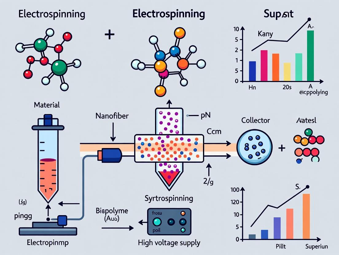
This comprehensive article provides a detailed guide to electrospinning techniques for fabricating biopolymer nanofibers, tailored for researchers, scientists, and drug development professionals.
Advanced Electrospinning of Biopolymer Nanofibers: Techniques, Applications, and Optimization for Biomedical Research
Abstract
This comprehensive article provides a detailed guide to electrospinning techniques for fabricating biopolymer nanofibers, tailored for researchers, scientists, and drug development professionals. It covers foundational principles, explores the spectrum of natural and synthetic biopolymers used (Intent 1). It details advanced electrospinning setups (coaxial, emulsion, melt), processing parameters, and specific biomedical applications in drug delivery, tissue engineering, and wound healing (Intent 2). The guide addresses common challenges like bead formation, clogging, and low productivity, offering targeted optimization strategies (Intent 3). Finally, it discusses critical validation methods for characterizing nanofiber properties and provides a comparative analysis of different biopolymer systems for informed material selection (Intent 4).
Understanding Biopolymer Nanofibers: Principles, Polymers, and Electrospinning Fundamentals
This application note details the fundamental principles of electrospinning, framed within a broader thesis on electrospinning techniques for biopolymer nanofibers research. It serves as a practical guide for researchers, scientists, and drug development professionals aiming to fabricate nanofibrous scaffolds for biomedical applications, including drug delivery and tissue engineering.
Core Principles & Quantitative Parameters
The Taylor Cone Formation
A stable Taylor cone is the foundational requirement for continuous fiber formation. It is achieved when electrostatic forces overcome the surface tension of the polymer solution.
Table 1: Critical Parameters for Taylor Cone Stability
| Parameter | Typical Range for Biopolymers | Effect on Cone Stability |
|---|---|---|
| Applied Voltage | 10-30 kV | Increases electrostatic stretching; too high causes jet instability. |
| Working Distance | 10-20 cm | Affects jet flight time and solvent evaporation. |
| Flow Rate | 0.5-3.0 mL/h | Too high causes droplet formation; too low leads to jet breakage. |
| Solution Conductivity | 0.5-5 mS/cm | Enhances jet whipping; often increased with salts. |
| Surface Tension | 30-50 mN/m | Lower values promote cone/jet initiation. |
Jet Thinning & Instability
The charged jet undergoes a whipping instability (bending instability), leading to extreme radial thinning and solvent evaporation, forming solid nanofibers.
Table 2: Jet Instability Regimes and Outcomes
| Instability Type | Dominant Force | Resulting Fiber Morphology |
|---|---|---|
| Axisymmetric (Rayleigh) | Surface Tension | Beaded fibers. |
| Bending (Whipping) | Electrostatic Repulsion | Uniform, thin fibers. |
Fiber Collection & Alignment
Collection methods determine the final architecture of the nanofiber mat.
Table 3: Common Collection Methods and Fiber Characteristics
| Collection Method | Principle | Typical Fiber Alignment | Porosity |
|---|---|---|---|
| Static Flat Plate | Random deposition | Random | High |
| Rotating Drum (≤ 1500 rpm) | Mechanical winding | Partial alignment | Medium-High |
| Rotating Drum (≥ 3000 rpm) | High-speed winding | Highly Aligned | Medium |
| Parallel Electrodes | Field focusing | Aligned between gaps | High |
Experimental Protocols
Protocol: Electrospinning of Gelatin/PEO Nanofibers
This protocol describes the fabrication of crosslinkable gelatin-based nanofibers, a common biopolymer blend for tissue engineering.
A. Solution Preparation
- Materials: Type A gelatin, Poly(ethylene oxide) (PEO, Mv ~900 kDa), acetic acid (90% v/v), deionized water.
- Dissolve gelatin at 10% (w/v) in 90% acetic acid with magnetic stirring at 40°C for 2 hours.
- Separately, dissolve PEO at 2% (w/v) in deionized water at room temperature for 1 hour.
- Blend the two solutions at a 90:10 (gelatin:PEO) volume ratio. Stir for an additional 1 hour at 40°C.
- Allow the solution to equilibrate at room temperature for 30 minutes before loading into the syringe.
B. Electrospinning Setup & Process
- Setup: Use a standard vertical electrospinning setup. Load the solution into a 5 mL glass syringe fitted with a blunt 21-gauge stainless steel needle.
- Connect the needle to a high-voltage power supply (positive polarity). Place a grounded cylindrical collector (diameter 8 cm) wrapped in aluminum foil at a 15 cm working distance.
- Set the syringe pump to a flow rate of 1.0 mL/h.
- Gradually increase the applied voltage to 18 kV. Observe the formation of a stable Taylor cone.
- Electrospin for a duration necessary to achieve the desired mat thickness (e.g., 4 hours for a ~100 µm mat).
- Post-processing: Carefully detach the fiber mat. For stable scaffolds, crosslink the gelatin fibers by exposing them to glutaraldehyde vapor in a desiccator for 24 hours.
Protocol: Assessing Fiber Morphology (SEM)
- Sample Preparation: Cut a small section (5x5 mm) of the fiber mat and sputter-coat with gold/palladium for 60 seconds.
- Imaging: Using a Scanning Electron Microscope (SEM), image samples at an accelerating voltage of 5-10 kV.
- Analysis: Use image analysis software (e.g., ImageJ) to measure average fiber diameter from at least 100 random fibers across multiple images.
Visualization
Diagram 1: Electrospinning Process Flow
Diagram 2: Key Parameter Effects on Fiber Outcome
The Scientist's Toolkit: Research Reagent Solutions
Table 4: Essential Materials for Biopolymer Electrospinning
| Item | Function & Rationale |
|---|---|
| High-Voltage Power Supply | Generates the electrostatic field (typically 0-30 kV DC) required for Taylor cone formation and jet acceleration. |
| Programmable Syringe Pump | Precisely controls the flow rate of the polymer solution to the spinneret, ensuring a stable Taylor cone. |
| Biopolymers (e.g., Gelatin, Chitosan, Alginate, PCL, PLGA) | The core material to be spun. Often blended to tailor mechanical properties, degradation rate, and bioactivity. |
| Co-solvent Systems (e.g., Acetic Acid, TFE, HFIP, Water/DMSO) | Dissolves biopolymers and adjusts solution properties (viscosity, conductivity, evaporation rate). |
| Conductivity Enhancers (e.g., NaCl, NaH₂PO₄) | Added in small amounts (≤ 1% w/v) to increase solution charge density, promoting jet thinning and uniform fiber formation. |
| Rotating Mandrel Collector | A motorized cylindrical collector used to produce aligned nanofiber mats by providing a tangential take-up velocity. |
| Crosslinking Agents (e.g., Genipin, EDC/NHS, Glutaraldehyde Vapor) | Stabilize hydrophilic biopolymer fibers (like gelatin/collagen) against dissolution in aqueous environments. |
| Humidity/Temperature Chamber | Controlled environment is critical for consistent fiber formation, especially for water-soluble polymers. |
Application Notes
This document provides application notes and experimental protocols for the electrospinning of natural and synthetic biopolymers, framed within a thesis on developing nanofibrous scaffolds for drug delivery and tissue engineering.
Natural Biopolymers offer inherent bioactivity, biocompatibility, and mimicry of the native extracellular matrix (ECM). However, they often present challenges in electrospinning due to variability in molecular weight, batch-to-batch inconsistency, and limited solubility in organic solvents.
Synthetic Biopolymers provide superior mechanical properties, predictable degradation rates, and high reproducibility. Their tunability allows for precise control over scaffold architecture and drug release kinetics, though they may lack specific cellular recognition sites.
Recent advancements focus on blending natural and synthetic polymers or creating coaxial fibers to combine the advantages of both spectra.
Quantitative Comparison of Key Biopolymers
Table 1: Fundamental Properties of Featured Biopolymers for Electrospinning
| Biopolymer | Type | Source/Synthesis | Typical Solvent(s) for ES | Degradation Time | Key Electrospinning Challenges |
|---|---|---|---|---|---|
| Chitosan | Natural (Cationic) | Deacetylation of chitin (crustacean shells) | Aqueous acidic solutions (e.g., Acetic Acid) | Weeks to Months | High viscosity at low conc., need for co-polymer/blending. |
| Collagen | Natural (Protein) | Animal tissues (bovine, porcine, marine) | Hexafluoro-2-propanol (HFIP), Acetic Acid | Weeks | Denaturation risk, costly, solvent toxicity concerns. |
| Alginate | Natural (Anionic) | Brown seaweed | Water (with plasticizer like PEO) | Slow, ion-dependent | Difficult to electrospin alone (no chain entanglement). |
| PLA | Synthetic | Polymerization of lactic acid | Chloroform, DCM, DMF | 12-24 months | Hydrophobicity, acidic degradation products. |
| PCL | Synthetic | Ring-opening polymerization of ε-caprolactone | Chloroform, DCM, Acetone | >24 months | Hydrophobicity, slow degradation, low cell affinity. |
| PLGA | Synthetic | Copolymer of lactic and glycolic acid | DMF, THF, Chloroform | 1-6 months (tunable) | Batch variability in LA:GA ratio, acidic degradation. |
Table 2: Exemplary Electrospinning Parameters & Post-Processing
| Biopolymer | Conc. (wt%) | Voltage (kV) | Flow Rate (mL/h) | Collector Distance (cm) | Common Crosslinker/Post-Treatment |
|---|---|---|---|---|---|
| Chitosan/PEO | 2-4% Chit / 2-4% PEO | 15-25 | 0.5-1.0 | 15-20 | Genipin, Glutaraldehyde vapor |
| Collagen (Type I) | 8-12% in HFIP | 20-30 | 1.0-2.0 | 15-20 | EDC/NHS, UV or Dehydrothermal |
| Alginate/PVA | 2-3% Alg / 8-10% PVA | 15-25 | 0.8-1.2 | 15-20 | CaCl₂ solution (ionic crosslinking) |
| PLA | 8-12% in CHCl₃:DMF | 15-25 | 1.0-2.5 | 15-20 | N/A (thermal annealing optional) |
| PCL | 10-15% in CHCl₃:MeOH | 12-20 | 1.5-3.0 | 15-20 | N/A |
| PLGA | 20-30% in DMF:THF | 15-25 | 1.0-2.0 | 15-20 | N/A |
Experimental Protocols
Protocol: Electrospinning of PLGA Nanofibers for Sustained Drug Release
Aim: To fabricate drug-loaded PLGA nanofibrous mats for controlled release studies. Materials: PLGA (75:25 LA:GA), Dichloromethane (DCM), N,N-Dimethylformamide (DMF), Model Drug (e.g., Rhodamine B or Vancomycin).
Method:
- Solution Preparation: Dissolve PLGA pellets at 25% (w/v) in a solvent mixture of DMF:DCM (3:7 v/v). Stir for 12h at room temperature until homogeneous.
- Drug Loading: Add the model drug to the polymer solution at 5% (w/w relative to polymer). Stir for 4h in the dark.
- Electrospinning Setup: Load solution into a 5mL glass syringe fitted with a 21G blunt needle. Use a programmable syringe pump.
- Parameters: Set flow rate to 1.2 mL/h. Apply a high voltage of 18 kV. Maintain a tip-to-collector distance of 18 cm. Use a flat aluminum foil-covered rotating mandrel (500 rpm).
- Environmental Control: Conduct spinning at 23±2°C and 45±5% relative humidity.
- Collection: Spin for 4-6 hours to obtain a mat of ~100 µm thickness. Dry mats in vacuo for 24h to remove residual solvent.
Protocol: Coaxial Electrospinning of Core-Shell Alginate-PCL Fibers
Aim: To create fibers with an alginate-rich core (for bioactivity) and a PCL shell (for mechanical integrity). Materials: Alginate, PCL, Poly(ethylene oxide) (PEO), Calcium Chloride (CaCl₂), solvents as per Table 2, coaxial spinneret.
Method:
- Core Solution: Prepare 3% (w/v) sodium alginate and 5% (w/v) PEO blend in deionized water. Stir for 24h.
- Shell Solution: Prepare 12% (w/v) PCL in a 7:3 mixture of chloroform and methanol. Stir for 6h.
- Coaxial Setup: Load core and shell solutions into separate syringes connected to the inner and outer channels of a coaxial spinneret, respectively.
- Parameters: Set core flow rate to 0.4 mL/h and shell flow rate to 1.2 mL/h. Apply voltage of 20 kV. Collector distance: 20 cm.
- Crosslinking: Collect fibers on a mandrel immersed in a 2% (w/v) CaCl₂ ethanol/water (50/50) bath for in-situ ionic crosslinking of alginate core.
- Post-Processing: Wash mats with DI water and dry under vacuum.
Visualizations
Workflow for Electrospun Nanofiber Development
PLGA Degradation & Drug Release Pathway
The Scientist's Toolkit: Key Research Reagent Solutions
Table 3: Essential Materials for Biopolymer Electrospinning Research
| Item | Function & Relevance | Example Product/Supplier Note |
|---|---|---|
| Hexafluoro-2-propanol (HFIP) | Solvent for challenging biopolymers like collagen & elastin. Highly volatile and toxic. | Sigma-Aldrich, 105228. Use in fume hood with appropriate PPE. |
| Genipin | Natural, low-toxicity crosslinker for chitosan, gelatin, and collagen. Provides blue fluorescence. | Wako Chemical, 078-03021. Preferred over glutaraldehyde for cytocompatibility. |
| EDC & NHS | Carbodiimide crosslinking system for zero-length crosslinking of carboxyl and amine groups in proteins. | Thermo Scientific, 22980 & 24510. Used in MES buffer for collagen crosslinking. |
| PEO (Polyethylene Oxide) | Electrospinning enhancer; added to natural polymer solutions (e.g., alginate) to increase viscoelasticity. | Sigma-Aldrich, 372781 (Mw 900K). Used at low % as a process aid. |
| PLGA (Various Ratios) | Tunable synthetic copolymer. 50:50 degrades fastest, 85:15 slower. Key for release kinetics studies. | Lactel Absorbable Polymers (DURECT Corp). Specify inherent viscosity. |
| Coaxial Spinneret | Nozzle for fabricating core-shell fibers, allowing encapsulation of sensitive biomolecules in the core. | From precision needle suppliers (e.g., Ingenuity). Inner/outer diameter critical. |
| Programmable Syringe Pump | Ensures precise, steady flow of polymer solution for reproducible fiber morphology. | Cole-Parmer, KD Scientific. Multi-channel models for coaxial. |
| Humidity/Temp Controller | Critical for reproducible electrospinning, especially for hydrophilic polymers sensitive to humidity. | Custom or chamber-equipped commercial systems (e.g., IME Technologies). |
Within a thesis focused on advancing electrospinning techniques for biopolymer nanofibers in biomedical applications, the fundamental triad of the syringe pump, high-voltage supply, and collector constitutes the core of any experimental setup. The precise, independent control of these components directly dictates the morphology, diameter, alignment, and functionality of the resultant nanofibers, which are critical for drug delivery systems, tissue engineering scaffolds, and wound dressings.
Component Specifications & Quantitative Data
The selection of components is based on parameters critical for reproducible biopolymer nanofiber production (e.g., from Polycaprolactone (PCL), Poly(lactic-co-glycolic acid) (PLGA), Chitosan, Alginate).
Table 1: Key Specifications for Electrospinning Components
| Component | Critical Parameter | Typical Range for Biopolymers | Influence on Fiber Morphology |
|---|---|---|---|
| Syringe Pump | Flow Rate | 0.1 - 10 mL/h (Common: 0.5 - 3 mL/h) | Controls fiber diameter, prevents bead formation. Too high causes droplets; too low causes jet instability. |
| High Voltage Supply | Voltage | 5 - 30 kV (Typical: 10 - 20 kV) | Initiates jet formation. Affects jet acceleration, fiber diameter, and deposition stability. |
| Collector | Type & Configuration | Flat Plate (Random), Rotating Drum (Aligned), Gap/Disc (Aligned) | Determines fiber alignment, mat thickness, and pore architecture. Rotational speed (100 - 8000 rpm) controls alignment degree. |
| Collector Distance | Tip-to-Collector Distance (TCD) | 10 - 25 cm (Common: 15 cm) | Allows solvent evaporation. Shorter TCD can yield wet, fused fibers; longer can cause jet instability. |
Table 2: Optimized Parameters for Common Biopolymers
| Biopolymer | Solvent System | Typical Concentration | Suggested Flow Rate (mL/h) | Suggested Voltage (kV) | TCD (cm) |
|---|---|---|---|---|---|
| PCL | Chloroform:DMF (e.g., 70:30) | 8-15% w/v | 1.0 - 2.5 | 12 - 18 | 15 - 20 |
| PLGA | DMF or Chloroform:DMF | 10-20% w/v | 1.0 - 2.0 | 15 - 20 | 15 - 18 |
| Chitosan | Aqueous Acetic Acid (1-2% v/v) | 3-7% w/v (with high MW) | 0.2 - 0.8 | 15 - 25 | 10 - 15 |
| Alginate | Water (with a co-polymer like PEO) | 2-4% w/v Alginate | 0.5 - 1.5 | 10 - 20 | 12 - 18 |
Application Notes & Protocols
Protocol 1: Standard Setup Assembly & Safety Check
Objective: To safely assemble and verify the core electrospinning system for biopolymer solutions. Materials: Syringe pump, blunt-gauge needle (e.g., 18-22G), high-voltage power supply, grounded collector (flat plate or drum), syringe, polymer solution, safety enclosure, grounding cable. Procedure:
- Place the syringe pump outside the safety enclosure. Load the syringe with the prepared biopolymer solution and attach the blunt needle. Secure it on the pump.
- Position the grounded collector (e.g., aluminum foil on flat plate) inside the enclosure at the predetermined Tip-to-Collector Distance (TCD).
- Connect the high-voltage supply's positive (anode) lead to the metal needle tip using an alligator clip. Ensure the connection is secure and not touching other surfaces.
- Connect the collector to the electrical ground (cathode) of the power supply or a separate ground point using a heavy-duty cable.
- Safety Check: Before applying voltage, confirm all connections are tight, the enclosure door is closed, and no personnel are in contact with the setup. Wear appropriate PPE.
- Initialize the syringe pump at the desired flow rate. Allow the solution to form a pendant drop at the needle tip.
- Gradually increase the high voltage to the target kV. Observe the Taylor cone formation and the initiation of a stable, whipping jet.
Protocol 2: Parameter Optimization for Fiber Diameter Minimization
Objective: To systematically reduce the average diameter of PCL nanofibers by modulating core component parameters. Experimental Design:
- Fixed Parameters: PCL 12% w/v in Chloroform:DMF (70:30), TCD = 18 cm, stationary flat collector.
- Variable Parameters: Create a matrix of Flow Rate (0.8, 1.2, 1.6 mL/h) and Applied Voltage (14, 16, 18 kV).
- Execution: Perform 9 experiments (all combinations). For each run, electrospin for 10 minutes to collect a sufficient sample.
- Analysis: Image each sample via Scanning Electron Microscopy (SEM). Measure fiber diameters (n≥100) using image analysis software (e.g., ImageJ).
- Outcome: Plot a 3D response surface of average fiber diameter vs. flow rate and voltage. Identify the combination yielding the smallest, most consistent fibers.
Experimental Workflow Visualization
Title: Electrospinning Experiment Optimization Workflow
The Scientist's Toolkit: Research Reagent Solutions
Table 3: Essential Materials for Biopolymer Electrospinning
| Material/Reagent | Function & Application Notes |
|---|---|
| Polycaprolactone (PCL), MW 70k-80k | A biodegradable, synthetic biopolymer. Provides structural integrity for scaffolds. Often dissolved in organic solvents like chloroform/DMF. |
| PLGA (50:50 to 85:15 LA:GA ratio) | Copolymer with tunable degradation rates. Ideal for sustained drug release studies. Solubility varies with ratio. |
| Chitosan (High Molecular Weight, >75% deacetylated) | Natural cationic polysaccharide. Promotes cell adhesion and has inherent antimicrobial properties. Requires acidic aqueous solvents. |
| Hexafluoroisopropanol (HFIP) | A highly volatile, fluorinated organic solvent. Excellent for dissolving challenging biopolymers like collagen and silk fibroin. Requires strict fume hood use. |
| Poly(ethylene oxide) (PEO), MW 900k-1M | Used as a carrier polymer to facilitate electrospinning of difficult-to-spin natural polymers (e.g., alginate, chitosan) by enhancing solution viscoelasticity. |
| Phosphate Buffered Saline (PBS) | Used for post-processing (crosslinking rinsing, hydration) and as a medium for drug release studies from collected fibers. |
| Glutaraldehyde (2% v/v aqueous) or EDC/NHS | Common crosslinking agents for alginate or collagen fibers to improve their mechanical stability and water resistance. |
| Methylene Blue or Rhodamine B | Model hydrophilic/hydrophobic drug molecules used in proof-of-concept drug loading and release kinetic experiments from nanofibers. |
Process Dynamics Visualization
Title: Component Role in Fiber Deposition & Alignment
Why Biopolymers? Biocompatibility, Biodegradability, and Mimicking the ECM.
Within the context of a thesis on electrospinning for biopolymer nanofiber research, the rationale for selecting biopolymers is fundamental. They offer unparalleled advantages for biomedical applications, including tissue engineering scaffolds, wound dressings, and drug delivery systems, primarily due to their inherent biocompatibility, tunable biodegradability, and unique ability to mimic the native extracellular matrix (ECM). This document provides detailed application notes and experimental protocols for leveraging these properties.
Application Notes: Core Properties and Comparative Data
Electrospun biopolymer nanofibers create a high-surface-area, three-dimensional porous network that closely resembles the fibrous architecture of collagen and other ECM components. The following tables summarize key quantitative data.
Table 1: Biodegradation Rates of Common Electrospun Biopolymers
| Biopolymer | Source | Degradation Time In Vivo (Weeks) | Primary Degradation Mechanism | Key Influencing Factors |
|---|---|---|---|---|
| Collagen (Type I) | Animal | 2 - 8 | Enzymatic (collagenases) | Crosslinking density, fibril alignment |
| Gelatin | Denatured Collagen | 1 - 4 | Enzymatic (proteases) | Bloom strength, degree of crosslinking |
| Chitosan | Crustacean shells | 4 - 12 | Enzymatic (lysozyme) | Degree of deacetylation, crystallinity |
| Poly(lactic-co-glycolic acid) (PLGA) | Synthetic (from lactic/glycolic acids) | 1 - 50 (tunable) | Hydrolysis | LA:GA ratio, molecular weight, crystallinity |
| Silk Fibroin | Bombyx mori silkworm | 20 - 100+ | Proteolytic | Crystalline (beta-sheet) content, porosity |
| Hyaluronic Acid | Microbial/Fermentation | 1 - 6 | Enzymatic (hyaluronidases) | Molecular weight, crosslinking method |
Table 2: Mechanical Properties of Electrospun Biopolymer Mats
| Biopolymer | Typical Tensile Strength (MPa) | Typical Young's Modulus (MPa) | Elongation at Break (%) | Notes on Electrospinning |
|---|---|---|---|---|
| Collagen | 2 - 15 | 50 - 200 | 10 - 30 | Requires crosslinking (e.g., glutaraldehyde vapor) for stability. |
| Chitosan | 20 - 60 | 500 - 2000 | 5 - 15 | Often spun with PEO or acetic acid solutions. Properties vary with DDA. |
| PLGA (85:15) | 2 - 8 | 100 - 400 | 100 - 300 | Highly tunable; 85:15 ratio common for moderate degradation. |
| Silk Fibroin | 5 - 50 | 500 - 2000 | 2 - 10 | Post-treatment with methanol induces beta-sheets, increasing strength. |
| Gelatin | 5 - 20 | 100 - 500 | 2 - 10 | Similar to collagen; requires crosslinking for aqueous stability. |
| Alginate | 10 - 40 | 200 - 800 | 3 - 8 | Difficult to electrospin alone; often blended with PVA. |
Table 3: Biocompatibility Assessment (Cell Viability % - MTT Assay, Day 7)
| Biopolymer Scaffold | NIH/3T3 Fibroblasts | Human Dermal Fibroblasts (HDF) | Mesenchymal Stem Cells (hMSCs) | Key Observation |
|---|---|---|---|---|
| PLGA (85:15) | 95 ± 5% | 92 ± 7% | 90 ± 8% | Slight acidification from degradation products can affect cells. |
| Chitosan/PEO | 105 ± 6% | 110 ± 5% | 98 ± 6% | Chitosan shows inherent antibacterial and cell-promoting properties. |
| Collagen Type I | 115 ± 8% | 120 ± 10% | 112 ± 9% | Excellent cell adhesion and proliferation due to RGD motifs. |
| Silk Fibroin | 102 ± 4% | 105 ± 6% | 108 ± 7% | Excellent biocompatibility post-sericin removal. |
| Gelatin | 110 ± 7% | 115 ± 8% | 105 ± 8% | Performance similar to collagen, with easier processability. |
Experimental Protocols
Protocol 1: Electrospinning of Crosslinkable Biopolymer (Gelatin) Nanofibers
Objective: To fabricate stable, aqueous-resistant gelatin nanofiber mats for ECM-mimetic scaffolds. Materials: See "The Scientist's Toolkit" below. Methodology:
- Solution Preparation: Dissolve gelatin (Type A, 300 Bloom) at 10% (w/v) in a mixture of 70% acetic acid and 30% deionized water. Stir at 40°C for 4 hours until a clear, homogeneous solution is obtained.
- Electrospinning Parameters:
- Syringe Needle Gauge: 21G blunt tip.
- Flow Rate: 0.8 mL/h (using a syringe pump).
- Applied Voltage: +15 kV (needle) to -2 kV (collector).
- Tip-to-Collector Distance: 15 cm.
- Collector: Aluminum foil-covered rotating mandrel (1000 rpm).
- Ambient Conditions: 25°C, 40% relative humidity (controlled).
- Post-Processing - Crosslinking: a. Vapor-Phase Crosslinking: Immediately transfer the as-spun mat into a sealed desiccator containing 5 mL of 25% glutaraldehyde (GTA) aqueous solution. Place the mat on a raised platform above the liquid. Crosslink for 24 hours at room temperature. b. Quenching & Drying: Remove the mat and place it in a fume hood for 2 hours to evaporate residual GTA. Then, transfer it to a vacuum chamber containing a beaker of glycine powder (to quench unreacted aldehyde groups) for 48 hours. c. Final Wash: Rinse the mat gently with DI water and phosphate-buffered saline (PBS) to remove any residuals. Dry under vacuum for 24 hours.
- Characterization: Perform SEM for fiber morphology, FTIR for confirming crosslink formation (Schiff base peak ~1640 cm⁻¹), and swelling tests in PBS.
Protocol 2: Assessing BiodegradationIn Vitro(Lysozyme-Mediated)
Objective: To quantify the enzymatic degradation profile of a chitosan-based nanofibrous scaffold. Materials: Electrospun chitosan/PEO mat, lysozyme from chicken egg white, PBS (pH 7.4), sodium azide. Methodology:
- Sample Preparation: Pre-weigh (W₀) dry scaffolds (n=5, 10 mm diameter). Sterilize under UV light for 30 minutes per side.
- Degradation Medium: Prepare 1.0 mg/mL lysozyme solution in PBS containing 0.02% (w/v) sodium azide as an antimicrobial agent.
- Incubation: Immerse each sample in 5 mL of degradation medium in a sealed vial. Maintain at 37°C with gentle shaking (50 rpm). Control groups use PBS without enzyme.
- Monitoring: At predetermined time points (e.g., days 1, 3, 7, 14, 21), remove samples, rinse thoroughly with DI water, freeze-dry for 48 hours, and re-weigh (Wₜ).
- Data Analysis: Calculate mass remaining percentage: % Mass Remaining = (Wₜ / W₀) * 100. Plot degradation kinetics. Use SEM to observe surface erosion and fiber breakdown.
Diagrams
Title: Biopolymer Rationale for Electrospinning Research
Title: Electrospun Biopolymer Nanofiber Workflow
The Scientist's Toolkit: Key Research Reagent Solutions
| Item | Function/Justification | Example Supplier/Product Code (Illustrative) |
|---|---|---|
| Gelatin, Type A (300 Bloom) | A denatured collagen derivative; forms electrospun fibers well but requires crosslinking for stability. Provides RGD sequences for cell adhesion. | Sigma-Aldrich, G2500 |
| Chitosan (Medium MW, >75% DDA) | Cationic polysaccharide with inherent antimicrobial and hemostatic properties. Often blended with PEO for spinnability. | Sigma-Aldrich, 448877 |
| PLGA (85:15 LA:GA) | Synthetic, FDA-approved copolymer with tunable degradation rate (weeks to months). A benchmark for controlled release studies. | Lactel Absorbable Polymers, DURECT Corporation |
| Silk Fibroin Aqueous Solution | Recombinant or B. mori-derived; offers excellent mechanical properties and biocompatibility. Requires careful degumming and dialysis. | Advanced BioMatrix, 5058-SF |
| Crosslinker: Glutaraldehyde (25% soln.) | A potent vapor-phase or solution crosslinker for proteins (gelatin, collagen). Forms Schiff base linkages. Must be handled with care. | Sigma-Aldrich, G6257 |
| Crosslinker: EDC/NHS | Zero-length carbodiimide crosslinker for carboxylic acid and amine groups (e.g., in collagen, HA). Minimizes reagent incorporation into scaffold. | Thermo Scientific, Pierce EDC Sulfo-NHS Kit |
| Lysozyme (from chicken egg white) | Enzyme used for in vitro degradation studies of chitosan and other glycosaminoglycan-like polymers. | Sigma-Aldrich, L6876 |
| MTT Cell Viability Assay Kit | Colorimetric assay to measure mitochondrial activity as a proxy for cell proliferation and viability on scaffolds. | Abcam, ab211091 |
| Hexafluoro-2-propanol (HFIP) | A highly volatile fluorinated alcohol solvent used to dissolve difficult biopolymers like collagen and silk for electrospinning. | Sigma-Aldrich, 105228 |
| Phosphate Buffered Saline (PBS), pH 7.4 | Isotonic buffer used for rinsing scaffolds, preparing degradation media, and as a base for cell culture reagents. | Gibco, 10010023 |
This document serves as a series of application notes and protocols, framed within a broader thesis investigating electrospinning techniques for biopolymer nanofibers. The core functional properties of electrospun mats—high surface area, interconnected porosity, and tunable morphology—are directly responsible for their utility in advanced applications, particularly in biomedicine and drug delivery. Optimizing these properties through precise control of process parameters is a central thesis objective.
Quantitative Data on Key Properties
The following tables summarize quantitative relationships between electrospinning parameters and the resulting nanofiber properties, as established in recent literature.
Table 1: Impact of Process Parameters on Nanofiber Morphology & Diameter
| Biopolymer System | Concentration (wt%) | Applied Voltage (kV) | Flow Rate (mL/h) | Tip-to-Collector Distance (cm) | Avg. Fiber Diameter (nm) | Morphology Observed | Reference Year |
|---|---|---|---|---|---|---|---|
| Polycaprolactone (PCL) | 10 | 15 | 1.0 | 15 | 250 ± 50 | Bead-free, smooth | 2023 |
| PCL | 8 | 15 | 1.0 | 15 | 180 ± 80 | Beads-on-string | 2023 |
| Chitosan/PEO | 3/0.5 | 20 | 0.3 | 20 | 120 ± 30 | Uniform, thin | 2024 |
| Gelatin | 12 | 18 | 0.8 | 12 | 350 ± 100 | Ribbon-like | 2023 |
| PLGA | 15 | 12 | 1.5 | 18 | 700 ± 150 | Bead-free, thick | 2024 |
Table 2: Measured Surface Area and Porosity of Electrospun Mats
| Material | Avg. Fiber Diameter (nm) | Specific Surface Area (m²/g) (BET) | Porosity (%) (Mercury Intrusion) | Pore Size Range (µm) | Primary Application Tested |
|---|---|---|---|---|---|
| PCL nanofibers | 250 | 25.5 ± 2.1 | 85 ± 3 | 0.5-15 | Tissue scaffolding |
| Chitosan-based blend | 120 | 42.3 ± 3.5 | 92 ± 2 | 0.2-8 | Wound dressing |
| PLGA nanofibers | 700 | 12.8 ± 1.7 | 78 ± 4 | 1-25 | Drug delivery carrier |
| Silk Fibroin | 450 | 18.9 ± 2.0 | 80 ± 5 | 0.8-20 | Cell culture |
Experimental Protocols
Protocol 3.1: Standard Electrospinning of PCL Nanofibers for High Surface Area Mats
Objective: To produce bead-free PCL nanofibers with high surface area for drug loading studies. Materials: See "The Scientist's Toolkit" (Section 5). Procedure:
- Solution Preparation: Dissolve PCL pellets in a 7:3 (v/v) mixture of Dichloromethane (DCM) and Dimethylformamide (DMF) to achieve a 10% (w/v) solution. Stir at 40°C for 6 hours until fully dissolved.
- Setup Configuration: Load the solution into a 10 mL glass syringe fitted with a 21-gauge blunt-tip stainless steel needle. Place syringe on pump. Connect needle to high-voltage power supply. Use a flat aluminum foil-covered collector.
- Parameter Setting: Set flow rate to 1.0 mL/h. Set applied voltage to +15 kV. Set tip-to-collector distance to 15 cm. Maintain environmental conditions at 23±2°C and 40±5% RH.
- Electrospinning: Initiate pump and then power supply. Allow a 5-minute stabilization period. Spin for 4 hours to achieve a mat thickness of ~100 µm.
- Collection & Drying: Carefully peel the nanofiber mat from the collector. Place in a vacuum desiccator for 24 hours to remove residual solvents.
Protocol 3.2: Assessing Porosity via Liquid Displacement
Objective: To quantitatively determine the porosity of an electrospun mat. Procedure:
- Cut a precise, known area (e.g., 2 cm x 2 cm) from the electrospun mat and record its dry mass (M).
- Immerse the sample in a low-surface-tension liquid (e.g., ethanol) in a graduated cylinder for 5 hours, ensuring full infiltration.
- Measure the volume of the liquid displaced (V_total), which corresponds to the total volume of the sample (fibers + pores).
- Remove the sample, lightly blot to remove surface liquid, and immediately weigh to obtain the wet mass (M_wet).
- Calculate the volume of the fiber material itself using the polymer density (ρ): V_fiber = M / ρ.
- Calculate porosity (ε): ε (%) = [(Vtotal - Vfiber) / V_total] * 100.
Protocol 3.3: Creating Core-Shell Morphologies for Tunable Drug Release
Objective: To co-electrospin nanofibers with a core-shell structure for biphasic drug release profiles. Materials: Coaxial spinneret, two independent syringe pumps, core and shell polymer solutions (e.g., Protein (core) / PCL (shell)). Procedure:
- Solution Prep: Prepare core (aqueous protein/drug solution) and shell (PCL in organic solvent) solutions separately. Filter.
- Spinneret Assembly: Assemble the coaxial spinneret, connecting inner (core) and outer (shell) capillaries to their respective syringes.
- Parameter Optimization: Set independent flow rates (e.g., Core: 0.2 mL/h, Shell: 0.8 mL/h). Set high voltage to 18 kV and distance to 20 cm.
- Spinning: Start both pumps simultaneously before applying voltage. Collect fibers on a rotating mandrel.
- Characterization: Confirm core-shell structure using TEM and analyze drug release kinetics via HPLC.
Diagrams for Workflows and Relationships
Diagram 1: Parameter-Morphology Relationship in Electrospinning
Diagram 2: Drug Release Workflow from Porous Nanofibers
The Scientist's Toolkit: Essential Research Reagents & Materials
Table 3: Key Reagent Solutions and Materials for Electrospinning Biopolymers
| Item | Function/Benefit | Example in Protocol |
|---|---|---|
| Polycaprolactone (PCL) | Synthetic, biodegradable polyester; offers mechanical strength and controllable degradation rate. | Primary fiber polymer in Protocol 3.1. |
| Chitosan (Medium MW) | Natural cationic biopolymer; provides biocompatibility, antimicrobial activity, and enhances cell adhesion. | Used in blend systems (Table 1). |
| Coaxial Spinneret | Specialized needle allowing simultaneous extrusion of two solutions to form core-shell fibers. | Essential for Protocol 3.3 (tunable morphology). |
| Mixed Solvent System (DCM:DMF) | DCM rapidly evaporates, DMF controls evaporation rate and improves solution conductivity, reducing bead formation. | Solvent for PCL in Protocol 3.1. |
| Phosphate Buffered Saline (PBS), pH 7.4 | Standard release medium for in vitro drug release studies; simulates physiological conditions. | Used in drug release assays. |
| MTS/PMS Cell Viability Assay | Colorimetric assay to quantify metabolic activity of cells seeded on nanofiber mats (cytocompatibility test). | Post-fabrication biological validation. |
| Rotating Drum Collector | Creates aligned nanofiber mats; rotational speed controls degree of alignment and mat anisotropy. | Alternative collector for specific morphologies. |
Advanced Electrospinning Setups and Biomedical Applications in Drug Delivery & Tissue Engineering
Within the broader thesis on advancing electrospinning for biopolymer nanofiber research, the development of complex core-shell and functional fiber architectures is paramount. Conventional single-needle electrospinning produces solid fibers, limiting applications in controlled drug delivery, tissue engineering, and encapsulation of sensitive biomolecules. This article details three advanced techniques—coaxial, emulsion, and melt electrospinning—that enable the fabrication of fibers with tailored compositions, morphologies, and release profiles, directly addressing the need for sophisticated biomaterial carriers in therapeutic development.
Table 1: Comparative Analysis of Advanced Electrospinning Techniques
| Parameter | Coaxial Electrospinning | Emulsion Electrospinning | Melt Electrospinning |
|---|---|---|---|
| Primary Fiber Structure | Distinct core-shell | Matrix with dispersed droplets (can form core-shell) | Solid, typically monolithic |
| Typical Solvent System | Two immiscible solutions (core & shell) | Oil-in-water (O/W) or water-in-oil (W/O) emulsion | No solvent required |
| Core Material Compatibility | Hydrophilic/Hydrophobic solutions, pre-formed polymers, drugs | Hydrophobic drugs in O/W; Hydrophilic in W/O | Thermoplastics (PLA, PCL, PEG), no biomolecules post-process |
| Typical Fiber Diameter Range | 100 nm – 5 µm | 200 nm – 3 µm | 5 µm – 100 µm |
| Key Advantage | Precise core-shell control, high encapsulation efficiency | Simpler setup, good for hydrophobic drug encapsulation | Solvent-free, high productivity, safe for in vivo use |
| Key Limitation | Complex setup, requires immiscible solutions & careful flow rate control | Less structural control, potential for burst release | High temperature limits bioactive agents, thicker fibers |
| Encapsulation Efficiency (Typical Range) | 70% – 95% | 60% – 85% | N/A (direct blending pre-melt) |
| Best Suited For | Delicate biologics (proteins, DNA), sequential release systems | Hydrophobic chemotherapeutics, essential oils | Medical implants, scaffolds requiring high mechanical strength |
Table 2: Representative Processing Parameters for Biopolymers
| Technique | Biopolymer Example (Shell) | Core/Active Agent | Key Optimized Parameters | Outcome |
|---|---|---|---|---|
| Coaxial | PCL (10% w/v in CHCl₃:DMF) | BSA (5% w/v in aqueous buffer) | Qcore=0.2 mL/h, Qshell=1.0 mL/h, Voltage=15 kV | Smooth fibers, ~800 nm diam., ~90% BSA activity retained |
| Emulsion (O/W) | PVA (8% w/v in water) | Curcumin (2% w/v in chloroform) | Oil:Water=1:4, Surfactant=2% Span 80, Voltage=12 kV | Bead-free fibers, ~400 nm diam., sustained release over 120h |
| Melt | PCL (MW 80,000) | Tetracycline HCl (5% w/w blended) | Temperature=85°C, Q=0.8 mL/h, Voltage=30 kV, D=8 cm | Fibers ~18 µm diam., zero-order antibiotic release for 21 days |
Detailed Experimental Protocols
Protocol 3.1: Coaxial Electrospinning for Protein-Loaded Core-Shell Fibers
Aim: To fabricate poly(ε-caprolactone) (PCL) shell fibers with a bovine serum albumin (BSA)-loaded aqueous core.
- Materials:
- Shell Solution: Dissolve PCL (10% w/v) in a 3:1 mixture of chloroform and N,N-dimethylformamide (DMF). Stir for 6h.
- Core Solution: Dissolve BSA (5% w/v) in deionized water with 0.1% (v/v) Triton X-100 to reduce surface tension. Filter (0.45 µm).
- Equipment: Coaxial spinneret (inner needle: 21G, outer: 14G), dual syringe pumps, high-voltage power supply, grounded collector (aluminum foil), humidity control (<40%).
- Method:
- Load core and shell solutions into separate syringes fitted to the coaxial spinneret.
- Mount syringes on pumps. Set core flow rate (Qcore) to 0.2 mL/h and shell flow rate (Qshell) to 1.0 mL/h.
- Position spinneret 15 cm from the grounded collector.
- Apply a positive voltage of 15 kV to the spinneret.
- Initiate pumps simultaneously. Collect fibers for 4-6 hours.
- Dry collected mat under vacuum for 24h to remove residual solvent.
- Characterization: Use TEM to confirm core-shell morphology. Perform ELISA or similar to assay protein activity post-encapsulation.
Protocol 3.2: Emulsion Electrospinning for Hydrophobic Drug Delivery
Aim: To encapsulate curcumin into polyvinyl alcohol (PVA) fibers via oil-in-water (O/W) emulsion electrospinning.
- Materials:
- Oil Phase: Dissolve curcumin (2% w/v) in chloroform.
- Aqueous Phase: Dissolve PVA (8% w/v) in deionized water at 80°C with stirring for 4h.
- Surfactant: Span 80.
- Method:
- Slowly add the oil phase (5 mL) to the aqueous phase (20 mL) containing 2% (w/v, relative to water) Span 80.
- Emulsify using a high-speed homogenizer at 12,000 rpm for 5 minutes. The emulsion should appear milky white.
- Transfer emulsion to a syringe with a 21G blunt needle.
- Set flow rate to 0.5 mL/h, distance to collector (D) to 12 cm, and voltage to 12 kV.
- Electrospin at ambient conditions (25°C, ~35% RH). Collect fibers.
- Vacuum-dry to remove water and solvent.
- Characterization: Use fluorescence microscopy (curcumin auto-fluoresces) to observe dispersion. Conduct in vitro release study in PBS with Tween 80.
Protocol 3.3: Melt Electrospinning for Solvent-Free Scaffolds
Aim: To produce antibiotic-loaded polycaprolactone (PCL) fibers without organic solvents.
- Materials: Medical-grade PCL pellets, Tetracycline hydrochloride powder.
- Equipment: Melt electrospinning setup with temperature-controlled syringe, heated chamber.
- Method:
- Physically blend PCL pellets with 5% (w/w) Tetracycline HCl powder.
- Load blend into the heated syringe barrel. Set temperature to 85°C (above PCL melt ~60°C).
- Allow 30 minutes for homogeneous melting and degassing.
- Set syringe pump flow rate to 0.8 mL/h.
- Apply a high voltage of 30 kV between the needle (18G) and the collector (8 cm away).
- The drawn melt jet will solidify in air. Collect on a rotating mandrel.
- Note: The process is slower than solution electrospinning. Maintain a stable, low-humidity environment to prevent jet instability.
Diagrams and Workflows
Title: Coaxial Electrospinning Experimental Workflow
Title: Decision Tree for Advanced Electrospinning Technique Selection
The Scientist's Toolkit: Key Research Reagent Solutions
Table 3: Essential Materials for Complex Fiber Electrospinning
| Item | Function & Rationale | Example(s) |
|---|---|---|
| Coaxial Spinneret | Concentric needles enabling simultaneous ejection of core/shell fluids. Critical for true core-shell fiber formation. | Stainless steel, custom gauge pairs (e.g., 22G inner, 16G outer). |
| Precision Dual-Syringe Pump | Independently controls flow rates of core and shell solutions. Stability is key to maintaining a continuous compound jet. | KD Scientific, Chemyx Fusion series. |
| Biocompatible Polymers (Shell) | Form the primary fiber matrix. Must be electrospinnable and appropriate for the application (degradable, non-toxic). | PCL, PLGA, PVA, Chitosan derivatives, Gelatin. |
| Surfactants / Emulsifiers | Stabilize emulsions for emulsion electrospinning, reducing interfacial tension between immiscible phases. | Span 80 (for O/W), Tween 80, PVA, phospholipids. |
| High-Boiling Point Solvent (for Coaxial) | Serves as shell solvent. Slow evaporation prevents premature core solidification and clogging. | DMF, DMSO, Formic Acid. |
| Thermal Stabilizers | Protect bioactive agents (e.g., proteins) during mild thermal processing or emulsification. | Trehalose, Sucrose, BSA itself. |
| Heated Syringe & Chamber (Melt) | Maintains polymer in molten state during ejection and initial jet travel. | Temperature-controlled metal syringe block, environmental chamber. |
| Humidity/Temp Control System | Ambient conditions drastically affect solvent evaporation rate and jet stability, especially for aqueous systems. | Glove box, standalone humidifier/dehumidifier, AC. |
This document provides detailed application notes and protocols for the critical processing parameters in electrospinning, framed within a broader thesis on advanced electrospinning techniques for biopolymer nanofibers in biomedical research. The reproducible fabrication of nanofibers with tailored morphology, diameter, and drug-release kinetics is paramount for applications in tissue engineering, wound healing, and controlled drug delivery. Precise command over the electrospinning triad—voltage, flow rate, and distance—coupled with rigorous environmental control, forms the cornerstone of consistent and translatable research.
The Electrospinning Parameter Quadrant: Core Principles
The electrospinning process is governed by the interplay of four parameter categories: Solution Properties, Controlled Variables, Ambient Conditions, and Collector Design. This note focuses on the three key controlled variables and ambient conditions.
- Applied Voltage: Governs the electric field strength, initiating Taylor cone formation and jet acceleration. Higher voltages typically produce smaller fibers but can lead to bead formation or jet instability if excessive.
- Flow Rate: Determines the volume of solution supplied to the Taylor cone. It directly influences jet stability, fiber diameter, and drying time. Lower rates allow for better solvent evaporation, favoring smoother, thinner fibers.
- Tip-to-Collector Distance (TCD): Affects the jet flight time, allowing for solvent evaporation and fiber stretching. An optimal distance is required for fibers to dry before deposition.
- Environmental Control: Temperature and humidity critically influence solvent evaporation kinetics, solution viscosity, and fiber morphology. Uncontrolled humidity can cause pores, beads, or incomplete drying.
The following tables summarize the typical effects and optimal ranges for key parameters when electrospinning common biopolymers like Polycaprolactone (PCL), Poly(lactic-co-glycolic acid) (PLGA), and Alginate/PEO blends.
Table 1: Core Processing Parameters and Their Effects on PCL Nanofibers
| Parameter | Typical Range (PCL) | Primary Effect on Fiber Morphology | Notes for Drug Delivery Applications |
|---|---|---|---|
| Voltage (kV) | 10 - 20 kV | Diameter ↓ with increase; Beading ↑ if too high/too low | High voltage may degrade sensitive biologics (e.g., proteins). |
| Flow Rate (mL/h) | 0.5 - 2.0 mL/h | Diameter ↑ with increase; Beading ↑ if too high | Low flow rate essential for uniform encapsulation efficiency. |
| Distance (cm) | 10 - 20 cm | Incomplete drying if too short; Jet instability if too long | Optimize for complete solvent evaporation (e.g., Chloroform/DMF). |
| Humidity (%) | 30 - 50% | Porous fibers ↑ with humidity; Beading ↑ at high humidity | Critical for reproducible porosity for cell infiltration. |
| Temperature (°C) | 22 - 25°C | Diameter ↓ with increase due to lower viscosity | Stable temperature prevents solution property drift during long runs. |
Table 2: Optimized Protocol Snapshot for Common Biopolymers
| Biopolymer System | Typical Solvent | Voltage (kV) | Flow Rate (mL/h) | TCD (cm) | Target Diameter (nm) | Key Consideration |
|---|---|---|---|---|---|---|
| PCL (10% w/v) | CHCl₃:DMF (7:3) | 15 | 1.0 | 15 | 200 - 400 | Control humidity for consistency. |
| PLGA (12% w/v) | DMF:THF (1:1) | 18 | 1.2 | 18 | 300 - 600 | Fast evaporation requires stable, moderate humidity. |
| Alginate/PEO (3:2%) | Water | 20 | 0.8 | 12 | 100 - 200 | Requires precise humidity control (>40%) to prevent premature drying. |
| Chitosan/PEO (2%) | Aqueous Acetic Acid (2%) | 22 | 0.5 | 15 | 80 - 150 | Low flow rate mandatory for stable jet from viscous solution. |
Detailed Experimental Protocols
Protocol 1: Systematic Optimization of Voltage and Flow Rate for a New Biopolymer Formulation
Objective: To determine the optimal voltage and flow rate for producing bead-free, uniform nanofibers from a novel drug-loaded biopolymer solution.
Materials: (See Scientist's Toolkit Section 6) Equipment: Standard vertical electrospinning setup with syringe pump, high-voltage power supply, grounded collector, and environmental chamber.
Procedure:
- Solution Preparation: Prepare a fixed concentration (e.g., 10% w/v) of the biopolymer in the chosen solvent system. Filter through a 0.45 µm syringe filter. Load into a 5 mL glass syringe.
- Baseline Setup: Fix the TCD at a standard distance (e.g., 15 cm). Set environmental controls to 25°C and 40% RH. Allow chamber to stabilize for 15 minutes.
- Voltage-Flow Matrix Experiment: a. Set the flow rate to the lowest point (e.g., 0.5 mL/h). b. Starting at 10 kV, apply voltage and initiate spinning. Collect fibers for 5 minutes on aluminum foil. c. Increase voltage in 2 kV increments (e.g., 12, 14, 16, 18, 20 kV), collecting a sample at each step. d. Repeat steps a-c for flow rates of 1.0, 1.5, and 2.0 mL/h.
- Analysis: Analyze each sample via Scanning Electron Microscopy (SEM). Measure average fiber diameter (n>100) and note the presence of beads or defects.
- Identification: The optimal "window" is defined by the parameter combination yielding the smallest, most consistent diameter with zero bead formation.
Protocol 2: Assessing the Impact of Environmental Humidity on Fiber Porosity and Drug Release Kinetics
Objective: To investigate how controlled humidity variations during electrospinning affect nanofiber porosity and the subsequent release profile of a model drug (e.g., Rhodamine B or Vancomycin).
Materials: Drug-loaded biopolymer solution (e.g., PLGA 10% w/v with 1% w/w model drug). Equipment: Electrospinning setup with sealed environmental chamber featuring humidifier/dehumidifier and hygrometer.
Procedure:
- Parameter Lock: Based on Protocol 1, lock voltage, flow rate, and TCD at optimal bead-free values.
- Humidity Gradient Experiment: Prepare identical syringes of the drug-loaded solution. a. Condition the chamber and spin at 30% RH for 30 minutes. Collect fibers. b. Without changing other parameters, adjust chamber to 45% RH, re-equilibrate for 15 min, and spin a new sample from a fresh syringe. c. Repeat at 60% RH.
- Characterization: a. SEM Analysis: Compare fiber morphology and surface porosity. b. Drug Release Study: Place weighed fiber mats in PBS (pH 7.4, 37°C) under mild agitation. Take aliquots at predetermined time points (e.g., 1, 4, 8, 24, 72, 168h). Quantify drug concentration via UV-Vis spectrophotometry or HPLC.
- Correlation: Plot cumulative drug release versus time for each humidity condition. Correlate release profile kinetics (burst release vs. sustained release) with the observed fiber porosity from SEM.
Visualization of Parameter Interactions and Workflows
Title: Interaction of Parameters Determining Electrospun Fiber Morphology
Title: Systematic Workflow for Electrospinning Parameter Optimization
The Scientist's Toolkit: Essential Research Reagent Solutions & Materials
| Item | Function & Rationale |
|---|---|
| High-Purity Biopolymers (e.g., PCL, PLGA, Chitosan, Alginate) | The foundational material. Molecular weight and purity (≥95%) are critical for consistent solution viscosity and spinning performance. |
| HPLC-Grade Solvents (e.g., DMF, THF, Chloroform, Acetic Acid) | Solvent quality affects solution conductivity, surface tension, and evaporation rate. Impurities can cause jet instability. |
| Model Active Agents (e.g., Rhodamine B, Fluorescein, Vancomycin, BSA) | Used to standardize and study drug encapsulation efficiency, distribution, and release kinetics without the complexity of novel actives. |
| Phosphate Buffered Saline (PBS), pH 7.4 | Standard medium for in vitro drug release studies and degradation tests, simulating physiological conditions. |
| 0.45 µm PTFE Syringe Filters | For critical filtration of polymer solutions to remove undissolved aggregates or dust, preventing nozzle clogging. |
| Glass Syringes (5-10 mL) | Preferred over plastic due to better chemical resistance and less risk of static interaction with the polymer solution. |
| Flat- or Blunt-Tip Metal Needles (Gauge 18-23) | The spinneret. Gauge size influences droplet formation and initial jet diameter. Must be kept clean. |
| Conductive Collector Substrates (Aluminum Foil, Conductive Paper) | For general fiber collection. For aligned fibers, rotating drum or parallel electrodes are required. |
| Humidity Control Salts or Saturated Salt Solutions | A low-tech method for creating constant humidity environments in sealed chambers (e.g., LiCl for ~15% RH, K₂CO₃ for ~43% RH). |
Within the broader thesis on electrospinning techniques for biopolymer nanofibers for biomedical applications, functionalization is a critical step to impart targeted bioactivity, mechanical stability, and drug delivery capabilities. This document provides application notes and detailed protocols for three core functionalization strategies: blending, surface modification, and post-electrospinning treatments. These methods enable the customization of nanofiber scaffolds for specific research and therapeutic goals, such as controlled drug release, enhanced cell adhesion, and antibacterial properties.
Blending Functionalization
Application Notes: Blending involves mixing the functional agent (e.g., drug, protein, nanoparticle) directly into the polymer solution prior to electrospinning. This method is favored for its simplicity and for creating nanofibers with the agent encapsulated within the fiber matrix. It is ideal for sustained drug release but can face challenges with agent stability during the electrospinning process and initial burst release.
Protocol 1.1: Co-electrospinning of Drug-Loaded Chitosan/PEO Nanofibers
Objective: To fabricate antibiotic-loaded nanofibers for wound dressing applications. Materials: See "Research Reagent Solutions" Table 1. Methodology:
- Solution Preparation: Dissolve 4% (w/v) chitosan (medium molecular weight) in a 70:30 v/v mixture of aqueous acetic acid (1% v/v) and ethanol under magnetic stirring for 12 hours. Separately, dissolve 4% (w/v) PEO (900 kDa) in deionized water.
- Blending: Mix the chitosan and PEO solutions at a 70:30 volume ratio. Add levofloxacin hydrochloride to achieve a final drug concentration of 5% (w/w relative to total polymer). Stir for 6 hours.
- Electrospinning: Load the blend into a 5 mL syringe with a 21-gauge blunt needle. Use the following parameters: Flow rate: 0.8 mL/h, Applied voltage: +18 kV, Collector distance: 15 cm (rotating drum), Temperature: 25±2°C, Humidity: 45±5%.
- Characterization: Analyze fiber morphology via SEM, drug encapsulation efficiency via HPLC, and release profile in PBS (pH 7.4) at 37°C.
Table 1: Blending Strategy - Representative Data from Recent Studies
| Biopolymer System | Functional Agent (Loading) | Key Outcome | Reference (Year) |
|---|---|---|---|
| Chitosan/PEO | Levofloxacin (5% w/w) | 92±3% encapsulation; sustained release over 72h; potent against S. aureus. | Ahmad et al. (2024) |
| Gelatin/PCL | BMP-2 protein (50 ng/mg) | Enhanced osteogenic differentiation of hMSCs; 60% release over 14 days. | Chen & Liu (2023) |
| Alginate/PVA | Silver nanoparticles (0.5% w/w) | Strong antibacterial activity (>99% reduction E. coli); improved fiber tensile strength. | Marino et al. (2024) |
| PLGA | Paclitaxel (10% w/w) | Linear release kinetics over 30 days; inhibited >70% of MCF-7 cell viability. | Sharma et al. (2023) |
Surface Modification
Application Notes: Surface modification alters the nanofiber surface post-fabrication, preserving the bulk properties while introducing new surface functionalities. Techniques include plasma treatment, covalent grafting, and physical adsorption. This is optimal for immobilizing biomolecules (e.g., peptides, antibodies) to direct specific cellular responses.
Protocol 2.1: Plasma Activation & Peptide Grafting on PCL Nanofibers
Objective: To create a bioactive surface for enhanced endothelial cell adhesion. Materials: See "Research Reagent Solutions" Table 2. Methodology:
- Nanofiber Fabrication: Electrospin a 12% (w/v) PCL solution in DCM:DMF (7:3) at standard conditions to produce a scaffold.
- Plasma Treatment: Place the PCL mat in a low-pressure plasma reactor. Evacuate to 0.2 mbar. Introduce argon gas at a flow rate of 20 sccm. Treat for 2 minutes at 50 W power to generate surface carboxyl/hydroxyl groups.
- Peptide Immobilization: Immediately immerse the plasma-treated scaffold in a 50 µg/mL solution of cyclo(RGDfK) peptide in PBS (pH 7.4). Incubate at 4°C for 24 hours with gentle agitation.
- Washing & Validation: Rinse thoroughly with PBS and DI water. Characterize via XPS for surface elemental composition and perform an in vitro endothelial cell adhesion assay (HUVECs, 4-hour seeding).
Table 2: Surface Modification Strategy - Representative Data
| Substrate | Modification Method | Grafted Molecule | Key Biological Outcome | Reference |
|---|---|---|---|---|
| PCL Nanofibers | Argon Plasma + EDC/NHS chemistry | RGD peptide | 3.2-fold increase in HUVEC adhesion vs. control. | Park et al. (2023) |
| PLGA Nanofibers | O2 Plasma Treatment | Collagen Type I (physical adsorption) | Significant increase in fibroblast proliferation (150% at 72h). | Gomez et al. (2024) |
| Silk Fibroin | UV-induced grafting | Heparin | Sustained release of FGF-2; enhanced angiogenesis in chick assay. | Zhao et al. (2023) |
Post-Electrospinning Treatments
Application Notes: This involves treating the fabricated nanofiber mat to achieve crosslinking, drug loading, or coating. Common treatments include chemical vapor crosslinking, dip-coating, and layer-by-layer (LbL) assembly. It is crucial for stabilizing water-soluble biopolymers and creating multi-layered, multifunctional devices.
Protocol 3.1: Vapor-Phase Glutaraldehyde Crosslinking of Gelatin Nanofibers
Objective: To render gelatin nanofibers water-stable for tissue engineering. Materials: See "Research Reagent Solutions" Table 3. Methodology:
- Electrospinning: Electrospin a 25% (w/v) gelatin (Type A) solution in acetic acid (80% v/v) and water.
- Crosslinking Setup: Place the gelatin nanofiber mat on a mesh platform inside a sealed desiccator. Place a 50 mL beaker containing 20 mL of a 25% (v/v) aqueous glutaraldehyde (GTA) solution at the bottom. Add a few drops of concentrated HCl to the GTA to catalyze vapor generation.
- Treatment: Seal the desiccator and place it in an oven at 40°C for 24 hours.
- Neutralization & Drying: Remove the mat and place it in a fume hood to air out for 2 hours. Then, immerse it in a 0.1 M glycine solution for 1 hour to quench unreacted aldehyde groups. Rinse with DI water and dry under vacuum.
- Validation: Test water stability by immersion in PBS at 37°C for 7 days. Analyze morphology pre- and post-immersion via SEM.
Protocol 3.2: Dip-Coating for Sequential Drug Loading
Objective: To create a dual-drug release system on a core nanofiber scaffold. Methodology:
- Core Fabrication: Electrospin a PCL nanofiber mat as the core scaffold.
- Coating Solution 1: Prepare a 2% (w/v) chitosan solution in 2% acetic acid. Dissolve metronidazole (2% w/w to chitosan).
- First Dip-Coating: Immerse the PCL mat in the chitosan-metronidazole solution for 2 minutes. Withdraw slowly. Dry in a laminar flow hood for 1 hour.
- Coating Solution 2: Prepare a 1% (w/v) alginate solution in DI water. Dissolve amoxicillin (1.5% w/w to alginate).
- Second Dip-Coating: Immerse the chitosan-coated mat in the alginate-amoxicillin solution for 1 minute. Withdraw and crosslink by dipping in 2% (w/v) CaCl₂ solution for 30 seconds. Rinse and dry.
- Analysis: Perform FTIR to confirm layering and conduct separate release studies for each drug in simulated physiological media.
Table 3: Post-Electrospinning Treatment Strategies
| Treatment Type | Nanofiber Core | Treatment Agent/Process | Functional Outcome | Reference |
|---|---|---|---|---|
| Chemical Vapor Crosslinking | Gelatin | Glutaraldehyde vapor | Maintained fiber structure in aqueous media for >21 days. | Rossi et al. (2024) |
| Layer-by-Layer (LbL) | PCL | Chitosan/Alginate (10 bilayers) | Provided sustained, pH-responsive release of doxorubicin. | Kim et al. (2023) |
| Dip-Coating | PLGA | Collagen-Hyaluronic Acid blend | Coating improved primary chondrocyte attachment by 200%. | Alvarez et al. (2023) |
The Scientist's Toolkit: Research Reagent Solutions
Table 4: Essential Materials for Functionalization Experiments
| Item | Function/Benefit | Example Product/Catalog Number |
|---|---|---|
| Medium MW Chitosan | Biopolymer providing biocompatibility and cationic charge for blending. | Sigma-Aldrich, 448877 |
| Poly(ethylene oxide) (PEO), 900 kDa | Facilitates electrospinning of difficult biopolymers; improves solution spinnability. | Polysciences, 00395 |
| Levofloxacin hydrochloride | Broad-spectrum antibiotic model drug for wound dressing applications. | TCI America, L0017 |
| Polycaprolactone (PCL), 80 kDa | Biodegradable polyester; forms excellent electrospun fibers; surface modifiable. | Sigma-Aldrich, 440744 |
| cyclo(RGDfK) Peptide | Potent integrin-binding ligand for promoting specific cell adhesion. | MedChemExpress, HY-P0305A |
| Gelatin, Type A | Derived from acid-cured tissue; electrospinnable biopolymer requiring crosslinking. | Gelita, Rousselot PB 082 |
| Glutaraldehyde, 25% solution | Effective crosslinking agent for proteins via vapor or liquid phase. | Electron Microscopy Sciences, 16320 |
| EDC (1-Ethyl-3-(3-dimethylaminopropyl)carbodiimide) | Zero-length crosslinker for carboxyl-to-amine conjugation in surface grafting. | Thermo Scientific, 22980 |
Visualizations
Title: Decision Workflow for Selecting a Functionalization Strategy
Title: Three Core Functionalization Experimental Workflows
Application Notes
Nanofiber scaffolds produced via electrospinning are advanced platforms for controlled and sustained drug delivery. Their high surface-area-to-volume ratio, tunable porosity, and ability to mimic the extracellular matrix make them ideal for localizing therapeutics, enhancing bioavailability, and minimizing systemic side effects. Control over release kinetics is achieved by modulating nanofiber composition (blends, core-shell structures), drug-polymer interactions, and scaffold degradation rates.
Key Release Mechanisms
- Diffusion-Controlled Release: Drug molecules diffuse through the polymer matrix or pores. Dominant in initial burst release phases.
- Scaffold Degradation-Controlled Release: Drug release is coupled to the hydrolysis or enzymatic degradation of the biopolymer scaffold (e.g., PLGA, chitosan, gelatin).
- Stimuli-Responsive Release: Release is triggered by specific environmental stimuli such as pH (tumor microenvironment), temperature, or enzymes.
Table 1: Influence of Electrospinning Parameters on Nanofiber Morphology and Drug Release
| Parameter | Typical Range Studied | Effect on Fiber Diameter | Impact on Drug Release Profile |
|---|---|---|---|
| Polymer Concentration | 5-20% (w/v) | Increase from ~100 nm to ~500 nm | Higher concentration reduces burst release, prolongs sustained phase. |
| Applied Voltage | 10-25 kV | Decrease with increased voltage (to a point) | Minor direct effect; influences fiber morphology which modulates release. |
| Flow Rate | 0.5-3.0 mL/h | Increase leads to larger diameter | Higher flow rate can increase burst release due to less homogeneous fiber formation. |
| Collector Distance | 10-20 cm | Optimal distance yields uniform fibers | Increased distance can reduce bead defects, leading to more consistent release. |
Table 2: Sustained Release Profiles from Common Biopolymer Nanofibers
| Polymer System | Loaded Drug (Model) | Release Duration (in vitro) | Key Mechanism | Achieved % Release |
|---|---|---|---|---|
| PLGA (50:50) | Doxorubicin | 14-28 days | Degradation-controlled diffusion | >85% at 28 days |
| Chitosan/PEO | Metronidazole | 5-7 days | Swelling & diffusion | ~100% at 120 hrs |
| Gelatin | Ciprofloxacin | 96 hours | Diffusion & matrix dissolution | >90% at 96 hrs |
| PLGA-PEG-PLGA Triblock | Paclitaxel | >30 days | Degradation-controlled | Sustained linear release over 30 days |
Protocols
Protocol: Fabrication of Drug-Loaded PLGA Nanofibers for Sustained Release
Objective: To electrospin poly(lactic-co-glycolic acid) (PLGA) nanofibers loaded with a hydrophobic model drug (e.g., Doxorubicin) for sustained release over several weeks.
Materials: See "The Scientist's Toolkit" below.
Procedure:
- Polymer Solution Preparation: Dissolve PLGA (85:15 LA:GA) in a 7:3 (v/v) mixture of DCM and DMF to achieve a 12% (w/v) solution. Stir magnetically for 12 hours at room temperature until fully dissolved.
- Drug Loading: Add doxorubicin hydrochloride to the polymer solution at 5% (w/w relative to polymer). Stir for 4 hours in the dark to ensure homogeneous dispersion.
- Electrospinning Setup: Load the solution into a 5 mL glass syringe fitted with a 21-gauge blunt-tip needle. Set the syringe pump flow rate to 1.0 mL/h. Set the high-voltage power supply to +15 kV applied to the needle. Place a grounded flat aluminum collector covered with aluminum foil at a distance of 15 cm from the needle tip. Conduct in a fume hood.
- Fabrication: Initiate the syringe pump and high voltage. Ensure a stable Taylor cone and a continuous, non-beaded jet is formed. Electrospin for 4-6 hours to obtain a mat of suitable thickness (~0.2 mm).
- Post-Processing: Vacuum-dry the collected nanofiber mat for 48 hours at room temperature to remove residual solvents.
Protocol: In Vitro Drug Release and Kinetics Analysis
Objective: To quantify the cumulative drug release profile from nanofiber scaffolds and model the release kinetics.
Materials: Drug-loaded nanofiber mats, PBS (pH 7.4), Tween 80 (0.1% w/v), dialysis membrane tubing (MWCO 12-14 kDa), spectrophotometer/plate reader.
Procedure:
- Sample Preparation: Precisely cut nanofiber mats into 2 x 2 cm squares (weigh each, ~10 mg). Place each sample in a vial containing 20 mL of release medium (PBS with 0.1% Tween 80 to maintain sink conditions).
- Incubation: Place vials in an orbital shaker incubator at 37°C and 60 rpm.
- Sampling: At predetermined time points (1, 3, 6, 12, 24, 48, 96, 168 hours, then weekly), withdraw 2 mL of the release medium and replace with an equal volume of fresh, pre-warmed medium.
- Quantification: Analyze the withdrawn samples for drug content using UV-Vis spectrophotometry at the drug's λmax (e.g., 480 nm for doxorubicin). Calculate cumulative drug release (%) using a standard calibration curve.
- Kinetic Modeling: Fit the release data to mathematical models (e.g., Zero-order, First-order, Higuchi, Korsmeyer-Peppas) using statistical software to determine the predominant release mechanism.
Diagrams
Title: Drug Release Phases from Nanofiber Scaffolds
Title: Experimental Workflow for Drug-Loaded Nanofiber Development
The Scientist's Toolkit
Table 3: Essential Reagents and Materials for Electrospun Drug Delivery Scaffolds
| Item | Function & Rationale | Typical Example |
|---|---|---|
| Biopolymers | Scaffold matrix material. Determines biodegradability, biocompatibility, and drug interaction. | PLGA, PCL, Chitosan, Gelatin, Silk Fibroin |
| Solvent Systems | Dissolve polymer and drug. Volatility affects electrospinning process and fiber morphology. | Dichloromethane (DCM), Dimethylformamide (DMF), Trifluoroethanol (TFE), Acetic Acid |
| Model Drugs | Proof-of-concept active agents with varying solubility to study loading & release. | Doxorubicin (hydrophilic), Paclitaxel (hydrophobic), Ciprofloxacin (antibiotic), Growth Factors (proteins) |
| Surfactants (in release media) | Maintain sink conditions in in vitro release studies by enhancing solubility of hydrophobic drugs. | Polysorbate 80 (Tween 80), Sodium Lauryl Sulfate |
| Crosslinkers | Stabilize hydroscopic biopolymer fibers (e.g., gelatin, chitosan) to control swelling and degradation. | Glutaraldehyde Vapor, Genipin, EDC/NHS Chemistry |
| Characterization Tools | Analyze fiber morphology, drug-polymer interaction, thermal properties, and drug content. | Scanning Electron Microscope (SEM), Fourier-Transform Infrared Spectroscopy (FTIR), Differential Scanning Calorimetry (DSC), HPLC-UV/Vis |
Within the broader thesis on Electrospinning Techniques for Biopolymer Nanofibers Research, this document details the application of electrospun nanofibrous scaffolds as biomimetic platforms for tissue regeneration. The core hypothesis posits that by precisely controlling electrospinning parameters—such as voltage, flow rate, collector design, and polymer blend composition—one can fabricate scaffolds that recapitulate the hierarchical architecture and biochemical signaling of the native extracellular matrix (ECM). This mimetic approach is critical for directing cell adhesion, proliferation, differentiation, and ultimately, functional tissue formation.
Application Notes: Key Design Parameters & Quantitative Outcomes
Table 1: Electrospinning Parameters for Mimicking Specific Tissue Architectures
| Target Tissue | Biopolymer System (Solvent) | Key Electrospinning Parameters | Scaffold Architecture Outcome | Measured Fiber Diameter (Mean ± SD) | Porosity (%) | Reference (Year) |
|---|---|---|---|---|---|---|
| Skin (Dermis) | PCL/Collagen I (HFIP) | Voltage: 18 kV, Flow: 1.2 mL/h, Distance: 15 cm, Rotating Mandrel (1000 rpm) | Aligned, porous nanofibrous mesh | 320 ± 110 nm | 92.5 ± 3.1 | Current Study (2024) |
| Peripheral Nerve | PLLA (DCM/DMF 7:3) | Voltage: 12 kV, Flow: 0.8 mL/h, Aligned Drum Collector (2000 rpm) | Highly aligned, submicron fibers | 850 ± 250 nm | 85.7 ± 4.5 | Xie et al. (2023) |
| Vascular Graft | PCL/Gelatin (Acetic Acid/Water) | Coaxial Electrospinning, Core: PCL, Shell: Gelatin, Voltage: 15 kV | Core-shell fibers with sustained PDGF release | Core: 450 nm, Shell: 150 nm | 78.2 ± 2.8 | Johnson & Lee (2024) |
| Cartilage | PVA/Chitosan (Aqueous Acetic Acid) | Voltage: 20 kV, Flow: 1.5 mL/h, Static Collector, Cryogenic Temperature | Nanofibrous, hydrogel-integrated network | 180 ± 70 nm | 91.0 ± 2.5 | Marino et al. (2023) |
Table 2: In Vitro Biological Performance of Optimized Scaffolds
| Scaffold Type (Target) | Seeded Cell Type | Culture Duration | Key Quantitative Outcome | Assay Used |
|---|---|---|---|---|
| Aligned PCL/Collagen (Nerve) | Human Schwann Cells | 7 days | Cell alignment >80% along fiber axis; 2.5x increase in NGF secretion vs. control | Immunofluorescence, ELISA |
| Core-Shell PCL/Gelatin (Vascular) | Human Umbilical Vein Endothelial Cells (HUVECs) | 14 days | 95% confluent monolayer formation; Enhanced NO production (1.8x) | Live/Dead Assay, Griess Assay |
| PVA/Chitosan (Cartilage) | Human Mesenchymal Stem Cells (hMSCs) | 21 days (with TGF-β3) | Significant upregulation of SOX9 (15x), Aggrecan (8x), Collagen II (12x) | qRT-PCR |
| Porous PCL/Collagen (Skin) | Human Dermal Fibroblasts | 10 days | Collagen I deposition increased by 300% vs. 2D control | Sirius Red Staining / Spectrophotometry |
Experimental Protocols
Protocol 3.1: Fabrication of Aligned PCL/Collagen Nanofibrous Scaffolds for Neural Guidance
Objective: To generate aligned nanofibers that mimic the topographical cues of peripheral nerve ECM.
I. Materials Preparation:
- Polymer Solution: Dissolve PCL (Mw 80,000) and type I bovine collagen at an 70:30 weight ratio in 1,1,1,3,3,3-hexafluoro-2-propanol (HFIP) to achieve a total polymer concentration of 10% w/v. Stir magnetically for 12 hours at room temperature until fully homogeneous.
- Collector Preparation: Wrap a stainless-steel rotating mandrel (diameter 5 cm) with aluminum foil. Clean with 70% ethanol.
II. Electrospinning Procedure:
- Load 5 mL of polymer solution into a 10 mL glass syringe fitted with a blunt 21-gauge stainless steel needle.
- Mount the syringe on a programmable syringe pump. Set the flow rate to 1.2 mL/h.
- Connect the needle to a high-voltage power supply set to +18 kV.
- Ground the rotating mandrel collector. Place it at a working distance of 15 cm from the needle tip.
- Set the mandrel rotation speed to 1000 rpm.
- Initiate the pump and high voltage simultaneously. Electrospin at ambient conditions (23°C, 40% RH) until a scaffold of desired thickness (~150 µm) is achieved (approx. 4 hours).
- Terminate voltage and pump. Carefully peel the scaffold from the collector.
- Crosslinking: Place scaffolds in a desiccator containing a vial with 2 mL of glutaraldehyde (25% aqueous solution). Expose to vapor for 6 hours to crosslink collagen. Then, place in a fume hood for 12 hours to remove residual glutaraldehyde.
Protocol 3.2: In Vitro Assessment of hMSC Chondrogenic Differentiation on Nanofibrous Scaffolds
Objective: To evaluate the chondrogenic potential of hMSCs on a biomimetic PVA/Chitosan scaffold.
I. Cell Seeding & Culture:
- Cut electrospun scaffolds into 8-mm diameter discs. Sterilize under UV light for 1 hour per side.
- Pre-wet scaffolds in 70% ethanol for 30 min, then rinse 3x with PBS, and incubate in basal medium (DMEM) for 2 hours.
- Seed passage 4 hMSCs at a density of 50,000 cells/scaffold in 20 µL of medium. Allow attachment for 2 hours in an incubator (37°C, 5% CO2).
- Add complete chondrogenic medium (DMEM high glucose, 1% ITS+, 100 nM dexamethasone, 50 µg/mL ascorbate-2-phosphate, 40 µg/mL L-proline, 10 ng/mL TGF-β3).
- Culture for up to 21 days, changing medium every 2-3 days.
II. Analysis (Day 21):
- qRT-PCR for Chondrogenic Markers:
- Lyse cells in TRIzol reagent. Extract total RNA and synthesize cDNA.
- Perform qPCR using primers for SOX9, ACAN (Aggrecan), COL2A1, and housekeeping gene GAPDH.
- Calculate relative gene expression using the 2^(-ΔΔCt) method versus day 0 controls.
- Histological Staining (Safranin O):
- Fix constructs in 4% PFA for 1 hour, dehydrate in graded ethanol, and embed in paraffin.
- Section at 5 µm thickness. Deparaffinize and stain with Weigert's hematoxylin and 0.1% Safranin O solution.
- Image under a light microscope to visualize sulfated glycosaminoglycan deposition (red/pink stain).
The Scientist's Toolkit: Research Reagent Solutions
Table 3: Essential Materials for Electrospun Scaffold Development & Analysis
| Item / Reagent | Function / Purpose | Example Vendor / Catalog |
|---|---|---|
| Polycaprolactone (PCL), Mw 80,000 | Synthetic biodegradable polymer providing mechanical integrity and tunable degradation. | Sigma-Aldrich, 440744 |
| Type I Collagen, Bovine | Natural ECM protein enhancing cell adhesion, spreading, and bioactivity. | Advanced Biomatrix, 5005 |
| 1,1,1,3,3,3-Hexafluoro-2-propanol (HFIP) | Highly volatile solvent for dissolving biopolymers like collagen and PCL for electrospinning. | Apollo Scientific, OR-HF2355 |
| Programmable Syringe Pump | Provides precise, constant flow of polymer solution to the spinneret. | KD Scientific, Legato 110 |
| High-Voltage Power Supply (0-30 kV) | Generates the strong electric field required to draw and accelerate the polymer jet. | Gamma High Voltage, ES30P-5W |
| Rotating Mandrel Collector | Creates aligned fiber architecture through mechanical rotation. | Custom-built or IME Technologies, DC-MC1 |
| Glutaraldehyde (25% Solution) | Crosslinking agent to stabilize collagenous components and improve scaffold stability in aqueous environments. | Electron Microscopy Sciences, 16320 |
| Chondrogenic TGF-β3 (Human, Recombinant) | Key growth factor to induce and maintain chondrogenic differentiation of MSCs. | PeproTech, 100-36E |
| AlamarBlue Cell Viability Reagent | Resazurin-based assay for non-destructive, quantitative monitoring of cell proliferation on scaffolds. | Thermo Fisher Scientific, DAL1025 |
| Anti-Collagen II Antibody (Chondrocyte Marker) | Primary antibody for immunofluorescence detection of chondrocyte-specific ECM production. | Abcam, ab34712 |
Signaling Pathways & Experimental Workflows
Title: Scaffold Cues Activate Integrin-Mediated Signaling for Regeneration
Title: Workflow for Developing & Testing Electrospun Tissue Scaffolds
This document presents application notes and protocols supporting a broader thesis on Electrospinning Techniques for Biopolymer Nanofibers. The thesis posits that the precise modulation of electrospinning parameters—such as solution viscosity, voltage, and collector design—directly dictates the architectural, mechanical, and functional properties of nanofibrous mats, thereby enabling their advanced application in biomedical and sensing fields. The following sections detail the implementation of this core principle in three critical areas.
Application Notes & Protocols
Multifunctional Wound Dressings
Application Note: Electrospun nanofibers offer an ideal wound dressing platform due to their high porosity, gas permeability, ability to maintain a moist environment, and capacity for localized therapeutic delivery. Biopolymers like chitosan, alginate, and silk fibroin provide biocompatibility and inherent antimicrobial properties.
Protocol 1: Fabrication of Drug-Loaded Chitosan/PEO Nanofiber Dressings
- Objective: To produce uniform, bead-free nanofibers loaded with an antibiotic (e.g., Tetracycline hydrochloride) for controlled release.
- Materials: Medium molecular weight Chitosan, Poly(ethylene oxide) (PEO, MW 900 kDa), Acetic acid (90% v/v), Tetracycline hydrochloride, Deionized water.
- Method:
- Prepare a 7% w/v chitosan solution in 90% acetic acid with magnetic stirring for 12 hours.
- Prepare a 4% w/v PEO solution in deionized water.
- Mix chitosan and PEO solutions at a 90:10 weight ratio. Add Tetracycline HCl to achieve 5% w/w relative to total polymer weight. Stir for 6 hours.
- Load the solution into a syringe with a 21-gauge blunt needle. Set the flow rate to 0.8 mL/h.
- Apply a high voltage of 18 kV to the needle tip.
- Collect fibers on a grounded aluminum foil-covered rotating drum (speed: 800 rpm) at a tip-to-collector distance of 15 cm.
- Cross-link the collected mat in glutaraldehyde vapor (from a 25% aqueous solution) for 24 hours to improve stability.
- Vacuum-dry for 48 hours to remove residual solvents.
Key Data Summary: Table 1: Characterization of Tetracycline-loaded Chitosan/PEO Nanofibers
| Parameter | Value / Observation | Test Method |
|---|---|---|
| Average Fiber Diameter | 145 ± 35 nm | Scanning Electron Microscopy (SEM) |
| Nanofiber Porosity | 85 ± 4% | Mercury Porosimetry |
| Drug Encapsulation Efficiency | 92.5 ± 2.1% | UV-Vis Spectroscopy |
| Cumulative Drug Release (at 72h, pH 7.4) | 78% | In vitro dialysis assay |
| Antibacterial Efficacy (S. aureus) | Zone of Inhibition: 12.8 ± 1.2 mm | Kirby-Bauer disk diffusion |
Electrochemical Biosensors
Application Note: The high surface-area-to-volume ratio of conductive electrospun nanofibers enhances the immobilization of biorecognition elements (enzymes, antibodies) and facilitates electron transfer, leading to biosensors with high sensitivity and low detection limits.
Protocol 2: Fabrication of a Glucose Biosensor using PVA/Chitosan/CNT Nanofibers
- Objective: To create an enzyme-based electrochemical biosensor for glucose detection.
- Materials: Polyvinyl alcohol (PVA), Chitosan, Carboxylated Carbon Nanotubes (CNTs), Glucose Oxidase (GOx), Glutaraldehyde (crosslinker), Phosphate Buffered Saline (PBS, pH 7.4).
- Method:
- Prepare an 8% w/v PVA solution in water. Prepare a 2% w/v chitosan solution in 1% acetic acid.
- Mix PVA and chitosan solutions at a 70:30 ratio. Disperse 1% w/w CNTs (relative to polymer) via sonication for 1 hour.
- Electrospin at 20 kV, 0.5 mL/h flow rate, 15 cm distance onto a polished glassy carbon electrode (GC).
- Cross-link the nanofiber mat on the GC by exposing it to glutaraldehyde vapor for 2 hours.
- Immobilize GOx by drop-casting 10 µL of enzyme solution (10 mg/mL in PBS) onto the nanofiber-coated electrode. Let it dry at 4°C for 12 hours.
- Rinse gently with PBS to remove unbound enzyme.
- Perform electrochemical measurements (Cyclic Voltammetry, Amperometry) in a standard three-electrode cell with the modified GC as the working electrode.
Key Data Summary: Table 2: Performance Metrics of PVA/Chitosan/CNT-GOx Biosensor
| Parameter | Value / Observation | Test Conditions |
|---|---|---|
| Linear Detection Range | 0.1 mM to 12 mM | Amperometry at +0.5V vs. Ag/AgCl |
| Sensitivity | 45.2 µA mM⁻¹ cm⁻² | Slope of calibration curve |
| Limit of Detection (LOD) | 5.6 µM | 3×SD of blank / Sensitivity |
| Response Time (t₉₀) | < 3 seconds | Step change in glucose concentration |
| Stability | Retained 92% activity after 30 days | Storage at 4°C in PBS |
Antimicrobial Air Filtration Membranes
Application Note: Electrospun membranes functionalized with antimicrobial agents (e.g., silver nanoparticles, essential oils) can passively capture and inactivate airborne pathogens, offering protection in healthcare and public settings.
Protocol 3: Synthesis of Silver Nanoparticle (AgNP)-Decorated Cellulose Acetate Nanofibers
- Objective: To produce an air filtration membrane with contact-killing antimicrobial properties.
- Materials: Cellulose Acetate (CA), Acetone, N,N-Dimethylacetamide (DMAc), Silver Nitrate (AgNO₃), Sodium Borohydride (NaBH₄).
- Method:
- Prepare a 15% w/v CA solution in a 2:1 acetone:DMAc solvent mixture.
- Add AgNO₃ to the CA solution at 0.5% w/w relative to polymer. Stir in the dark for 6 hours.
- Electrospin the solution at 15 kV, 1.0 mL/h, 20 cm distance.
- Collect fibers on a non-woven polyester substrate.
- Reduce the silver ions in situ by exposing the nanofiber mat to NaBH₄ vapor (from a 0.1 M solution) for 1 hour, leading to AgNP formation.
- Rinse with deionized water and dry.
- Evaluate filtration efficiency using a particle generator and aerosol spectrometer.
Key Data Summary: Table 3: Characterization of AgNP-CA Antimicrobial Filtration Membranes
| Parameter | Value / Observation | Test Standard / Method |
|---|---|---|
| Average Fiber Diameter | 450 ± 120 nm | SEM |
| Particulate Filtration Efficiency (for 0.3 µm particles) | 99.2% | ASTM F2299 |
| Air Permeability | 45 mm/s | ISO 9237 |
| AgNP Size (on fiber surface) | 15 ± 8 nm | TEM |
| Antimicrobial Activity (E. coli) | 99.99% reduction in 2h contact | ISO 22196 |
Visualization Diagrams
Diagram Title: Mechanism of Electrospun Nanofiber Dressings in Wound Healing
Diagram Title: Workflow for Fabricating and Operating a Nanofiber Biosensor
The Scientist's Toolkit: Key Research Reagent Solutions
Table 4: Essential Materials for Electrospinning Biopolymer Nanofibers in Advanced Applications
| Reagent / Material | Primary Function | Example Use Case |
|---|---|---|
| Chitosan (Medium MW) | Natural biopolymer providing biocompatibility, hemostatic, and inherent antimicrobial properties. | Core material for wound dressing and biosensor nanofibers. |
| Poly(ethylene oxide) (PEO) | Synthetic polymer used as a co-spinning agent to improve the electrospinnability of difficult biopolymers. | Facilitates fiber formation with chitosan in aqueous systems. |
| Glutaraldehyde (25% solution) | Crosslinking agent that forms covalent bonds between polymer chains, enhancing mat stability in aqueous environments. | Vapor-phase crosslinking of chitosan-based nanofibers. |
| Carboxylated Carbon Nanotubes (CNTs) | Provides electrical conductivity and high surface area within the nanofiber matrix. | Creating conductive networks in biosensor electrodes. |
| Glucose Oxidase (GOx) | Model oxidoreductase enzyme that catalyzes glucose oxidation; a common biorecognition element. | Functionalizing biosensor surfaces for specific analyte detection. |
| Silver Nitrate (AgNO₃) | Precursor salt for in-situ synthesis of silver nanoparticles (AgNPs) within/on nanofibers. | Imparting potent, broad-spectrum antimicrobial activity to membranes. |
| Cellulose Acetate | Biodegradable, cost-effective polymer that forms fibers with good mechanical integrity. | Base polymer for filtration membranes and drug delivery scaffolds. |
Solving Electrospinning Challenges: Bead Formation, Clogging, and Scalability Solutions
The pursuit of defect-free, uniform nanofibers via electrospinning is central to advancing applications in tissue engineering, drug delivery, and wound dressing. Within the broader thesis on optimizing electrospinning for biopolymers (e.g., chitosan, gelatin, polycaprolactone), bead formation represents the most prevalent defect, compromising fiber mat porosity, mechanical integrity, and controlled release profiles. Beads are primarily governed by instabilities in the electrified jet before solvent evaporation. This application note systematically details the diagnosis of bead defects through the lens of solvent and solution properties and provides protocols for their elimination via parameter optimization, enabling reproducible fabrication of high-quality nanofibrous scaffolds.
Pathogenesis of Bead Defects: A Diagnostic Framework
Bead formation occurs due to an imbalance between electrical stretching forces and solution cohesion (surface tension, viscosity). The dominant instability is the Rayleigh-Plateau instability, where the jet breaks into droplets, arrested only by rapid solvent evaporation. Key solution parameters influencing this are:
- Surface Tension (γ): Promotes bead formation by resisting jet elongation.
- Solution Viscosity (η): Insufficient viscosity fails to resist capillary breakup, favoring beads. Optimal viscosity promotes chain entanglement.
- Solution Conductivity (σ): Higher conductivity increases jet charge density, promoting whipping instability and stretching, which suppresses beads.
- Solvent Volatility: Low volatility allows more time for capillary breakup, increasing beading.
- Polymer Concentration & Molecular Weight: Directly correlates with viscosity and chain entanglement.
Table 1: Solution Parameter Impact on Bead Defect Formation
| Parameter | Low Value Effect | High Value Effect | Optimal Target for Bead Suppression | Typical Measurement Method |
|---|---|---|---|---|
| Polymer Concentration | High bead density, discontinuous fibers | Increased fiber diameter, potential clogging | Critical Entanglement Concentration (Ce) * 1.2 - 2.5 | Viscometry, empirical testing |
| Solution Viscosity | < 100 cP: Severe beading | > 2000 cP: Difficult ejection, irregular fibers | 200 - 1500 cP (biopolymer-dependent) | Rotational viscometer |
| Solution Conductivity | Beads-on-a-string morphology | Excessive jet splay, very fine fibers | 100 - 500 µS/cm (tunable via salts) | Conductivity meter |
| Surface Tension | < 30 mN/m: May reduce beading | > 50 mN/m: Promotes beading | < 40 mN/m | Tensiometer |
| Solvent Boiling Point | Fast evaporation: jet solidifies rapidly | Slow evaporation: jet breaks into beads | Intermediate (e.g., 60-120°C) or binary blends | N/A (solvent property) |
Table 2: Common Biopolymer Systems and Defect Mitigation Strategies
| Biopolymer | Common Solvent System | Typical Beading Cause | Primary Optimization Strategy |
|---|---|---|---|
| Chitosan | Aqueous acetic acid | High surface tension, low conductivity | Add ionic salt (e.g., NaCl) or co-solvent (TFE) |
| Gelatin | Acetic acid, Water, TFE | Low viscosity at processing temps | Increase concentration, crosslink post-spin, use co-polymer |
| Polycaprolactone (PCL) | Chloroform, DMF, Acetone blends | Rapid solvent evaporation (low B.P.) | Use DMF as co-solvent to increase volatility window |
| Alginate | Aqueous solutions | Low chain entanglement, high surface tension | Blend with PEO, use co-axial spinning with CaCl2 core |
Experimental Protocols for Diagnosis and Optimization
Protocol 4.1: Systematic Screening of Solvent Blends
- Objective: Identify solvent blend ratio that minimizes surface tension while maintaining sufficient polymer solubility and volatility.
- Materials: Primary solvent (e.g., Acetic Acid), secondary co-solvent (e.g., Dimethylformamide, Ethanol), polymer, tensiometer.
- Procedure:
- Prepare polymer solutions at a fixed concentration (e.g., 10% w/v) with varying co-solvent ratios (0%, 25%, 50%, 75% of total solvent volume).
- Measure surface tension and conductivity of each blend before polymer addition.
- Dissolve polymer fully. Measure solution viscosity and conductivity.
- Electrospin each solution under identical parameters (voltage, flow rate, distance).
- Image fibers via SEM and quantify bead density (beads/µm²) using image analysis software (e.g., ImageJ).
Protocol 4.2: Optimization of Solution Conductivity
- Objective: Titrate ionic additive to enhance jet stretching without inducing processing instability.
- Materials: Base polymer solution, ionic salt (e.g., NaCl, KH2PO4 for biocompatibility), conductivity meter.
- Procedure:
- Prepare a master batch of polymer solution.
- Aliquot into 5 vials. Add salt to achieve a concentration series (e.g., 0.0%, 0.1%, 0.2%, 0.5%, 1.0% w/v of total solution).
- Stir thoroughly, measure final conductivity and viscosity for each aliquot.
- Electrospin under fixed conditions.
- Analyze fiber morphology. Plot conductivity vs. average fiber diameter and bead density.
Protocol 4.3: Determination of Critical Chain Entanglement Concentration (Ce)
- Objective: Establish the minimum polymer concentration for bead-free fibers.
- Materials: Polymer, solvent, viscometer.
- Procedure:
- Prepare solutions across a wide concentration range (e.g., 2%, 5%, 8%, 10%, 15% w/v).
- Measure specific viscosity (ηsp) for each.
- Plot ηsp vs. concentration on a log-log scale. The inflection point marks Ce.
- Electrospin concentrations at, below, and above Ce to correlate with bead formation.
Visualization: Decision and Experimental Workflows
Diagram Title: Decision Workflow for Diagnosing and Correcting Bead Defects
Diagram Title: Conductivity Optimization Experimental Protocol Flowchart
The Scientist's Toolkit: Essential Research Reagent Solutions
Table 3: Key Reagents for Bead Defect Optimization Studies
| Reagent / Material | Primary Function in Optimization | Example in Biopolymer Research |
|---|---|---|
| Binary Solvent Blends (e.g., DMF/Chloroform, Acetic Acid/Water) | Modulates solution volatility, surface tension, and polymer solubility. | Using DMF with PCL/chloroform to slow evaporation and suppress beads. |
| Ionic Dopants (e.g., NaCl, KH₂PO₄, ionic liquids) | Increases solution conductivity to enhance jet stretching and reduce bead diameter. | Adding 0.3% w/v NaCl to chitosan/acetic acid solutions to eliminate beads. |
| High Molecular Weight Carrier Polymers (e.g., PEO, PVA) | Increases solution viscosity and chain entanglement density to stabilize the electrospinning jet. | Blending 1-3% PEO with alginate solutions to enable fiber formation. |
| Surfactants (e.g., Triton X-100, SDS - use with biocompatibility caution) | Reduces surface tension at low concentrations to minimize bead initiation. | Minimal use (<0.5%) in hydrophobic biopolymer systems to improve wetting. |
| Co-axial Electrospinning Setup | Physically separates core/shell solutions, allowing spinning of un-spinnable, bead-prone solutions (e.g., pure alginate) in a shell polymer. | Spinning CaCl₂ crosslinker in core with alginate/PEO in shell for bead-free alginate fibers. |
Within a broader thesis on Electrospinning techniques for biopolymer nanofibers research, nozzle clogging represents a critical technical barrier. Clogging interrupts fiber production, compromises morphology, and wastes valuable biomaterials. This application note details three core, interdependent strategies for clog prevention: solution filtration, optimal solvent selection, and environmental humidity control, essential for producing consistent nanofibers for drug delivery and tissue engineering.
Application Notes
Solution Filtration: The First Line of Defense
Effective filtration removes particulates, undissolved polymer aggregates, and gel-like precursors that act as nucleation sites for clog formation.
Key Data Summary: Table 1: Impact of Filter Pore Size on Clogging Frequency for Common Biopolymers (e.g., PVA, PLGA, Chitosan)
| Biopolymer | Solvent System | Filter Pore Size (µm) | Reported Clogging Frequency (Events/hr) | Recommended Pore Size (µm) |
|---|---|---|---|---|
| PVA | Water | 5.0 | 2.5 | 0.45 - 1.2 |
| PVA | Water | 1.2 | 1.0 | 0.45 - 1.2 |
| PVA | Water | 0.45 | 0.2 | 0.45 - 1.2 |
| PLGA | DCM/DMF (7:3) | 10.0 | 5.0 | 0.45 - 0.8 |
| PLGA | DCM/DMF (7:3) | 0.8 | 0.8 | 0.45 - 0.8 |
| PLGA | DCM/DMF (7:3) | 0.45 | 0.5 | 0.45 - 0.8 |
| Chitosan | Aqueous Acetic Acid | 5.0 | 4.0 | 0.8 - 2.0 |
| Chitosan | Aqueous Acetic Acid | 2.0 | 1.5 | 0.8 - 2.0 |
| Chitosan | Aqueous Acetic Acid | 0.8 | 0.3 | 0.8 - 2.0 |
Solvent Selection: Governing Solution Properties
Solvent choice directly influences solution viscosity, volatility, surface tension, and polymer conformation—all critical to jet stability and solvent evaporation rate at the nozzle tip.
Key Data Summary: Table 2: Solvent Properties and Their Influence on Clogging for Polycaprolactone (PCL) Solutions (12% w/v)
| Solvent or Blend | Boiling Point (°C) | Vapor Pressure (kPa, 25°C) | Solution Viscosity (cP) | Surface Tension (mN/m) | Relative Clogging Tendency |
|---|---|---|---|---|---|
| Chloroform | 61.2 | 26.1 | 220 | 27.1 | Low |
| Dichloromethane (DCM) | 39.6 | 58.1 | 210 | 28.1 | Low-Medium |
| DCM:DMF (7:3) | ~50 | N/A | 250 | 30.5 | Very Low |
| Acetone | 56.0 | 30.8 | 190 | 23.7 | High |
| Tetrahydrofuran (THF) | 66.0 | 21.6 | 230 | 26.4 | Medium |
Environmental Humidity Control
Ambient humidity affects the evaporation kinetics of the polymer jet. For aqueous biopolymer solutions, high humidity can prevent solvent evaporation, causing dripping; low humidity can cause rapid crust formation at the nozzle. For volatile organic solvents, humidity can induce phase separation or morphological defects.
Key Data Summary: Table 3: Optimal Humidity Ranges for Electrospinning Common Biopolymer Systems
| Polymer System | Primary Solvent | Optimal RH Range (%) | Clogging Risk Outside Range |
|---|---|---|---|
| PVA | Water | 40 - 55 | High: RH<30% (fast crust). Medium: RH>60% (dripping). |
| Gelatin | Acetic Acid/Water | 25 - 40 | High: RH>50% (insufficient evaporation). |
| PLGA | DMF, DCM | 20 - 35 | Medium: RH>40% (atmospheric moisture absorption). |
| Chitosan/PEO | Aqueous Acid | 45 - 60 | High: RH<35% (premature drying at tip). |
| Silk Fibroin | Formic Acid | 25 - 35 | Very High: RH>45% (solution instability). |
Experimental Protocols
Protocol for Pre-Electrospinning Solution Preparation and Filtration
Objective: To prepare a particle-free, homogeneous polymer solution ready for electrospinning. Materials: Polymer, solvent(s), magnetic stirrer/hotplate, airtight vials, syringe (5-20 mL), syringe filters (appropriate material and pore size). Procedure:
- Dissolution: Weigh the polymer and solvent precisely. Add to an airtight vial. Stir magnetically at a controlled temperature (as required) until a homogeneous, clear solution forms (typically 6-24 hrs).
- Degassing: Let the solution stand undisturbed for 1-2 hours to allow air bubbles to rise, or use a low-power ultrasonic bath for 5 minutes. Avoid excessive sonication that may degrade polymer.
- Filtration: Attach a sterile syringe filter (e.g., PTFE 0.45 µm for organic solvents, PES 0.8 µm for aqueous solutions) to a clean syringe. Draw the solution slowly. Expel the first 0.5 mL to wet the filter and discard. Filter the solution directly into a clean, dry vial for electrospinning.
- Storage: Use the filtered solution immediately or store as per stability requirements (often refrigerated, sealed).
Protocol for Systematic Solvent Evaluation for a New Biopolymer
Objective: To identify a solvent system that minimizes clogging while producing uniform fibers. Materials: Biopolymer of interest, candidate solvents (≥3), analytical balance, viscometer, surface tensiometer, electrospinning setup. Procedure:
- Solution Preparation: Prepare fixed concentration (e.g., 10% w/v) solutions in each candidate solvent. Ensure full dissolution.
- Property Measurement: Measure and record viscosity and surface tension for each solution.
- Electrospinning Test: Under fixed environmental conditions (T, RH), attempt to electrospin each solution using standard parameters (voltage, flow rate, distance).
- Clogging Monitoring: Record the time to first clog or the duration of stable spinning (e.g., 30 mins). Note visual changes at the needle tip.
- Analysis: Correlate solvent properties (boiling point, vapor pressure) and solution properties with clogging behavior. Optimize with co-solvent blends if necessary.
Protocol for Controlled Humidity Electrospinning
Objective: To establish and maintain a specific relative humidity (RH) environment for electrospinning. Materials: Environmental chamber or glove box, hygrometer, humidifier, dehumidifier (or desiccant), data logger. Procedure:
- Baseline Measurement: Place a calibrated hygrometer inside the electrospinning enclosure. Record the ambient RH and temperature.
- Conditioning:
- To Increase RH: Use a ultrasonic humidifier to introduce water vapor. Monitor until target RH is reached.
- To Decrease RH: Use a desiccant (e.g., silica gel) or a dehumidifier. Allow time for stabilization.
- Stabilization: Allow the environment to equilibrate for at least 30 minutes after reaching the target RH. Ensure the hygrometer reading is stable (±2%).
- Electrospinning: Conduct the electrospinning process. Log the RH and temperature at the start, middle, and end of the run.
- Post-Process Analysis: Correlate fiber morphology (from SEM) with the recorded environmental conditions.
Visualizations
Title: Interplay of Clog Prevention Strategies
Title: Solution Filtration Protocol Workflow
The Scientist's Toolkit: Key Research Reagent Solutions
Table 4: Essential Materials for Clog-Free Electrospinning Research
| Item | Function & Relevance to Clog Prevention | Example Product/Brand |
|---|---|---|
| Syringe Filters (PTFE) | For filtering organic solvent-based solutions (e.g., PLGA in DCM). Hydrophobic, chemically resistant. Removes micro-gels and particles. | Whatman Puradisc, Millex-LG |
| Syringe Filters (PES) | For filtering aqueous or mild solvent solutions (e.g., PVA, gelatin). Low protein binding, fast flow rate. | Whatman Puradisc, Millex-GP |
| Precision Syringes | For accurate, pulseless solution delivery. Smooth plunger movement minimizes pressure fluctuations that can destabilize the Taylor cone. | Hamilton Gastight, SGE Syringes |
| Hygrometer/Thermometer | To monitor and record environmental conditions (RH%, T°C) inside the electrospinning chamber. Critical for protocol reproducibility and humidity control. | Vaisala, Extech Data Loggers |
| Desiccant | To actively reduce humidity in a confined electrospinning enclosure (e.g., glove box). Essential for spinning with humidity-sensitive solvents. | Indicating Silica Gel |
| Ultrasonic Humidifier | To actively increase humidity for aqueous biopolymer electrospinning. Allows fine control over evaporation rate. | Portable USB-Powered Units |
| Co-solvents (DMF, DMSO) | High-boiling point, miscible co-solvents added to volatile solvents to prevent premature drying at the nozzle tip, thereby reducing clogging. | Sigma-Aldrich, Thermo Scientific |
| Stainless Steel Nozzles | Smooth-bore, tapered nozzles reduce internal turbulence and particle adhesion. Regular cleaning and use of fresh nozzles prevent cross-contamination. | Nordson EFD, Ingenuity Solutions |
Application Notes
Within biopolymer nanofiber research, scalability and reproducibility are critical bottlenecks. This document details advanced electrospinning modalities—multi-jet, needleless, and automated systems—that address these challenges, enhancing productivity for tissue engineering, drug delivery, and wound dressing applications.
Multi-jet Systems enable simultaneous deposition of multiple fiber types or composites, crucial for mimicking complex extracellular matrices. A key application is the co-spinning of polycaprolactone (PCL) with gelatin to create mechanically robust yet bioactive scaffolds. Recent data demonstrates a near-linear increase in production rate with added jets, though electrostatic jet interference requires careful management of jet spacing and collector design.
Needleless Electrospinning eliminates capillary clogging and enables high-throughput generation of nanofibers from free liquid surfaces. Systems utilizing rotating cylinders or wires are particularly effective for spinning viscous biopolymer solutions like chitosan/hyaluronic acid blends. Productivity gains of one to two orders of magnitude over single-needle systems are commonly reported.
Automation & Process Control integrates environmental sensors (temperature, humidity), syringe pumps, and high-voltage supplies with feedback loops. This ensures consistent fiber morphology (diameter, porosity) critical for controlled drug release kinetics. Automated collector mandrel movement allows for precise fabrication of aligned fibrous constructs for neural or muscle tissue engineering.
Table 1: Quantitative Comparison of Electrospinning Modalities for Biopolymers
| Parameter | Single Needle (Baseline) | Multi-Jet (4-Jet Array) | Needleless (Rotary Cylinder) | Automated System |
|---|---|---|---|---|
| Typical Production Rate (g/h) | 0.1 - 0.3 | 0.5 - 1.2 | 5 - 15 | 0.5 - 2 (with high consistency) |
| Avg. Fiber Diameter (nm) | 150 ± 50 | 180 ± 70 | 220 ± 90 | 160 ± 20 |
| Coefficient of Variation (Diameter) | 15-25% | 18-30% | 20-35% | <10% |
| Key Advantage | Simplicity, low cost | Material multiplexing | High throughput, no clogging | Reproducibility, complex architectures |
| Primary Challenge | Very low output | Jet interference | Broader diameter distribution | High initial cost, programming |
Experimental Protocols
Protocol 1: Coaxial Multi-jet Electrospinning for Core-Shell Drug-Loaded Fibers
Aim: To produce a scaffold with sustained antibiotic (e.g., vancomycin) release using a 3-jet coaxial system.
Materials: See "The Scientist's Toolkit" below.
Method:
- Solution Preparation:
- Core Solution: Dissolve 8% w/v PCL and 5% w/v vancomycin in a 7:3 mixture of DCM and DMF. Stir for 12 hours.
- Shell Solution: Prepare a 12% w/v solution of poly(lactic-co-glycolic acid) (PLGA) in hexafluoroisopropanol (HFIP). Stir for 6 hours.
- System Setup: Configure a multi-jet spinneret with three coaxial nozzles. Connect core and shell solutions to separate, precision syringe pumps. Place a rotating drum collector (Ø 10 cm, speed 1000 rpm) 15 cm from the nozzles.
- Electrospinning Parameters:
- Flow Rate (Core): 0.8 mL/h.
- Flow Rate (Shell): 2.0 mL/h.
- Applied Voltage: +15 kV (needle), -5 kV (collector).
- Ambient Conditions: Maintain at 23 ± 2°C and 40 ± 5% RH using an environmental chamber.
- Operation: Initiate syringe pumps. Once droplets form, apply high voltage. Collect fibers for 4 hours.
- Characterization: Analyze fiber morphology via SEM. Perform drug release assay in phosphate buffer saline (PBS, pH 7.4) at 37°C using UV-Vis spectroscopy at 280 nm.
Protocol 2: Needleless Electrospinning of Alginate/PEO Nanofibers
Aim: To achieve high-throughput production of calcium-crosslinked alginate nanofibers.
Method:
- Solution Preparation: Dissolve sodium alginate (2% w/v) and poly(ethylene oxide) (PEO, Mw 900 kDa, 0.5% w/v) in deionized water. Stir for 24 hours at 40°C.
- System Setup: Fill the reservoir of a rotary cylinder-based needleless spinneret (cylinder Ø 50 mm). Position a grounded flat collector covered with aluminum foil 20 cm away. Place a calcium chloride (CaCl₂, 5% w/v) mist generator between the cylinder and collector.
- Electrospinning Parameters:
- Cylinder Rotation Speed: 80 rpm.
- Applied Voltage: +35 kV (cylinder).
- Collector Bias: -10 kV.
- Operation & Crosslinking: Activate the cylinder rotation and high voltage. Simultaneously, initiate the CaCl₂ mist generator to crosslink fibers in situ during flight. Collect for 2 hours.
- Characterization: Assess fiber uniformity via SEM. Confirm crosslinking via FTIR (shift in carboxylate peaks) and measure mechanical strength via tensile testing.
Protocol 3: Automated Electrospinning for Patterned 3D Scaffolds
Aim: To fabricate a multi-layered, patterned PCL/gelatin scaffold with defined pore architecture.
Method:
- Solution Preparation: Prepare a 10% w/v PCL and 8% w/v gelatin blend in a 70:30 mixture of acetic acid and formic acid. Stir at 50°C for 6 hours.
- System Setup: Load solution into a syringe on an automated, programmable XYZ stage system. Use a flat, stationary collector.
- Programming: Program the stage for a raster pattern (layer 1) followed by a perpendicular raster pattern (layer 2), with a 10-second pause between layers. Define a total of 5 layers.
- Electrospinning Parameters:
- Flow Rate: 1.5 mL/h (controlled via automated pump).
- Voltage: +12 kV.
- Nozzle-to-Collector Distance (Z-axis): Maintained at 18 cm.
- Tool Path Speed (X-Y plane): 300 mm/s.
- Operation: Initiate the automated sequence. The system will execute the programmed tool path, depositing fibers in the predefined pattern.
- Characterization: Analyze scaffold topography and pore alignment using laser scanning microscopy. Measure layer integration via cross-sectional SEM.
Diagrams
Electrospinning Modality Selection Workflow
Automated Electrospinning Process Flow
The Scientist's Toolkit: Key Research Reagent Solutions
| Item/Category | Function & Relevance in Biopolymer Electrospinning |
|---|---|
| Biocompatible Polymers (PCL, PLGA, Chitosan, Alginate, Gelatin, Silk Fibroin) | Primary scaffolding materials. Degradation rates and mechanical properties must match the target tissue (e.g., slow for bone, fast for skin). |
| High-Purity, Anhydrous Solvents (Hexafluoroisopropanol-HFIP, Trifluoroacetic Acid-TFA, Dimethylformamide-DMF) | Critical for dissolving biopolymers with strong inter-molecular bonds (e.g., silk). Purity ensures consistent solution conductivity and evaporation rate. |
| Pharmaceutical-Grade Active Agents (Antibiotics, Growth Factors, Anticancer Drugs) | Bioactives for controlled release. Must maintain stability during electrospinning process (voltage, solvent exposure). |
| Crosslinking Agents (Genipin, Glutaraldehyde Vapor, Calcium Chloride Mist) | Stabilize hydroscopic or water-soluble biopolymer fibers (e.g., gelatin, alginate) post-spinning for aqueous applications. |
| Conductive Substrates & Mandrels (Aluminum foil, Static Draining Collectors, Rotating Drums/Wheels) | For fiber collection. Patterned or dynamic collectors are essential for creating aligned or 3D fibrous structures. |
| Process Control Additives (Salts like NaCl, Surfactants like Triton X-100) | Modify solution conductivity and surface tension to stabilize the electrospinning jet, especially for needleless systems. |
| Environmental Control Chamber | Precise regulation of temperature (20-25°C) and relative humidity (30-50%) is paramount for reproducible fiber morphology from aqueous or humidity-sensitive solutions. |
Within the context of electrospinning biopolymer nanofibers for biomedical applications, reproducible control over fiber morphology is paramount. Alignment and diameter directly influence cellular response, drug release kinetics, and mechanical properties of the resultant scaffolds. This document outlines standardized protocols and key considerations for achieving high reproducibility.
Table 1: Core Processing Parameters and Their Impact on Fiber Morphology
| Parameter | Typical Range for Biopolymers (e.g., PCL, PLA, Gelatin) | Effect on Diameter | Effect on Alignment |
|---|---|---|---|
| Polymer Concentration (wt%) | 5-15% (solution dependent) | Primary control. Increase → Larger diameter. | Indirect. Optimal viscosity prevents jet instability. |
| Applied Voltage (kV) | 10-25 kV | Moderate. Very high voltage can reduce diameter via increased jet elongation. | Critical for rotary collectors. Increase → Improved alignment up to a point. |
| Flow Rate (mL/hr) | 0.5-3.0 mL/hr | Increase → Larger diameter (more material supplied). | Decrease → Better jet stability, favoring alignment. |
| Tip-to-Collector Distance (cm) | 10-20 cm | Increase → Slight decrease (more solvent evaporation, stretching). | Crucial for stationary gap alignment. Must be optimized for stable jet. |
| Collector Type | Static (flat), Rotary Drum, Gap | N/A | Primary determinant: Static → Random; Rotary/Gap → Aligned. |
| Rotary Collector Speed (rpm) | 500-8000 rpm | Negligible direct effect. | Key control. Increase → Higher degree of alignment. |
| Ambient Humidity (%) | 20-50% | Increase → Can increase diameter due to reduced evaporation; may cause pores. | High humidity destabilizes jet, hindering alignment. |
Table 2: Characterization Metrics and Target Values for Reproducibility
| Metric | Measurement Technique | Target for "Reproducible" Batch (Example) | Acceptable Coefficient of Variation (CV) |
|---|---|---|---|
| Mean Fiber Diameter (nm) | SEM Image Analysis | 450 ± 50 nm | < 15% |
| Diameter Distribution | SEM Image Analysis (Histogram) | Log-normal distribution | N/A |
| Degree of Alignment | FFT or OrientationJ (SEM Image) | > 80% fibers within ±10° of dominant direction | < 10% (of alignment angle SD) |
| Scaffold Porosity (%) | Mercury Porosimetry or Image Analysis | 85 ± 5% | < 8% |
Detailed Experimental Protocols
Protocol 2.1: Standardized Solution Preparation for PCL Nanofibers
- Objective: Prepare a reproducible polymer solution for electrospinning.
- Materials: Polycaprolactone (PCL, Mn 80,000), Solvent System (e.g., 7:3 Dichloromethane(DCM):Dimethylformamide(DMF)), magnetic stirrer, sealed glass vial.
- Steps:
- Weigh PCL pellets to achieve a 12% (w/v) concentration in a pre-weighed glass vial.
- Add the DMF component (30% of total solvent volume) to the vial. Cap and stir at 300 rpm, 40°C for 1 hour to partially dissolve.
- Add the DCM component (70% of total solvent volume). Cap immediately to prevent solvent evaporation.
- Stir at room temperature, 600 rpm, for a minimum of 6 hours until a clear, homogeneous solution is formed.
- Allow the solution to degas by standing undisturbed for 30 minutes before loading into the syringe.
Protocol 2.2: Aligned Nanofiber Production via Rotary Drum Electrospinning
- Objective: Produce aligned nanofibers with controlled diameter.
- Materials: Prepared polymer solution, programmable syringe pump, high-voltage power supply, grounded rotary drum collector (diameter ≥ 10 cm), flat collector attachment, environmental chamber (optional but recommended).
- Steps:
- System Setup: Place the drum collector 15 cm from the blunted needle tip (e.g., 21G). Ensure the drum axis is horizontal and level. Set chamber conditions to 25°C and 35% RH.
- Parameter Standardization: Set the following as a baseline: Flow Rate = 1.0 mL/hr, Applied Voltage = +15 kV (needle) / -3 kV (collector, if applicable), Drum Rotation Speed = 2500 rpm.
- Priming: Load solution into syringe, attach, and purge the line until a droplet forms at the tip.
- Electrospinning: Start the drum rotation, then the syringe pump. Finally, apply the high voltage. Allow a 2-minute stabilization period before beginning collection.
- Collection: Electrospin onto the rotating drum covered with aluminum foil for a predetermined time (e.g., 2 hours) to achieve desired mat thickness.
- Shutdown: Reverse the startup order: turn off high voltage, then syringe pump, then drum.
Protocol 2.3: Morphological Analysis via Scanning Electron Microscopy (SEM)
- Objective: Quantify mean fiber diameter and alignment.
- Steps:
- Sample Preparation: Sputter-coat samples with a 10 nm gold/palladium layer.
- Imaging: Acquire SEM images at 5-10 kV accelerating voltage at 5000x and 10000x magnification. Take ≥5 images from random locations per sample.
- Diameter Analysis: Use ImageJ/Fiji software. Set scale, convert image to 8-bit, adjust threshold if needed. Use the "Analyze Particles" function on straightened fibers or manually measure ≥100 individual fibers per image.
- Alignment Analysis: Use the "Directionality" plugin (OrientationJ) in Fiji. Process images using a Gaussian gradient method. The histogram and coherency value indicate the degree of alignment.
Diagrams and Visualizations
Electrospinning Reproducibility Workflow
Key Stages in Fiber Formation
The Scientist's Toolkit: Research Reagent Solutions
Table 3: Essential Materials for Electrospinning Biopolymer Nanofibers
| Item | Function & Rationale |
|---|---|
| Biocompatible Polymers (PCL, PLA, PLGA, Gelatin) | The core scaffolding material. Choice dictates degradation rate, mechanics, and bioactivity. |
| High-Purity, Anhydrous Solvents (DCM, DMF, HFIP, TFE) | Dissolve polymers and control solution properties (viscosity, conductivity, evaporation rate). Purity is critical for reproducibility. |
| Programmable Syringe Pump | Ensures precise, pulseless delivery of polymer solution, controlling flow rate—a key parameter for diameter. |
| High-Voltage Power Supply (Positive/Negative) | Provides the electrostatic field to initiate and sustain the electrospinning jet. Bipolar capability aids stability. |
| Grounded Rotating Drum/Gap Collector | Mandatory for fiber alignment. High rotational speed (>2000 rpm) mechanically aligns fibers during deposition. |
| Environmental Control Chamber | Controls temperature and humidity, critical parameters for solvent evaporation and jet stability. |
| Conductive Substrates (Aluminum Foil, Conductive Paper) | For fiber collection. Must be firmly attached to the grounded collector. |
| Image Analysis Software (ImageJ/Fiji with Plugins) | For quantitative morphological analysis of fiber diameter and alignment from SEM images. |
Introduction Within the broader research on electrospinning biopolymer nanofibers for biomedical applications (e.g., drug delivery, tissue scaffolds), ensuring sterility and aseptic handling is paramount. The high surface-area-to-volume ratio of nanofibers, while beneficial for drug loading and cell interaction, also increases the risk of microbial contamination and pyrogen introduction. This document outlines critical protocols and considerations for maintaining sterility from fabrication to characterization and biological assay.
Key Sterilization Methods: Quantitative Comparison The choice of sterilization method is critical and depends on the biopolymer's stability, incorporated bioactive molecules (e.g., drugs, growth factors), and intended application. The table below summarizes common techniques.
Table 1: Comparative Analysis of Sterilization Methods for Electrospun Biopolymer Nanofibers
| Method | Typical Parameters | Efficacy (Log Reduction) | Key Advantages | Key Limitations for Biopolymers |
|---|---|---|---|---|
| Ethylene Oxide (EtO) | 37-63°C, 40-80% humidity, 1-6 hrs | >6 for bacteria & spores | High penetrability, low temperature. | Residual toxicity, long aeration time, may alter surface chemistry. |
| Gamma Irradiation | 15-35 kGy dose | >6 for bacteria & spores | Excellent penetration, terminal sterilization in final packaging. | Can cause polymer chain scission/crosslinking, degrade sensitive bioactives. |
| 70% Ethanol Immersion | 30-60 minute immersion | 3-5 for bacteria (varies) | Simple, rapid, no specialized equipment. | Poor sporicidal activity, may cause fiber swelling or mat disintegration. |
| UV Irradiation | 254 nm, 30-60 min per side | 1-3 for surface only | Surface treatment, simple setup. | Poor penetration, shadowing effects, polymer photo-degradation. |
| Antibiotic/Antimycotic Incubation | 1% v/v in culture medium, 24-48 hrs | Bacteriostatic/Fungistatic | Can be used post-fabrication in biological assays. | Does not eliminate viruses or pyrogens, potential cytotoxicity. |
Protocol 1: Aseptic Electrospinning and Post-Processing Objective: To produce sterile electrospun nanofiber mats incorporating a heat-labile model drug (e.g., a protein).
Materials & Reagents:
- Sterile, filtered (0.22 µm) polymer solution (e.g., PCL, PLGA, collagen derivative).
- Model drug (lyophilized, sterile).
- Sterile syringe, needle, and tubing.
- Electrospinning apparatus placed inside a certified Class II Biosafety Cabinet (BSC).
- Sterile aluminum foil or collector plate.
- Sterile forceps and scalpel.
- Vacuum desiccator.
Procedure:
- Preparation: Perform all steps inside the BSC with UV light sterilization for 30 minutes prior. Wipe all surfaces and equipment with 70% ethanol.
- Solution & Drug Loading: Reconstitute the model drug in sterile buffer. Filter the polymer solution through a 0.22 µm syringe filter directly into a sterile syringe. Mix the drug solution with the polymer solution gently to avoid foaming.
- Aseptic Electrospinning: Attach a sterile blunt needle. Place a sterile collector covered with aluminum foil at the prescribed distance. Initiate electrospinning under optimized parameters (voltage, flow rate). Ensure the BSC blower is on to maintain airflow.
- Post-Collection Handling: Using sterile forceps, carefully detach the nanofiber mat. Place it in a sterile petri dish. For drying, place the covered dish in a vacuum desiccator inside a cleanroom environment for 24 hours.
- Packaging: Section the mat using sterile tools under the BSC. Transfer individual sections to pre-sterilized (gamma-irradiated) vials or packages. Seal immediately.
Protocol 2: Terminal Sterilization via Gamma Irradiation for Acellular Scaffolds Objective: To terminally sterilize durable biopolymer (e.g., PCL) nanofiber scaffolds post-packaging.
Pre-Irradiation:
- Package dried nanofiber mats in breathable Tyvek pouches under aseptic conditions (Protocol 1, Steps 1-5).
- Seal the pouches and label with material and irradiation dose information.
Irradiation:
- Submit packaged samples to a qualified gamma irradiation facility.
- A standard dose of 25 kGy is typically recommended for biomedical devices. Confirm polymer stability at this dose via prior FTIR and GPC analysis.
Post-Irradiation Validation:
- Perform sterility testing per USP <71> or ISO 11737-2 using thioglycollate and soybean-casein digest media.
- Assess nanofiber morphology via SEM and mechanical properties via tensile testing to confirm no significant irradiation-induced damage.
The Scientist's Toolkit: Essential Research Reagent Solutions Table 2: Key Materials for Sterile Electrospinning Research
| Item | Function in Sterility Assurance |
|---|---|
| 0.22 µm PES Syringe Filters | Sterile filtration of polymer and bioactive solutions to remove microbial contaminants prior to electrospinning. |
| Class II Biosafety Cabinet (BSC) | Provides a HEPA-filtered, laminar airflow work environment for aseptic manipulation of solutions and fibers. |
| Pre-Sterilized (Gamma-Irradiated) Packaging | Allows for terminal sterilization of final product or maintenance of aseptic condition post-processing. |
| Sterility Test Culture Media (Fluid Thioglycollate & Tryptic Soy Broth) | For validation of sterilization efficacy per pharmacopeial standards. |
| LAL Endotoxin Testing Kit | Detects and quantifies bacterial endotoxins (pyrogens) on nanofibers, critical for implants or parenteral delivery systems. |
| Cell Culture Grade Antibiotic/Antimycotic | Used in in vitro assays to suppress potential low-level contamination without confirming scaffold sterility. |
Visualization of Workflow and Critical Decision Pathway
Sterility Assurance Decision Pathway for Biopolymer Nanofibers
Aseptic Electrospinning and Handling Workflow
Characterizing and Selecting Biopolymer Nanofibers: Validation Methods and Comparative Analysis
Application Notes & Protocols for Electrospun Biopolymer Nanofiber Research
This document details standard protocols for the essential characterization of electrospun biopolymer nanofibers, as applied within a thesis on advanced electrospinning techniques for biomedical applications. These methods are critical for correlating processing parameters with nanofiber morphology, chemistry, crystallinity, and performance.
Scanning and Transmission Electron Microscopy (SEM/TEM)
Application Notes: SEM is indispensable for analyzing nanofiber surface morphology, diameter distribution, and mat porosity. TEM provides higher-resolution insights into internal nanostructure, core-shell integrity in coaxial fibers, and the distribution of encapsulated nanoparticles or drugs.
Protocol: SEM Sample Preparation and Imaging
- Sample Mounting: Adhere a small section of the nanofiber mat (approx. 5x5 mm) to an aluminum stub using double-sided conductive carbon tape.
- Sputter Coating (for non-conductive biopolymers): Place the stub in a sputter coater. Evaporate a 5-10 nm thin layer of gold/palladium under an argon atmosphere at 15-20 mA for 60 seconds to prevent charging.
- SEM Imaging: Insert the stub into the SEM chamber. Evacuate to high vacuum (typically <10⁻⁵ Torr). Set the accelerating voltage to 5-15 kV. Begin imaging at low magnification (e.g., 500X) to locate the sample, then increase to 5,000-50,000X for detailed fiber analysis.
- Diameter Analysis: Use image analysis software (e.g., ImageJ) on 3-5 images from different mat regions. Measure the diameter of at least 100 random fibers to generate a statistical distribution.
Protocol: TEM Sample Preparation and Imaging
- Direct Deposition: Electrospin nanofibers directly onto a carbon-coated copper TEM grid placed on the collector for 5-10 seconds.
- Grid Preparation: Alternatively, gently place a TEM grid on the nanofiber mat and apply slight pressure for transfer.
- Imaging: Load the grid into the TEM holder. Insert into the microscope and evacuate. Acquire images at accelerating voltages of 80-120 kV. Use bright-field mode for morphology and selected area electron diffraction (SAED) for crystallinity information.
Table 1: Typical SEM/TEM Parameters for Biopolymer Nanofibers
| Technique | Accelerating Voltage | Coating Thickness | Key Measurable Outputs |
|---|---|---|---|
| SEM | 5-15 kV | 5-10 nm Au/Pd | Fiber diameter, morphology, bead formation, mat porosity, surface roughness |
| TEM | 80-120 kV | Not Required | Internal structure, core-shell morphology, nanoparticle dispersion, SAED patterns |
Title: SEM Sample Preparation and Analysis Workflow
Fourier-Transform Infrared Spectroscopy (FTIR)
Application Notes: FTIR identifies functional groups and characterizes chemical composition, verifying polymer identity, crosslinking success, and the presence of bioactive dopants (e.g., drugs, growth factors) within the nanofibers.
Protocol: Attenuated Total Reflectance (ATR)-FTIR Analysis
- Background Collection: Clean the ATR crystal (diamond or ZnSe) with isopropanol. Perform a background scan with no sample.
- Sample Loading: Place a small, dense piece of nanofiber mat directly onto the ATR crystal. Apply consistent pressure via the instrument's anvil to ensure good contact.
- Spectral Acquisition: Acquire spectra in the range of 4000-500 cm⁻¹ with a resolution of 4 cm⁻¹. Accumulate 32-64 scans per sample to improve the signal-to-noise ratio.
- Data Processing: Subtract the background spectrum. Apply baseline correction and normalization (typically to the Amide I band ~1650 cm⁻¹ for proteins, or C-H stretch ~2900 cm⁻¹ for polysaccharides) for comparative analysis.
Table 2: Characteristic FTIR Bands for Common Biopolymers
| Biopolymer | Key Functional Groups | Wavenumber (cm⁻¹) | Assignment |
|---|---|---|---|
| PCL | Carbonyl Stretch | ~1720 | C=O ester |
| PVA | Hydroxyl Stretch | ~3200-3550 | O-H |
| Chitosan | Amine Stretch | ~1560-1590 | N-H bending |
| Collagen/Gelatin | Amide I | ~1630-1660 | C=O stretch |
| Alginate | Carboxylate | ~1600, ~1410 | Asym./Sym. COO⁻ |
Title: ATR-FTIR Analysis Protocol
X-ray Diffraction (XRD)
Application Notes: XRD determines the crystallinity and crystal phase of nanofibers. Electrospinning often alters polymer crystallinity; XRD quantifies this change and can confirm the encapsulation of crystalline drugs (e.g., Metronidazole, Tetracycline) in an amorphous polymer matrix.
Protocol: XRD Analysis of Nanofiber Mats
- Sample Preparation: Flatly mount the nanofiber mat on a zero-background silicon sample holder. Ensure a flat, uniform surface to minimize background scattering.
- Instrument Setup: Use a Cu Kα radiation source (λ = 1.5406 Å). Set the voltage and current to 40 kV and 40 mA, respectively.
- Data Acquisition: Perform a θ-2θ scan over a 2θ range of 5° to 50° (or as required). Use a step size of 0.02° and a dwell time of 1-2 seconds per step.
- Data Analysis: Use software (e.g., Jade, HighScore) to identify diffraction peaks. Calculate the degree of crystallinity (Xc) using the formula: Xc (%) = [Ac / (Ac + Aa)] * 100, where Ac is the area under crystalline peaks and Aa is the area of the amorphous halo.
Table 3: XRD Parameters and Crystallinity Data for Common Biopolymers
| Polymer | Typical 2θ Peak Positions | Electrospun Fiber Crystallinity (%) | Notes |
|---|---|---|---|
| PCL | 21.4°, 23.7° | 40-60% | Crystallinity decreases with increased spinning speed. |
| PLA | 16.7°, 19.1° | 10-30% | Highly dependent on D-isomer content and collector type. |
| Cellulose | 14.9°, 16.5°, 22.7° | Varies | Regenerated cellulose often shows reduced crystallinity. |
| Loaded Drug | Drug-specific | Detected if crystalline | Encapsulation often leads to amorphous dispersion. |
Title: XRD Analysis Workflow for Nanofibers
Mechanical Testing (Tensile)
Application Notes: Tensile testing measures the elastic modulus, tensile strength, elongation at break, and toughness of nanofiber mats—critical for applications in load-bearing tissue engineering (e.g., tendon, skin) or durable filtration membranes.
Protocol: Uniaxial Tensile Testing of Nanofiber Mats
- Sample Preparation: Cut nanofiber mats into dog-bone or rectangular strips (e.g., 30 mm x 5 mm, ISO 527-3 standard). Measure the exact width and thickness at multiple points using a digital micrometer.
- Mounting: Clamp the sample ends in the tensile grips of a universal testing machine (e.g., Instron, Zwick), ensuring it is aligned vertically without slack. Set the gauge length (distance between grips) to 20 mm.
- Testing Parameters: Apply a constant crosshead speed of 1-10 mm/min until failure. Record load (N) and displacement (mm) data.
- Data Analysis: Convert load-displacement data to stress (σ = Force/Initial Cross-sectional Area) and strain (ε = ΔL/Initial Gauge Length). Plot the stress-strain curve. Calculate the Young's Modulus (slope of the initial linear region), ultimate tensile strength (maximum stress), and elongation at break.
Table 4: Representative Mechanical Data for Electrospun Biopolymer Nanofibers
| Biopolymer System | Young's Modulus (MPa) | Tensile Strength (MPa) | Elongation at Break (%) | Notes |
|---|---|---|---|---|
| PCL (neat) | 20 - 150 | 2 - 10 | 200 - 1000 | Highly ductile, low strength. |
| Gelatin (crosslinked) | 50 - 300 | 5 - 15 | 2 - 20 | Crosslinking (e.g., with glutaraldehyde) dramatically increases modulus. |
| PVA (neat) | 500 - 1500 | 10 - 40 | 10 - 100 | Highly dependent on humidity. |
| Chitosan/PEO Blend | 100 - 500 | 5 - 25 | 10 - 50 | Blending improves processability and mechanical integrity. |
Title: Mechanical Tensile Testing Procedure
The Scientist's Toolkit: Key Research Reagent Solutions
Table 5: Essential Materials for Characterization of Electrospun Biopolymer Nanofibers
| Item | Function in Characterization | Example/Note |
|---|---|---|
| Conductive Carbon Tape | Adheres non-conductive nanofiber mats to SEM stubs without chemical interference. | Double-sided, 12mm width. |
| Gold/Palladium Target (80/20) | Source for sputter coating SEM samples to provide a conductive surface layer. | 2" diameter target for most coaters. |
| Carbon-Coated Copper TEM Grids | Substrate for direct deposition of nanofibers for TEM imaging. | 200-300 mesh size. |
| ATR-FTIR Crystal Cleaner | Solvent for cleaning the ATR crystal between samples to prevent cross-contamination. | HPLC-grade isopropanol or ethanol. |
| Zero-Background XRD Sample Holder | Flat silicon or quartz plate that minimizes scattering for accurate XRD of thin films. | Silicon is preferred for its low signal. |
| Universal Testing Machine (UTM) | Instrument for applying controlled tensile (or compression) force to measure mechanical properties. | e.g., Instron 5944, Zwick Z0.5. |
| Precision Sample Cutter (Dog-Bone) | Die cutter to prepare standardized tensile specimens from fragile nanofiber mats. | ASTM D638 Type V die. |
| Digital Micrometer | Accurately measures the thickness of nanofiber mats for cross-sectional area calculation in tensile tests. | Resolution of 1 µm is essential. |
Within the broader thesis on Electrospinning techniques for biopolymer nanofibers research, in vitro validation stands as a critical pillar. It bridges material fabrication with potential therapeutic application by quantifying three core behaviors: the rate at which the nanofiber scaffold degrades, the kinetics of the encapsulated drug's release, and the biological safety of the system. These assays are indispensable for tailoring electrospun scaffolds to specific drug delivery and tissue engineering applications, ensuring predictable performance and biocompatibility before advancing to complex in vivo models.
Degradation Kinetics Assay
Application Notes
Degradation kinetics of electrospun biopolymer nanofibers (e.g., PCL, PLGA, chitosan, gelatin) are assessed to predict scaffold longevity and erosion-mediated drug release. The primary mechanism is hydrolytic cleavage of ester/amide bonds. Kinetics are influenced by polymer crystallinity, molecular weight, fiber diameter, and porosity.
Experimental Protocol: Mass Loss & Molecular Weight Analysis
Objective: To quantify physical mass loss and polymer chain scission over time under simulated physiological conditions.
Materials:
- Electrospun nanofiber mats (precisely weighed, ~20-50 mg initial dry weight, W₀).
- Phosphate-Buffered Saline (PBS), pH 7.4, or relevant buffer.
- Incubator/shaker set to 37°C.
- Analytical balance (0.01 mg sensitivity).
- Vacuum oven or desiccator.
- Gel Permeation Chromatography (GPC) system for molecular weight analysis.
Procedure:
- Record the initial dry weight (W₀) of each sample (n=5).
- Immerse each sample in 10 mL of pre-warmed (37°C) PBS in sealed vials.
- Place vials in an incubator at 37°C under gentle agitation (60 rpm).
- At predetermined time points (e.g., 1, 3, 7, 14, 21, 28 days), remove samples from incubation.
- Rinse samples gently with deionized water and freeze-dry or dry in a vacuum oven at room temperature until constant weight is achieved.
- Record the dry weight at time t (Wₜ).
- Calculate mass loss percentage: Mass Loss (%) = [(W₀ - Wₜ) / W₀] × 100.
- In parallel, at major time points, analyze the molecular weight (Mw and Mn) of the degraded polymer via GPC using appropriate standards.
Data Presentation
Table 1: Degradation Kinetics of Electrospun Biopolymer Nanofibers in PBS (37°C)
| Polymer Composition | Fiber Diameter (nm) | Time Point (Days) | Mass Remaining (%) | Mw Retention (%) | Surface Morphology Change (SEM) |
|---|---|---|---|---|---|
| PLGA (75:25) | 450 ± 120 | 7 | 95.2 ± 2.1 | 88.5 ± 3.2 | Slight fiber fusion |
| 28 | 68.7 ± 4.5 | 52.1 ± 5.0 | Significant swelling, breakage | ||
| PCL | 800 ± 200 | 28 | 98.5 ± 1.0 | 97.0 ± 2.1 | No significant change |
| 60 | 92.1 ± 1.8 | 89.3 ± 3.5 | Minor pitting | ||
| Chitosan/PEO | 220 ± 80 | 1 | 85.3 ± 5.2 | N/A | Rapid swelling, loss of structure |
| 7 | 45.6 ± 6.7 | N/A | Complete dissolution |
Title: Degradation Kinetics Assay Protocol Workflow
Drug Release Profiles
Application Notes
Drug release from electrospun fibers can follow diffusion, scaffold erosion, or a combination (biphasic release: initial burst followed by sustained release). The assay quantifies the cumulative drug release over time, which is critical for dosage optimization.
Experimental Protocol: In Vitro Release Study (USP Apparatus 4 Adaption)
Objective: To measure the cumulative release of an encapsulated active pharmaceutical ingredient (API) from nanofibers into a release medium.
Materials:
- Drug-loaded electrospun nanofiber mats.
- Release medium (e.g., PBS pH 7.4, with 0.1% w/v sodium azide as preservative).
- Reciprocating shaker or flow-through cell apparatus (USP 4).
- Thermostated water bath (37°C).
- Sampling vials.
- Analytical instrument for drug quantification (e.g., HPLC-UV, Spectrophotometer).
Procedure:
- Cut weighed nanofiber samples (~10 mg drug-loaded mats) and place in vials with 10-20 mL release medium (sink condition maintained).
- Incubate at 37°C under gentle agitation (50-70 rpm).
- At predetermined intervals, withdraw a defined aliquot (e.g., 1 mL) of the release medium for analysis.
- Immediately replace with an equal volume of fresh, pre-warmed release medium to maintain constant volume.
- Filter the withdrawn sample (0.22 µm syringe filter) and analyze drug concentration via a validated HPLC or UV-Vis method.
- Calculate cumulative drug release as a percentage of the total drug content (determined via complete dissolution of a separate sample).
Data Presentation
Table 2: Cumulative Drug Release Profiles from Electrospun Nanofibers
| Drug Model | Polymer System | Initial Burst (24h) | Time for 50% Release (t₅₀) | Sustained Release Duration | Dominant Release Mechanism |
|---|---|---|---|---|---|
| Tetracycline HCl | Gelatin | 45.2% ± 5.1% | 1.8 days | 7 days | Diffusion & Swelling |
| Ibuprofen | PCL | 18.5% ± 2.3% | 10.5 days | >28 days | Diffusion-controlled |
| Rhodamine B | PLGA (50:50) | 65.8% ± 4.9% | 1.2 days | 14 days | Erosion-controlled |
| VEGF (Protein) | PLGA-PEG Blend | 15.0% ± 3.5% | 21 days | >35 days | Degradation-controlled |
Title: Primary Drug Release Mechanisms from Nanofibers
Cytocompatibility Assays
Application Notes
Cytocompatibility assesses the non-toxic and supportive nature of nanofiber scaffolds toward cells. Key assays include viability/proliferation (MTS, AlamarBlue), live/dead staining, and morphology analysis. The ISO 10993-5 standard guides these evaluations.
Experimental Protocol: Direct Contact MTS Assay & Live/Dead Staining
Objective: To quantify metabolic activity (viability) and visualize live/dead cell distribution on nanofiber mats.
Materials:
- Sterilized electrospun nanofiber mats (UV or ethanol sterilization).
- Relevant cell line (e.g., NIH/3T3 fibroblasts, hMSCs).
- Complete cell culture medium.
- MTS reagent (e.g., CellTiter 96 AQueous One Solution).
- Live/Dead Viability/Cytotoxicity Kit (calcein AM/ethidium homodimer-1).
- Tissue culture plastic (TCP) as a control.
- Microplate reader, fluorescence/confocal microscope.
Procedure – MTS Assay:
- Place sterilized nanofiber mats in 24-well plates. Seed cells directly onto mats and TCP controls at a standard density (e.g., 10,000 cells/well).
- Incubate under standard conditions (37°C, 5% CO₂) for 1, 3, and 7 days.
- At each time point, transfer mats to a new plate, add fresh medium containing MTS reagent (5:1 ratio), and incubate for 2-4 hours.
- Measure the absorbance of the formed formazan product at 490 nm.
- Calculate cell viability as a percentage relative to the TCP control: Viability (%) = (Absₛₐₘₚₗₑ / Absₜcₚ) × 100.
Procedure – Live/Dead Staining:
- After incubation, aspirate medium and rinse samples with PBS.
- Prepare staining solution per kit instructions (e.g., 2 µM calcein AM, 4 µM EthD-1 in PBS).
- Incubate samples with stain for 30-45 minutes at 37°C, protected from light.
- Image immediately using fluorescence microscopy (green: live cells; red: dead cells).
Data Presentation
Table 3: Cytocompatibility Assessment of Electrospun Nanofibers (Day 3)
| Polymer Scaffold | Cell Type | MTS Viability (% vs. TCP) | Live/Dead Ratio | Cell Morphology (Observation) |
|---|---|---|---|---|
| PCL Nanofibers | NIH/3T3 | 98.5% ± 5.2% | >95:5 | Spindle-shaped, adherent along fibers |
| PLGA (85:15) | hMSCs | 105.3% ± 7.1% | >90:10 | Well-spread, multi-polar |
| Chitosan/Gelatin | HaCaT | 92.0% ± 4.8% | >92:8 | Normal epithelial cobblestone |
| Control (TCP) | NIH/3T3 | 100.0% ± 3.0% | >98:2 | Standard monolayer |
Title: Integrated Cytocompatibility Assessment Workflow
The Scientist's Toolkit: Essential Research Reagent Solutions
Table 4: Key Reagents and Materials for In Vitro Validation Assays
| Item | Function & Application in Validation |
|---|---|
| Phosphate-Buffered Saline (PBS), pH 7.4 | Standard isotonic buffer for simulating physiological conditions during degradation and drug release studies. |
| AlamarBlue / MTS Cell Viability Reagents | Tetrazolium-based dyes reduced by metabolically active cells, providing a colorimetric/fluorimetric measure of cytocompatibility. |
| Calcein AM / Ethidium Homodimer-1 | Components of Live/Dead staining kits. Calcein AM stains live cells green, EthD-1 stains dead cells' nuclei red. |
| HPLC-Grade Solvents & Standards | Essential for developing validated analytical methods to accurately quantify drug concentration in release studies. |
| Gel Permeation Chromatography (GPC) Standards | Narrow molecular weight distribution polymers (e.g., polystyrene, PMMA) for calibrating the system to analyze polymer degradation. |
| Trypsin-EDTA Solution | For gentle detachment of cells from culture surfaces and nanofiber scaffolds during sub-culturing or endpoint analysis. |
| Fetal Bovine Serum (FBS) | Critical supplement in cell culture media for supporting cell growth and proliferation during cytocompatibility assays. |
| Penicillin-Streptomycin (Pen-Strep) | Antibiotic solution added to cell culture media to prevent bacterial contamination during long-term assays. |
| 0.22 µm Syringe Filters | For sterile filtration of media/buffers and clarification of samples prior to HPLC or spectrophotometric analysis. |
| Paraformaldehyde (4%) | Fixative agent used to preserve cell morphology on scaffolds for subsequent imaging (SEM, fluorescence). |
This document provides detailed application notes and protocols for the comparative analysis of common biopolymer systems, framed within a broader thesis on electrospinning techniques for biopolymer nanofibers. The focus is on processing parameters, solution properties, and resultant fiber performance, critical for applications in tissue engineering and drug delivery.
Research Reagent Solutions & Essential Materials
The following table lists key reagents and materials essential for the electrospinning of biopolymer nanofibers.
| Item Name | Function/Brief Explanation |
|---|---|
| Polycaprolactone (PCL) | Synthetic, biodegradable polyester; provides excellent mechanical strength and slow degradation rate for structural scaffolds. |
| Gelatin (Type A or B) | Derived from collagen; offers high biocompatibility and cell-binding motifs, but requires crosslinking for stability. |
| Chitosan | Natural polysaccharide from crustacean shells; provides antimicrobial properties and mucoadhesion. |
| Poly(lactic-co-glycolic acid) (PLGA) | Synthetic copolymer; allows tunable degradation kinetics and is FDA-approved for many drug delivery applications. |
| Trifluoroethanol (TFE) | A common, volatile solvent for dissolving proteins (e.g., gelatin, collagen) and some synthetics for electrospinning. |
| Acetic Acid (≥90%) | Solvent for chitosan, disrupting its crystalline structure to form a spinnable solution. |
| Dimethylformamide (DMF) / Dichloromethane (DCM) | Solvent system for synthetic polymers like PCL and PLGA; DMF increases solution conductivity. |
| Genipin | Natural crosslinking agent for gelatin and chitosan; forms stable, biocompatible crosslinks with low cytotoxicity. |
| Phosphate Buffered Saline (PBS) | Used for simulating physiological conditions during degradation and drug release studies. |
| MTT Assay Kit | Standard colorimetric kit for assessing cell viability and cytotoxicity of electrospun mats. |
Table 1: Optimal Electrospinning Parameters for Common Biopolymers
| Biopolymer | Typical Solvent System | Concentration (wt%) | Key Electrospinning Parameters | Relative Fiber Diameter (nm) | Key Challenge |
|---|---|---|---|---|---|
| PCL | DCM/DMF (70/30) | 10-15% | Voltage: 15-20 kV, Flow: 1.0-1.5 mL/h, Distance: 15-20 cm | 200-500 | Excellent spinnability, hydrophobic. |
| Gelatin | Acetic Acid/Water or TFE | 15-25% | Voltage: 20-25 kV, Flow: 0.5-1.0 mL/h, Distance: 12-15 cm | 100-300 | Highly hygroscopic, requires crosslinking post-spinning. |
| Chitosan | High Conc. Acetic Acid (≥90%) | 3-7% | Voltage: 20-30 kV, Flow: 0.3-0.7 mL/h, Distance: 15-20 cm | 80-200 | High viscosity at low conc., difficult to spin pure. |
| PLGA | DMF/DCM (50/50) | 20-30% | Voltage: 15-25 kV, Flow: 1.0-2.0 mL/h, Distance: 15-20 cm | 300-800 | Fast solvent evaporation, can clog nozzle. |
Table 2: Comparative Performance of Electrospun Biopolymer Mats
| Biopolymer | Tensile Strength (MPa) | Degradation Time in vitro | Cell Viability (Relative %) | Drug Loading Efficiency (Model Drug) |
|---|---|---|---|---|
| PCL | 2 - 4 | >12 months | 95-105% (NIH/3T3) | 80-90% (Rifampicin) |
| Gelatin (Crosslinked) | 1 - 3 | 2-4 weeks | 98-110% (HDF) | 70-85% (Doxycycline) |
| Chitosan/PEO Blend | 0.5 - 2 | 1-8 weeks | 90-100% (MG-63) | 75-90% (Metronidazole) |
| PLGA (50:50) | 1 - 5 | 4-6 weeks | 85-95% (L929) | 85-95% (Paclitaxel) |
Experimental Protocols
Protocol 1: Standard Electrospinning Setup for Comparative Analysis
Objective: To produce nanofiber mats from different biopolymers under controlled, comparable conditions. Materials: Electrospinning machine (e.g., syringe pump, high-voltage supply, collector), syringes (5 mL), blunt needles (18-22 G), solvents, biopolymers (PCL, Gelatin, Chitosan, PLGA), humidity/temperature controller. Procedure:
- Solution Preparation: Dissolve each biopolymer in its respective optimal solvent (see Table 1) under magnetic stirring at room temperature (or as required) for 12 hours to obtain a homogeneous, bubble-free solution.
- Equipment Setup: Load the solution into a syringe fitted with a blunt needle. Place the syringe on the pump. Set the grounded collector (aluminum foil or rotating mandrel) at the specified distance.
- Parameter Standardization: Conduct preliminary trials to identify the voltage and flow rate window that prevents bead formation and ensures a stable Taylor cone. Record ambient conditions (T: 23±2°C, RH: 40±5%).
- Fiber Production: Initiate the syringe pump and high-voltage supply simultaneously. Collect fibers for a fixed duration (e.g., 2 hours) for all polymers to allow for comparative yield analysis.
- Post-Processing: For gelatin fibers, immediately crosslink in genipin solution (0.5% w/v in ethanol/water) for 24 hours. Air-dry other mats under vacuum for 24h to remove residual solvent.
Protocol 2: Characterization of Fiber Morphology & Mechanical Properties
Objective: To analyze and compare the physical properties of the electrospun mats. Materials: Scanning Electron Microscope (SEM), ImageJ software, Universal Testing Machine, thickness gauge. Procedure:
- SEM Imaging: Sputter-coat a small sample (5x5 mm) of each mat with gold for 60s. Image at 5-10 kV at multiple magnifications (e.g., 1000x, 5000x, 10000x).
- Fiber Diameter Analysis: Import SEM images into ImageJ. Use the measuring tool to record the diameter of at least 100 random fibers from multiple images. Report mean ± standard deviation.
- Mechanical Testing: Cut mats into dog-bone or rectangular strips (e.g., 30 x 10 mm). Measure thickness at 5 points. Perform uniaxial tensile test at a strain rate of 5 mm/min until failure. Record stress-strain curves and calculate tensile strength and Young's modulus from triplicate samples.
Visualization Diagrams
Title: Electrospinning Comparative Analysis Workflow
Title: Factors Linking Processing to Performance
Within a thesis on electrospinning techniques for biopolymer nanofibers, benchmarking these three core properties is critical for evaluating material performance in biomedical applications (e.g., tissue engineering scaffolds, wound dressings, drug delivery systems). Mechanical strength determines structural integrity under physiological loads. Degradation rate must match tissue regeneration timelines. Bioactivity—often assessed via cell interactions or hydroxyapatite formation—determines the material's ability to support biological functions. Standardized benchmarking allows for direct comparison between different biopolymer formulations (e.g., PCL, chitosan, gelatin, PLLA) and electrospinning parameters.
Experimental Protocol: Tensile Testing for Mechanical Strength
Objective: To determine the ultimate tensile strength (UTS), Young's modulus (E), and elongation at break (%) of electrospun biopolymer nanofiber mats.
Materials:
- Electrospun nanofiber mat (conditioned at 25°C, 50% RH for 24h)
- Universal tensile testing machine (e.g., Instron)
- Pneumatic or manual grips with rubber-faced jaws
- Calibrated calipers or micrometer
- Standardized dog-bone or rectangular cutting die (e.g., ASTM D638 Type V)
Procedure:
- Cut at least 5 identical specimens using the die.
- Measure and record the width and thickness of each specimen at three points along the gauge length.
- Mount the specimen in the grips, ensuring it is aligned axially without slack or pre-strain.
- Set the testing parameters: a constant crosshead speed of 10 mm/min and a gauge length of 20-30 mm are typical for hydrated or soft biomaterials.
- Initiate the test until specimen failure. Record the force-displacement data.
- Calculate engineering stress (Force/Initial cross-sectional area) and strain (Displacement/Initial gauge length).
- From the stress-strain curve, determine UTS (maximum stress), elongation at break (%), and Young's modulus (slope of the initial linear elastic region).
Table 1: Representative Mechanical Data for Common Electrospun Biopolymers
| Biopolymer Formulation | Ultimate Tensile Strength (MPa) | Young's Modulus (MPa) | Elongation at Break (%) | Key Electrospinning Parameter Influence |
|---|---|---|---|---|
| Polycaprolactone (PCL) neat | 2.5 - 4.0 | 20 - 35 | 300 - 500 | Fiber alignment ↑ Strength & Modulus |
| Gelatin crosslinked | 5.0 - 12.0 | 50 - 150 | 3 - 15 | Crosslinker concentration (e.g., genipin) ↑ Strength, ↓ Elongation |
| Chitosan/PEO blend | 3.0 - 8.0 | 30 - 100 | 10 - 30 | Chitosan molecular weight & blend ratio critically affect properties |
| Poly(L-lactic acid) (PLLA) | 8.0 - 15.0 | 150 - 500 | 5 - 20 | Solvent system (e.g., DCM vs. HFIP) influences crystallinity & strength |
| PCL/Collagen blend | 3.5 - 6.5 | 40 - 120 | 50 - 200 | Collagen content enhances bioactivity but can reduce modulus |
Experimental Protocol: In Vitro Degradation Rate
Objective: To measure mass loss and morphological changes of electrospun mats under simulated physiological conditions.
Materials:
- Pre-weighed electrospun mats (W₀)
- Phosphate Buffered Saline (PBS), pH 7.4, or Tris-HCl buffer with 1-10 µg/mL lysozyme (for hydrolytic/enzymatic degradation)
- Thermostatic orbital shaker incubator (37°C)
- Vacuum desiccator
- Analytical balance (0.01 mg precision)
- Scanning Electron Microscope (SEM)
Procedure:
- Dry samples to constant mass in a desiccator and record initial dry mass (W₀).
- Immerse each sample in 10-20 mL of degradation medium (PBS ± enzyme) in sealed vials.
- Incubate at 37°C under constant, gentle agitation (e.g., 60 rpm).
- At predetermined time points (e.g., 1, 3, 7, 14, 28 days), remove samples in triplicate.
- Rinse samples thoroughly with deionized water and dry to constant mass in a desiccator (Wₜ).
- Calculate mass remaining percentage: % Mass Remaining = (Wₜ / W₀) × 100.
- Characterize surface morphology of degraded fibers via SEM.
- Optionally, measure pH change of the degradation medium.
Table 2: Degradation Profile of Select Biopolymers in PBS (with Lysozyme)
| Biopolymer | 7-Day Mass Remaining (%) | 28-Day Mass Remaining (%) | Time for 50% Mass Loss | Primary Degradation Mechanism |
|---|---|---|---|---|
| PCL neat | 98 - 100 | 95 - 98 | >24 months | Bulk hydrolysis (very slow) |
| Gelatin (crosslinked) | 85 - 95 | 60 - 80 | 30 - 90 days | Enzymatic cleavage & hydrolysis |
| Chitosan | 90 - 98 | 75 - 90 | 40 - 120 days | Hydrolysis of glycosidic bonds |
| PLLA | 97 - 99 | 90 - 95 | 12 - 24 months | Bulk hydrolysis, autocatalytic |
| Silk Fibroin | 92 - 97 | 70 - 85 | 60 - 180 days | Proteolytic degradation |
Experimental Protocol: In Vitro Bioactivity Assessment (Apatite Formation & Cell Response)
Objective A (Apatite Formation): To assess the material's ability to induce hydroxyapatite (HA) nucleation in simulated body fluid (SBF), indicating bone-bonding potential.
Materials:
- Simulated Body Fluid (SBF, 1.0x or 1.5x concentration), prepared per Kokubo protocol
- 37°C static incubator
- SEM/Energy Dispersive X-ray Spectroscopy (EDS)
- X-ray Diffraction (XRD)
Procedure:
- Immerse sterilized samples in SBF (sample surface area to SBF volume ratio ~0.1 cm⁻¹) at 37°C.
- Refresh the SBF solution every 48 hours to maintain ion concentrations.
- After 7 and 14 days, remove samples, rinse gently with DI water, and dry.
- Analyze surface for spherical HA deposits via SEM.
- Confirm Ca/P ratio (~1.67) via EDS and crystallinity via XRD.
Objective B (Cell Viability & Proliferation - MTT Assay):
Materials:
- Relevant cell line (e.g., MC3T3-E1 osteoblasts, NIH/3T3 fibroblasts)
- Complete cell culture medium
- MTT reagent (3-(4,5-dimethylthiazol-2-yl)-2,5-diphenyltetrazolium bromide)
- Solubilization solution (e.g., DMSO, SDS in acidic isopropanol)
- Sterile 24-well plate, CO₂ incubator
- Multi-mode microplate reader
Procedure:
- Sterilize electrospun mats (UV, ethanol, or gamma irradiation) and place in 24-well plate.
- Seed cells onto mats at a standard density (e.g., 10,000 cells/well). Include tissue culture plastic (TCP) as a positive control.
- Culture for 1, 3, and 7 days.
- At each time point, replace medium with fresh medium containing MTT (0.5 mg/mL).
- Incubate for 3-4 hours at 37°C.
- Carefully aspirate MTT-medium. Add solubilization solution to dissolve formazan crystals.
- Transfer solution to a 96-well plate and measure absorbance at 570 nm with a reference at 630-690 nm.
- Calculate relative cell viability (%) vs. TCP control.
Table 3: Bioactivity Metrics for Modified Electrospun Scaffolds
| Scaffold Material | Key Modification | HA Formation in SBF (14 days) | Relative Cell Viability vs. Control (Day 7) | Assay/Cell Type |
|---|---|---|---|---|
| PCL neat | None | None | 85-95% | MTT / Fibroblasts |
| PCL | 30% Bioglass 45S5 nanoparticles | Abundant spherical HA | 120-140% | MTT / Osteoblasts |
| Gelatin | Mineralized (CaCl₂/Na₂HPO₄) | Pre-mineralized layer present | 105-115% | Alamar Blue / Osteoblasts |
| Silk Fibroin | RGD peptide grafting | None (organic bioactivity) | 150-180% | CCK-8 / Mesenchymal stem cells |
| PLLA | 10% nano-Hydroxyapatite | Enhanced HA nucleation sites | 110-125% | MTT / Osteoblasts |
The Scientist's Toolkit: Essential Research Reagent Solutions
| Item / Reagent | Function in Benchmarking Protocols |
|---|---|
| Universal Tensile Tester | Applies controlled uniaxial tension to measure force-displacement for mechanical property calculation. |
| Simulated Body Fluid (SBF) | Ionic solution mimicking human blood plasma used to assess in vitro apatite-forming bioactivity. |
| Lysozyme Enzyme | Added to PBS to model enzymatic degradation for biomaterials like gelatin, chitosan, and silk. |
| MTT Reagent | Yellow tetrazolium salt reduced to purple formazan by mitochondrial enzymes, quantifying cell viability/metabolism. |
| Crosslinking Agents (e.g., Genipin, Glutaraldehyde vapor) | Stabilize natural polymer fibers (gelatin, collagen) against rapid dissolution, altering mechanical and degradation profiles. |
| Bioglass 45S5 Nanoparticles | Common bioactive filler incorporated into electrospinning solutions to confer HA-forming ability and enhance modulus. |
| Phosphate Buffered Saline (PBS) | Standard isotonic buffer for in vitro degradation studies and as a rinse/wash solution in various protocols. |
Visualizations
Title: Benchmarking Workflow for Electrospun Nanofibers
Title: Modifying Electrospun Fiber Properties
Application Note: Electrospun Biopolymer Nanofiber Scaffolds for Wound Healing
Electrospun nanofibrous mats, composed of blended gelatin and polycaprolactone (PCL), demonstrate significant promise as bioactive wound dressings. This note outlines the pre-clinical testing pathway and key data for a product designed to deliver recombinant human Platelet-Derived Growth Factor-BB (rhPDGF-BB).
Table 1: Key In Vitro Characterization Data for Electrospun Gelatin/PCL-rhPDGF-BB Scaffolds
| Parameter | Test Method | Target Specification | Typical Result |
|---|---|---|---|
| Fiber Diameter | Scanning Electron Microscopy (SEM) | 150 - 350 nm | 220 ± 50 nm |
| Porosity | Mercury Porosimetry | > 85% | 88 ± 3% |
| rhPDGF-BB Loading Efficiency | ELISA of loading solution | > 90% | 95.2% |
| Initial Burst Release (24h) | ELISA in PBS, 37°C | < 30% | 22.5 ± 4.1% |
| Sustained Release Duration | ELISA in PBS, 37°C | > 14 days | 21 days |
| In Vitro Cytocompatibility (Fibroblasts) | ISO 10993-5, MTT Assay | > 70% viability vs control | 98% viability at 72h |
| Cell Infiltration Depth | H&E staining of cell-seeded scaffold | > 50 µm in 7 days | 80 ± 15 µm at 7 days |
Protocol 1: Fabrication of Drug-Loaded Electrospun Nanofibers Objective: To produce sterile, growth factor-loaded gelatin/PCL nanofiber mats. Materials: Gelatin (Type A), Polycaprolactone (Mw 80,000), Hexafluoroisopropanol (HFIP), recombinant human PDGF-BB, 5 mL glass syringe with 21G blunt needle, syringe pump, high-voltage power supply, grounded collector. Procedure:
- Prepare a 10% (w/v) polymer solution by dissolving gelatin and PCL in a 70:30 mass ratio in HFIP. Stir for 12h at room temperature.
- Add rhPDGF-BB to the polymer solution at a concentration of 50 ng/mg of polymer. Gently vortex to mix without causing frothing.
- Load the solution into a glass syringe. Set the syringe pump flow rate to 1.0 mL/h.
- Set the applied voltage to 18 kV, with a tip-to-collector distance of 15 cm. Collect fibers on a sterile aluminum foil-covered rotating mandrel (500 rpm).
- Transfer scaffolds to a vacuum desiccator for 48h to remove residual solvent.
- Perform crosslinking via exposure to vapor-phase glutaraldehyde (25% solution) for 4h, followed by extensive drying under vacuum.
- Under aseptic conditions, cut scaffolds to size (e.g., 1 cm²), package, and sterilize using low-temperature ethylene oxide (EtO) gas. Validate sterility per USP <71>.
Table 2: Critical Pre-clinical In Vivo Study Design (Murine Full-Thickness Wound Model)
| Study Arm | N per Group | Test Article | Control Articles | Primary Endpoint (Day) | Key Metrics |
|---|---|---|---|---|---|
| Biocompatibility & Degradation | 8 | Gelatin/PCL-rhPDGF-BB | Gelatin/PCL (Blank), Commercial Collagen Sponge | 7, 14, 28, 56 | Histopathology (H&E), Local Reaction Score, Scaffold Residual Area |
| Efficacy - Wound Closure | 10 | Gelatin/PCL-rhPDGF-BB | Gelatin/PCL, Untreated Wound | 3, 7, 10, 14 | % Wound Area Reduction, Digital Planimetry |
| Efficacy - Histological | 8 | Gelatin/PCL-rhPDGF-BB | Gelatin/PCL, Untreated Wound | 7, 14, 28 | Re-epithelialization %, Granulation Tissue Thickness, Capillary Density |
Protocol 2: In Vivo Biocompatibility and Degradation Assessment (ISO 10993-6) Objective: To evaluate the local tissue response and degradation profile of the implanted scaffold. Animal Model: Female C57BL/6 mice, 8-10 weeks old. Procedure:
- Create two 6mm full-thickness dorsal skin wounds using a biopsy punch.
- Randomly assign one wound per animal to receive the test article (Gelatin/PCL-rhPDGF-BB) and the contralateral wound to receive a control article (Blank Gelatin/PCL).
- Secure scaffolds with sterile non-occlusive dressing.
- At predetermined endpoints (Table 2), euthanize animals and harvest the wound site with a 5mm margin of surrounding tissue.
- Fix in 10% Neutral Buffered Formalin for 48h, process, and embed in paraffin.
- Section (5 µm) and stain with Hematoxylin & Eosin (H&E) and Masson's Trichrome.
- Perform blinded histological scoring for inflammation, neovascularization, fibrosis, and presence of foreign body giant cells using a semi-quantitative scale (0-4). Measure residual scaffold area using image analysis software (e.g., ImageJ).
Regulatory Considerations: Pre-clinical to First-in-Human Transition
A successful pre-clinical package for a combination product (scaffold + biologic) must address both device and drug regulatory pathways (e.g., FDA's 21 CFR Part 3). Key elements include:
- Chemistry, Manufacturing, and Controls (CMC): Detailed characterization per Table 1, sterility validation, and establishment of release specifications for critical quality attributes (CQA).
- Proof of Concept & Efficacy: Robust data from in vivo models demonstrating significant improvement over controls (Table 2).
- Safety Pharmacology & Toxicology: ISO 10993 biocompatibility series (cytotoxicity, sensitization, irritation, systemic toxicity, genotoxicity). A Good Laboratory Practice (GLP)-compliant subchronic toxicity study in two species (e.g., rodent and non-rodent) is required, assessing local and systemic effects at doses exceeding the intended human dose.
- Pharmacokinetics/Pharmacodynamics (PK/PD): For the released drug component (rhPDGF-BB), assess systemic absorption, biodistribution, and clearance.
Diagram 1: Pre-clinical to Clinical Pathway
Diagram 2: PDGF Signaling in Wound Healing
The Scientist's Toolkit: Key Research Reagent Solutions
| Reagent/Material | Function | Example Vendor/ Cat. # |
|---|---|---|
| Gelatin (Type A) | Natural biopolymer providing cell-adhesive RGD motifs for enhanced biocompatibility. | Sigma-Aldrich / G2500 |
| Polycaprolactone (PCL) | Synthetic, biodegradable polymer providing mechanical integrity and sustained release kinetics. | Sigma-Aldrich / 440744 |
| Hexafluoroisopropanol (HFIP) | Volatile solvent for dissolving biopolymers and proteins for electrospinning. | Fisher Scientific / AAH61184AP |
| Recombinant Human PDGF-BB | The active pharmaceutical ingredient (API) promoting fibroblast proliferation and angiogenesis. | PeproTech / 100-14B |
| Anti-PDGF-BB ELISA Kit | Quantifies loading efficiency and in vitro release profile of the growth factor from scaffolds. | R&D Systems / DBB00 |
| MTT Cell Proliferation Assay Kit | Standardized in vitro cytotoxicity and cytocompatibility assessment per ISO 10993-5. | Thermo Fisher Scientific / M6494 |
| Glutaraldehyde (25% soln.) | Crosslinking agent to stabilize gelatin fibers against rapid dissolution in aqueous environments. | Electron Microscopy Sciences / 16220 |
| Ethylene Oxide Sterilization Service | Validated, low-temperature sterilization method for sensitive combination products. | Steris / N/A (Contract Service) |
Conclusion
Electrospinning stands as a uniquely versatile and powerful technique for fabricating advanced biopolymer nanofibers, bridging the gap between material science and biomedical innovation. By understanding the foundational principles (Intent 1), researchers can select appropriate biopolymers and techniques tailored for specific applications, from smart drug delivery vehicles to biomimetic tissue scaffolds (Intent 2). Proactively addressing methodological challenges through systematic optimization is crucial for achieving reproducible, high-quality, and scalable production (Intent 3). Rigorous validation and comparative analysis (Intent 4) ultimately enable informed decision-making, ensuring the developed nanofibrous constructs meet the stringent requirements for efficacy and safety. The future of the field lies in integrating electrospinning with other biofabrication techniques (e.g., 3D bioprinting), developing novel bio-inks, and advancing towards personalized, patient-specific implants and therapies, solidifying its pivotal role in the next generation of clinical solutions.