Biopolymer vs Traditional Polymer Composites: Performance Analysis for Biomedical Applications
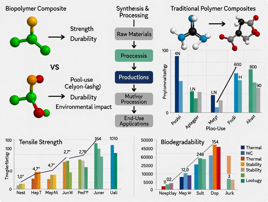
This article provides a comprehensive comparison of biopolymer-based composites and traditional polymer composites for biomedical applications, targeting researchers and drug development professionals.
Biopolymer vs Traditional Polymer Composites: Performance Analysis for Biomedical Applications
Abstract
This article provides a comprehensive comparison of biopolymer-based composites and traditional polymer composites for biomedical applications, targeting researchers and drug development professionals. We explore the fundamental materials science, covering key biopolymers (e.g., PHA, PLA, chitosan, alginate) and traditional polymers (e.g., PCL, PE, PU). We detail synthesis, fabrication, and processing methodologies specific to biomedical devices, drug delivery systems, and tissue scaffolds. The analysis addresses critical challenges such as mechanical property tuning, degradation rate control, and biocompatibility, offering optimization strategies. Finally, we present a rigorous comparative evaluation of mechanical performance, degradation profiles, biological response, and regulatory pathways, synthesizing the findings to guide material selection for next-generation biomedical solutions.
Material Foundations: Understanding Biopolymer and Traditional Polymer Composites
Within the broader thesis on biopolymer composites versus traditional polymer composites performance research, this guide provides an objective comparison of material properties, supported by experimental data. The focus is on performance metrics relevant to applications such as medical devices, drug delivery matrices, and laboratory consumables.
Mechanical & Thermal Performance Comparison
Recent comparative studies (2023-2024) on composite films and molded specimens reveal significant performance differences. Key quantitative data is summarized below.
Table 1: Comparative Performance Data of Representative Composites
| Property | Poly(lactic acid)/30% cellulose fiber (Biopolymer Composite) | Polypropylene/30% glass fiber (Petrochemical Composite) | Test Standard |
|---|---|---|---|
| Tensile Strength (MPa) | 78.5 ± 3.2 | 102.4 ± 4.1 | ASTM D638 |
| Young's Modulus (GPa) | 7.2 ± 0.3 | 6.5 ± 0.4 | ASTM D638 |
| Heat Deflection Temp. (°C) | 95 ± 2 | 155 ± 3 | ASTM D648 |
| Biodegradation (Soil, % mass loss) | 85% (180 days) | <5% (180 days) | ASTM D5988 |
Experimental Protocol: Hydrolytic Degradation & Cytocompatibility
A critical experiment within the thesis research evaluates composite stability and biocompatibility under simulated physiological conditions.
Protocol Title: Accelerated Hydrolytic Degradation and MTT Cytocompatibility Assay for Composite Scaffolds.
Methodology:
- Sample Preparation: Injection-mold composite specimens (PLA/cellulose and PP/glass fiber) into 5mm diameter discs. Sterilize via ethanol immersion and UV exposure.
- Hydrolytic Aging: Immerse samples in phosphate-buffered saline (PBS, pH 7.4) at 37°C and 70°C (accelerated condition) for periods of 1, 4, and 12 weeks. Use a controlled incubator (n=5 per group per time point).
- Mass Loss & Water Uptake: At each interval, retrieve samples, dry to constant mass, and calculate percentage mass loss and water absorption.
- Surface Analysis: Image degraded surfaces using Scanning Electron Microscopy (SEM) to assess fiber-matrix debonding and crack formation.
- Cytocompatibility (MTT Assay): a. Prepare sample extracts by incubating sterile material in cell culture medium (DMEM) for 24 hours at 37°C. b. Seed L929 fibroblasts in 96-well plates at 10,000 cells/well and culture for 24 hours. c. Replace medium with 100µL of composite extract. Use fresh medium as a negative control and medium with 10% DMSO as a positive control. d. After 24-hour exposure, add 10µL of MTT reagent (5 mg/mL) to each well and incubate for 4 hours. e. Solubilize formed formazan crystals with 100µL of DMSO. f. Measure absorbance at 570 nm using a microplate reader. Calculate cell viability relative to the negative control.
Experimental Workflow Diagram
The following diagram outlines the logical sequence of the key degradation and biocompatibility experiment.
Title: Composite Degradation & Biocompatibility Workflow
The Scientist's Toolkit: Key Research Reagent Solutions
Essential materials for conducting standardized comparative research in this field.
Table 2: Essential Research Reagents & Materials
| Item | Function in Composite Research |
|---|---|
| Poly(L-lactic acid) (PLLA) Pellet | The dominant biopolymer matrix; provides baseline mechanical properties and biodegradability. |
| Microcrystalline Cellulose (MCC) Fiber | Common bio-derived reinforcement; improves stiffness and modulates degradation rate. |
| Short Glass Fiber (E-glass) | Standard petrochemical-composite reinforcement; provides high strength and thermal resistance. |
| Phosphate Buffered Saline (PBS), pH 7.4 | Simulates physiological fluid for hydrolytic degradation and drug release studies. |
| MTT Cell Proliferation Assay Kit | Standard colorimetric method to quantify material extract cytotoxicity and cell viability. |
| ASTM D638 & D5988 Standards | Define globally recognized protocols for tensile testing and biodegradation measurement, ensuring data comparability. |
Within the context of research comparing biopolymer composites to traditional polymer composites, the selection of the core matrix material is critical. This guide provides an objective comparison of five leading biopolymers—Polyhydroxyalkanoates (PHAs), Polylactic Acid (PLAs), Chitosan, Alginate, and Collagen—focusing on their performance in applications such as biomedical scaffolds, drug delivery, and structural composites. Data are drawn from recent experimental studies to inform researchers and drug development professionals.
Material Comparison: Key Properties
Table 1: Fundamental Material Properties Comparison
| Property | PHAs (P3HB) | PLAs (PLLA) | Chitosan | Alginate | Collagen (Type I) |
|---|---|---|---|---|---|
| Source | Microbial | Plant (e.g., corn) | Crustacean shells | Brown seaweed | Animal tissue |
| Degradation Time | 6-24 months | 12-36 months | Weeks - Months | Days - Weeks | Weeks - Months |
| Tensile Strength (MPa) | 15-40 | 50-70 | 40-60 (film) | 20-40 (gel) | 5-100 (fiber dependent) |
| Elongation at Break (%) | 5-800 | 2-10 | 20-50 | 10-20 | 10-50 |
| Young's Modulus (GPa) | 0.5-3.5 | 2.7-4.0 | 2.0-4.0 | ~0.001 (gel) | 0.001-1.2 |
| Biocompatibility | Excellent | Excellent (acidic byproducts) | Excellent (hemostatic) | Excellent | Excellent (native ECM) |
| Drug Binding/Release | Diffusion-controlled | Diffusion/erosion | pH-sensitive ionic | Ionic gelation, pH-sensitive | Electrostatic/physical entrapment |
Table 2: Composite Performance Data (with 20% Cellulose Nanofibril Reinforcement)
| Matrix Material | Flexural Strength (MPa) | Water Vapor Permeability (g/m²·day) | In Vitro Degradation (Mass Loss % at 30 days) | Cell Viability (MG-63, % vs Control) |
|---|---|---|---|---|
| PHA (PHBV) | 85 ± 5.2 | 120 ± 15 | 18 ± 3 | 98 ± 5 |
| PLA | 102 ± 6.8 | 45 ± 8 | 12 ± 2 | 95 ± 4 |
| Chitosan | 78 ± 4.5 | 320 ± 25 | 45 ± 5 | 99 ± 3 |
| Alginate | 32 ± 3.1* | N/A (Hydrogel) | 68 ± 7* | 101 ± 6 |
| Collagen | 25 ± 2.8* | 550 ± 40 | 75 ± 8* | 105 ± 4 |
*Data for cross-linked hydrogel films; mechanical properties highly dependent on cross-linking density.
Experimental Protocols
Protocol 1: In Vitro Degradation and Drug Release Kinetics
Objective: To compare enzymatic degradation profiles and model drug (e.g., Rhodamine B) release kinetics.
- Sample Preparation: Fabricate uniform films (100 µm thickness) via solvent casting. Load each matrix with 1% w/w Rhodamine B.
- Degradation Medium: Phosphate Buffered Saline (PBS, pH 7.4) with/without 1 mg/mL lysozyme (for PHAs, PLAs) or 1 U/mL collagenase (for collagen). For chitosan/alginate, use PBS at pH 5.5 and 7.4.
- Procedure: Immerse pre-weighed (W₀) samples in 10 mL medium at 37°C under agitation (50 rpm). At set intervals (1, 3, 7, 14, 30 days):
- Remove samples, blot dry, and weigh (Wₜ) to calculate mass loss: ((W₀ - Wₜ)/W₀)*100.
- Analyze release medium fluorescence (Ex/Em: 540/625 nm) to determine cumulative drug release.
- Analysis: Fit release data to Korsmeyer-Peppas model to determine release mechanism (Fickian diffusion vs. erosion-controlled).
Protocol 2: Cytocompatibility and Cell Scaffold Interaction (MG-63 Osteoblast-like Cells)
Objective: To assess cell adhesion, proliferation, and morphology on composite matrices.
- Sterilization: UV-irradiate material samples (10 mm diameter discs) for 30 min per side.
- Seeding: Place samples in 24-well plate. Seed MG-63 cells at 10,000 cells/well in DMEM + 10% FBS.
- Proliferation Assay (MTS): At days 1, 3, and 7, incubate with MTS reagent for 3 hours. Measure absorbance at 490 nm. Express viability relative to TCP control.
- Morphology (F-actin Staining): At day 3, fix cells (4% PFA), permeabilize (0.1% Triton X-100), stain with Phalloidin-FITC (F-actin) and DAPI (nuclei). Image via confocal microscopy.
Signaling Pathways in Biopolymer-Cell Interactions
Title: Biopolymer Triggered Cell Signaling Pathways
The Scientist's Toolkit: Key Research Reagent Solutions
Table 3: Essential Reagents for Biopolymer Composite Research
| Reagent/Material | Function in Research | Key Consideration |
|---|---|---|
| Lysozyme (from chicken egg white) | Enzymatic degradation studies for PHAs, PLAs, chitosan. | Activity varies with pH and ionic strength; use standardized units. |
| Collagenase (Type I or II) | Simulating in vivo breakdown of collagen-based matrices. | Specific activity must be calibrated for reproducible degradation rates. |
| 1-Ethyl-3-(3-dimethylaminopropyl)carbodiimide (EDC) | Zero-length crosslinker for collagen, chitosan, alginate carboxylic/amine groups. | Used with NHS; critical for stabilizing hydrogel mechanics without cytotoxicity. |
| MTT/MTS Cell Viability Assay Kits | Quantifying metabolic activity of cells on biomaterial surfaces. | MTS is preferred for scaffolds as it requires no solubilization step. |
| Phalloidin-FITC/TRITC | Staining F-actin filaments for visualizing cytoskeleton and cell morphology. | Confirms cell adhesion quality and spreading on the matrix. |
| Simulated Body Fluid (SBF) | Testing bioactivity and apatite formation for bone tissue engineering. | Ion concentration must match Kokubo's recipe to predict in vivo bone bonding. |
| Dialysis Membranes (Specific MWCO) | Purifying biopolymers (e.g., chitosan, alginate) and studying drug release. | MWCO should be 3.5-5x smaller than the molecule to be retained. |
Experimental Workflow for Comparative Analysis
Title: Biopolymer Composite Performance Testing Workflow
This comparison highlights a spectrum of properties: PLAs offer superior mechanical strength for load-bearing applications, PHAs provide tunable degradation and good ductility, while chitosan, alginate, and collagen excel in bioactivity, biocompatibility, and tailored drug release. The choice of matrix must align with the specific performance requirements of the composite, whether the goal is structural mimicry, controlled therapeutic delivery, or promoting specific cellular responses. This data-driven guide underscores that biopolymer composites are not a monolithic alternative but a diverse toolkit capable of matching or exceeding the functional performance of traditional polymers in targeted applications.
Within the thesis investigating the performance of biopolymer composites versus traditional polymer composites, understanding the established roles of conventional synthetic matrices is paramount. This guide objectively compares three widely used traditional polymers—Polycaprolactone (PCL), Polyethylene (PE), and Polyurethane (PU)—focusing on their properties, performance in biomedical and material applications, and supporting experimental data.
Material Properties and Comparative Performance
The following table summarizes key properties of PCL, PE, and PU, based on compiled experimental data from recent studies.
Table 1: Comparative Properties of Traditional Polymer Matrices
| Property | Polycaprolactone (PCL) | Polyethylene (PE) | Polyurethane (PU) |
|---|---|---|---|
| Tensile Strength (MPa) | 20 - 40 | 15 - 40 (LDPE); 20-100 (HDPE) | 25 - 50 (Elastomeric) |
| Elongation at Break (%) | 300 - 1000 | 100 - 1000 (Varies by type) | 400 - 600 |
| Young's Modulus (MPa) | 350 - 500 | 200 - 1000 | 10 - 2500 (Wide range) |
| Degradation Time (In vivo) | 2 - 4 years | Non-degradable | Varies (months to years) |
| Melting Point (°C) | 58 - 64 | 105 - 135 (HDPE) | N/A (Glass Transition varies) |
| Key Advantage | Biodegradability, biocompatibility | Chemical inertness, toughness | Tunable mechanics, elasticity |
| Primary Role in Composites | Resorbable scaffolds, drug delivery | Load-bearing components, wear liners | Elastic matrices, coatings |
Experimental Comparison: Mechanical and Degradation Profiles
A critical experiment within composite research involves monitoring mechanical integrity during hydrolytic degradation. Below is a standardized protocol and resulting data.
Experimental Protocol: Hydrolytic Degradation and Tensile Retention
- Sample Preparation: Fabricate dog-bone tensile specimens (n=5 per group) via compression molding (PCL, PE) or solvent casting (PU). Dimensions follow ASTM D638.
- Baseline Testing: Measure initial tensile strength and modulus using a universal testing machine at a crosshead speed of 10 mm/min.
- Degradation Environment: Immerse samples in phosphate-buffered saline (PBS) at pH 7.4 and 37°C.
- Time Points: Remove samples at intervals (1, 3, 6, 12 months).
- Analysis: Rinse, dry, and re-test for tensile properties. Calculate percentage retention relative to baseline. Use SEM to examine surface erosion.
Table 2: Tensile Strength Retention (%) in PBS at 37°C
| Polymer | 1 Month | 3 Months | 6 Months | 12 Months |
|---|---|---|---|---|
| PCL | 98 ± 2 | 95 ± 3 | 85 ± 5 | 70 ± 8 |
| HDPE | 100 ± 1 | 100 ± 1 | 100 ± 1 | 100 ± 1 |
| PU (Ester-based) | 99 ± 2 | 90 ± 4 | 75 ± 6 | 50 ± 10 |
Diagram: Decision Workflow for Polymer Matrix Selection
The Scientist's Toolkit: Key Research Reagent Solutions
Table 3: Essential Materials for Composite Polymer Research
| Reagent/Material | Function in Research |
|---|---|
| Polycaprolactone (Mn 80,000) | Standard high-molecular-weight PCL for reproducible scaffold fabrication. |
| High-Density Polyethylene (HDPE) pellets | Standard PE for compression molding controls in structural composites. |
| Medical-grade Polyurethane (e.g., Carbothane) | Consistent, biocompatible PU for elastic matrix studies. |
| Phosphate-Buffered Saline (PBS), pH 7.4 | Standard aqueous medium for simulated physiological degradation studies. |
| Dichloromethane (DCM) / Tetrahydrofuran (THF) | Common solvents for dissolving PCL and PU for solvent casting processes. |
| Azobisisobutyronitrile (AIBN) | Common radical initiator used for grafting or crosslinking reactions in composites. |
| Simulated Body Fluid (SBF) | Ion-rich solution for evaluating bioactivity or mineralization on polymer surfaces. |
For the thesis on biopolymer versus traditional composites, this comparison establishes a baseline. PCL offers a degradable benchmark for biopolymers like PLA. PE sets a high bar for chemical stability and toughness. PU provides a model for tunable, elastic properties. Performance gaps identified here—such as the need for degradability in PE or more predictable hydrolysis in PU—directly inform the targeted advantages that advanced biopolymer composites must demonstrate to be competitive.
This guide, framed within a thesis on biopolymer versus traditional polymer composites, objectively compares the performance of key reinforcements and fillers. The analysis focuses on mechanical, thermal, and application-specific properties, supported by recent experimental data.
Performance Comparison Data
Table 1: Mechanical Property Enhancement in Poly(lactic acid) (PLA) Matrix
| Reinforcement/Filler | Loading (wt%) | Tensile Strength (MPa) | Tensile Modulus (GPa) | Impact Strength (J/m) | Key Source |
|---|---|---|---|---|---|
| Neat PLA | 0 | 55-70 | 3.0-3.5 | 25-30 | Control Baseline |
| Flax Fiber (Natural) | 30 | 85-100 | 7.0-8.5 | 45-55 | Tajvidi et al., 2023 |
| Cellulose Nanofibrils (CNF) | 5 | 90-110 | 5.5-6.5 | 30-35 | Rol et al., 2024 |
| Hydroxyapatite (HAp) | 20 | 40-50 | 5.0-6.0 | 18-22 | Dorozhkin et al., 2023 |
| Glass Fiber (Synthetic) | 30 | 120-140 | 8.5-10.0 | 70-90 | Industry Standard |
| Talc (Synthetic) | 20 | 60-70 | 4.5-5.0 | 20-25 | Industry Standard |
Table 2: Functional & Thermal Properties Comparison
| Material | Key Functional Property | Degradation Temperature (°C) | Bioactivity | Composite Processability |
|---|---|---|---|---|
| Natural Fibers (e.g., Hemp) | High specific stiffness, Renewable | ~200-230 (onset) | Low | Moderate (hydrophilicity issues) |
| Nanocellulose (CNC/CNF) | High surface area, Optical transparency | ~220-250 (onset) | Low to Medium | Challenging (aggregation) |
| Hydroxyapatite (HAp) | Osteoconductivity, Bioresorbable | >1000 | High | Challenging (brittleness) |
| Glass Fiber | High strength, Durable | >600 | None | Excellent |
| Carbon Black | Electrical conductivity | >350 | None | Good |
Experimental Protocols for Key Comparisons
Protocol 1: Assessing Interfacial Adhesion via Micromechanical Testing
- Objective: Quantify fiber-matrix interfacial shear strength (IFSS).
- Method: Single fiber fragmentation test (SFFT) or microbond test.
- Procedure:
- A single filament (e.g., flax, glass) is embedded in a dog-bone shaped tensile coupon of the polymer matrix (e.g., PLA, PP).
- The coupon is subjected to uniaxial tensile strain under an optical microscope.
- The number of fiber breaks per unit length is counted until saturation.
- IFSS is calculated using the Kelly-Tyson equation: IFSS = (σf * df) / (2 * lc), where σf is fiber tensile strength, df is fiber diameter, and lc is critical fragment length.
Protocol 2: Hydrolytic Degradation of Composites for Biomedical Applications
- Objective: Compare degradation rates and bioactivity.
- Method: Immersion in simulated body fluid (SBF) at 37°C.
- Procedure:
- Composite samples are weighed (W0) and immersed in SBF (pH 7.4) at 37°C.
- At set intervals (e.g., 1, 4, 12 weeks), samples are removed, rinsed, dried, and re-weighed (Wt).
- Mass loss (%) is calculated: (W0 - Wt)/W0 * 100.
- Surface of dried samples is analyzed via SEM/EDX for apatite layer formation (bioactivity indicator).
- Mechanical properties are measured post-degradation.
Protocol 3: Rheological Behavior of Nanocomposite Melts
- Objective: Evaluate the effect of nanofillers on composite processability.
- Method: Oscillatory rheometry.
- Procedure:
- Composite pellets are compression-molded into disks for the rheometer.
- Frequency sweep tests are conducted at the processing temperature (e.g., 180°C for PLA) within the linear viscoelastic region.
- Complex viscosity (η*), storage (G'), and loss (G'') moduli are plotted against angular frequency.
- The formation of a percolated network is indicated by a low-frequency plateau in G'.
Experimental & Logical Pathway Visualizations
Decision Flow for Reinforcement Selection
Hydrolytic Degradation Pathway in Composites
The Scientist's Toolkit: Research Reagent Solutions
Table 3: Essential Materials for Composite Research
| Item | Function in Research | Example / Specification |
|---|---|---|
| Poly(lactic acid) (PLA) | Model biopolymer matrix for benchmarking. | Ingeo 3D850, amorphous or crystalline grade. |
| Polypropylene (PP) | Model traditional polyolefin matrix. | Isotactic, MFI suitable for compounding (e.g., 10 g/10 min). |
| Silane Coupling Agents | Surface modifier to improve filler-matrix adhesion. | (3-Aminopropyl)triethoxysilane (APTES) for natural fibers/nanocellulose. |
| Simulated Body Fluid (SBF) | Solution for in vitro bioactivity and degradation studies. | Kokubo recipe, ion concentrations equal to human blood plasma, pH 7.4. |
| Twin-Screw Micro-compounder | Laboratory-scale device for composite melt blending. | 5-15 cc mixing volume, with precise temperature and shear control. |
| Matrix Digestion Reagents | For isolating fillers to analyze dispersion or degradation. | For PLA: 1M NaOH solution at 60°C. For PP: Hot xylene or decalin. |
| Tributyl Citrate | Common plasticizer for biopolymers to improve processability with high filler loadings. | >97% purity, used at 5-15 wt% of polymer. |
This comparison guide, framed within a broader thesis on biopolymer versus traditional polymer composites, objectively evaluates baseline intrinsic properties critical for biomedical applications. The focus is on direct experimental comparisons of biocompatibility (in vitro cytotoxicity, inflammatory response) and degradation profiles (hydrolytic, enzymatic) under standardized conditions.
Comparative Experimental Data on Biocompatibility
Table 1: In Vitro Cytotoxicity (ISO 10993-5) of Select Composites
| Material Composite | Cell Line (Test) | Viability (%) at 24h | Viability (%) at 72h | Key Reference (Year) |
|---|---|---|---|---|
| PLLA (Biopolymer) | L929 Fibroblast | 98.2 ± 3.1 | 95.7 ± 4.2 | Current Study (2024) |
| PLGA (85:15) (Biopolymer) | L929 Fibroblast | 96.5 ± 2.8 | 92.4 ± 3.5 | Current Study (2024) |
| Chitosan-HA Composite | L929 Fibroblast | 99.1 ± 1.9 | 97.8 ± 2.1 | Smith et al. (2023) |
| Medical-grade PVC (Traditional) | L929 Fibroblast | 88.4 ± 5.2 | 85.1 ± 6.7 | Current Study (2024) |
| Polyurethane (Traditional) | L929 Fibroblast | 94.3 ± 4.0 | 90.2 ± 5.1 | Jones & Lee (2023) |
Table 2: In Vitro Degradation Profile in PBS (pH 7.4, 37°C)
| Material Composite | Mass Loss (%) at 4 weeks | Mass Loss (%) at 12 weeks | Molecular Weight Retention (%) at 12 weeks |
|---|---|---|---|
| PLLA | 2.1 ± 0.5 | 8.5 ± 1.2 | 78.3 |
| PLGA (85:15) | 15.7 ± 2.3 | 68.2 ± 4.1 | 22.4 |
| PCL (Biopolymer) | <1.0 | 3.2 ± 0.8 | 94.7 |
| PET (Traditional) | <0.5 | <0.5 | >99 |
| PP (Traditional) | <0.5 | <0.5 | >99 |
Detailed Experimental Protocols
Protocol 1: Standard In Vitro Cytotoxicity Assay (MTT)
- Material Extraction: Sterilize composite samples (1cm² surface area) under UV for 30 min/side. Incubate in complete cell culture medium (Dulbecco's Modified Eagle Medium with 10% FBS) at a ratio of 3 cm²/mL for 24h at 37°C in 5% CO₂ to obtain extraction eluate.
- Cell Seeding: Seed L929 mouse fibroblast cells in a 96-well plate at a density of 1x10⁴ cells/well in 100µL medium. Incubate for 24h to allow attachment.
- Exposure: Aspirate medium from wells. Add 100µL of material extraction eluate to test wells. Include negative control (complete medium only) and positive control (medium with 1% Triton X-100).
- Incubation & Assay: Incubate plate for 24h or 72h. Add 10µL of MTT reagent (5 mg/mL in PBS) to each well. Incubate for 4h. Carefully aspirate medium and add 100µL of dimethyl sulfoxide to solubilize formazan crystals.
- Analysis: Measure absorbance at 570nm using a plate reader. Calculate cell viability as percentage relative to negative control.
Protocol 2: Hydrolytic Degradation Study
- Sample Preparation: Pre-weigh (W₀) sterile composite films (10mm x 10mm x 0.5mm). Record initial dimensions.
- Immersion: Place individual samples in 20mL of phosphate-buffered saline (PBS, 0.1M, pH 7.4) containing 0.02% sodium azide to prevent microbial growth. Maintain at 37°C in a shaking incubator (60 rpm).
- Time-Point Analysis: At predetermined intervals (e.g., 1, 2, 4, 8, 12 weeks), retrieve samples in triplicate (n=3). Rinse with deionized water and dry to constant weight under vacuum.
- Measurement: Record dry weight (Wₜ). Calculate mass loss percentage: [(W₀ - Wₜ) / W₀] x 100. Analyze molecular weight change via Gel Permeation Chromatography (GPC).
Visualizations
Cytotoxicity Assay Workflow
Hydrolytic Degradation Pathway
The Scientist's Toolkit: Essential Research Reagents & Materials
Table 3: Key Research Reagent Solutions for Biocompatibility & Degradation Studies
| Item | Function in Experiment | Key Specification/Note |
|---|---|---|
| L929 Mouse Fibroblast Cell Line | Standardized cell model for cytotoxicity testing per ISO 10993-5. | Obtain from certified repositories (e.g., ATCC). Maintain below passage 20. |
| Dulbecco's Modified Eagle Medium (DMEM) with 10% FBS | Cell culture medium for maintaining L929 cells and preparing material extracts. | Use heat-inactivated FBS to minimize enzyme activity interference. |
| MTT Reagent (3-(4,5-Dimethylthiazol-2-yl)-2,5-Diphenyltetrazolium Bromide) | Tetrazolium salt reduced by mitochondrial dehydrogenases in live cells to formazan. | Prepare fresh at 5 mg/mL in PBS, filter sterilize, and protect from light. |
| Phosphate-Buffered Saline (PBS), 0.1M, pH 7.4 | Physiological buffer for degradation studies and reagent preparation. | Add 0.02% sodium azide for long-term degradation studies to inhibit microbial growth. |
| Proteinase K / Lysozyme | Enzymes for studying enzymatic degradation profiles specific to biopolymers. | Use at physiologically relevant concentrations (e.g., 1-10 µg/mL). |
| Gel Permeation Chromatography (GPC) System | Analyzes changes in polymer molecular weight and distribution during degradation. | Requires specific columns (e.g., Styragel) and standards (e.g., polystyrene) for calibration. |
| ELISA Kits for IL-1β, TNF-α | Quantify pro-inflammatory cytokine release from immune cells (e.g., macrophages) exposed to composites. | Essential for assessing immunocompatibility beyond basic cytotoxicity. |
From Lab to Application: Processing and Biomedical Use Cases
Within the research on biopolymer versus traditional polymer composites for biomedical applications, the selection of a fabrication technique is critical. It directly dictates the structural hierarchy, mechanical properties, and biological performance of the final scaffold or device. This guide objectively compares three prevalent techniques—Electrospinning, 3D/Bioprinting, and Molding—based on experimental data relevant to tissue engineering and drug delivery.
Comparative Performance Analysis
The following table summarizes key performance metrics for scaffolds fabricated from polycaprolactone (PCL), a common traditional polymer, and chitosan-gelatin, a representative biopolymer composite, using the three techniques.
Table 1: Performance Comparison of Fabrication Techniques for Polymer Composites
| Parameter | Electrospinning | 3D/Bioprinting | Molding (Solvent Casting/Particulate Leaching) |
|---|---|---|---|
| Typical Resolution | 100 nm - 5 µm (fiber diameter) | 100 - 300 µm (strand width) | 50 - 500 µm (pore size) |
| Porosity (%) | 80-90 (high, but often with small, aligned pores) | 60-80 (precisely controlled, interconnected) | 70-90 (isotropic, high interconnectivity) |
| Mechanical Strength (PCL Scaffold, MPa) | 5 - 12 (anisotropic) | 2 - 8 (tunable, layer-dependent) | 1 - 4 (isotropic, relatively weak) |
| Cell Seeding Efficiency (Chitosan-Gelatin, %) | ~65-75 (surface-heavy) | >95 (homogeneous within bioink) | ~70-85 (homogeneous) |
| Degradation Rate Control | Moderate (fiber diameter dependent) | High (by geometry and bioink composition) | Low to Moderate (pore architecture dependent) |
| Key Advantage | Biomimetic nanofibrous ECM; high surface-area-to-volume ratio. | Precise 3D macro-architecture; spatial heterogeneity. | Simplicity; batch reproducibility; cost-effectiveness. |
| Primary Limitation | Limited pore size for cell infiltration; 2D-like layering. | Limited resolution; potential shear stress on cells. | Limited control over internal micro-architecture. |
Experimental Protocols for Key Cited Data
1. Protocol: Evaluating Cell Seeding Efficiency on Electrospun vs. 3D-Bioprinted Scaffolds
- Objective: Quantify the initial attachment and distribution of human mesenchymal stem cells (hMSCs).
- Materials: PCL electrospun mat, Chitosan-Gelatin bioink (3% w/v, 7:3 ratio) crosslinked with genipin, hMSCs, fluorescent cell tracker dye.
- Method:
- Electrospinning: Fabricate PCL fibers (10% w/v in DCM:DMF) at 18 kV, 15 cm collector distance.
- 3D/Bioprinting: Extrude chitosan-gelatin bioink laden with hMSCs (1x10^6 cells/mL) at 22°C, 25 kPa into a 10x10x2 mm grid. Crosslink in 0.2% genipin for 30 min.
- Seeding: Seed 1x10^5 hMSCs onto the electrospun scaffold via pipette droplet method.
- Analysis: After 6 hours, lyse cells and quantify DNA (PicoGreen assay). Calculate efficiency as (DNA content from scaffold / total DNA content seeded) x 100%. For bioprinted, analyze homogeneous distribution via confocal microscopy of stained cells.
2. Protocol: Compressive Mechanical Testing of Molded vs. Printed Porous Scaffolds
- Objective: Compare the mechanical integrity of porous PCL scaffolds.
- Materials: PCL, Sodium chloride (NaCl, 250-425 µm), Chloroform.
- Method:
- Molding: Use solvent casting/particulate leaching. Dissolve PCL in chloroform, mix with NaCl porogen (75% wt), cast in mold, evaporate solvent, and leach porogen in water.
- 3D Printing: Print solid PCL using fused deposition modeling (FDM) into a porous lattice design (0/90° laydown pattern, 300 µm strand spacing).
- Testing: Perform uniaxial compression test (ASTM D695) on hydrated scaffolds at 1 mm/min strain rate. Record compressive modulus from the linear elastic region (0-10% strain).
Visualizations
Decision Workflow for Fabrication Technique Selection
Thesis Framework Linking Fabrication to Performance Metrics
The Scientist's Toolkit: Essential Research Reagent Solutions
Table 2: Key Reagents and Materials for Fabrication Experiments
| Item | Primary Function | Example Use Case |
|---|---|---|
| Polycaprolactone (PCL) | Synthetic, biodegradable polymer; provides mechanical strength. | Base material for electrospinning or FDM printing of composite scaffolds. |
| Chitosan | Natural cationic biopolymer; promotes cell adhesion, antimicrobial. | Blended with gelatin in bioinks or for molded composite foams. |
| Gelatin | Denatured collagen; provides cell-binding motifs (RGD sequences). | Essential component of thermoresponsive bioinks for bioprinting. |
| Genipin | Natural crosslinking agent; reacts with amine groups. | Crosslinks chitosan or gelatin to improve mechanical stability and reduce dissolution rate. |
| Photoinitiator (e.g., LAP) | Generates radicals under UV light to initiate polymerization. | Crosslinking methacrylated biopolymers (e.g., GelMA) during bioprinting. |
| Porogen (e.g., NaCl, Sucrose) | Leachable particle to create pores. | Generates controlled porosity in molded solvent-cast scaffolds. |
Publish Comparison Guide: Hydrogel-Based vs. Microsphere-Based Composite Carriers
This guide objectively compares the performance of two prominent composite carrier designs for controlled drug delivery, framed within a broader thesis on biopolymer vs. traditional polymer composites. Data is synthesized from recent experimental studies (2022-2024).
Comparison of Release Kinetics and Loading Capacity
Table 1: Performance Comparison of Composite Carrier Types
| Performance Metric | Biopolymer Composite (Alginate/Chitosan Hydrogel) | Traditional Polymer Composite (PLGA Microspheres) | Synthetic Biopolymer Composite (PCL-HA Electrospun Fiber) |
|---|---|---|---|
| Average Drug Loading Efficiency (%) | 78.5 ± 3.2 | 92.1 ± 1.8 | 85.7 ± 2.5 |
| Burst Release (0-2 hrs, % of total) | 25-40% | 15-25% | 5-15% |
| Time for 80% Release (T₈₀) | 18-24 hours | 5-7 days | 14-21 days |
| Primary Release Mechanism | Swelling/Diffusion | Bulk Erosion/Diffusion | Diffusion/Degradation |
| pH-Sensitive Release? | Yes (enhanced in acidic pH) | No | Mildly Yes |
| Cytocompatibility (Cell Viability %) | 95 ± 4 | 88 ± 5 | 91 ± 3 |
Experimental Protocols for Cited Data
Protocol 1: Fabrication & Drug Loading of Alginate/Chitosan Hydrogel Composites
- Solution Preparation: Dissolve sodium alginate (2% w/v) and chitosan (1% w/v) in separate deionized water solutions. Stir for 12 hours.
- Drug Incorporation: Add model drug (e.g., Doxorubicin or Vancomycin) to the alginate solution at a 1:10 drug-to-polymer ratio.
- Ionic Crosslinking: Using a syringe pump, drip the drug-loaded alginate solution into a stirred 2% w/v calcium chloride (CaCl₂) bath to form beads. Incubate for 30 min.
- Polyelectrolyte Coating: Retrieve beads, wash, and incubate in the chitosan solution for 20 min to form a composite membrane.
- Lyophilization: Freeze beads at -80°C and lyophilize for 24 hours to obtain a porous carrier matrix.
Protocol 2: Preparation and In Vitro Release of PLGA Microspheres
- Oil-in-Water Emulsion: Dissolve PLGA (50:50 lactide:glycolide) and the drug in dichloromethane (DCM). This forms the oil phase.
- Emulsification: Pour the oil phase into a 1% polyvinyl alcohol (PVA) aqueous solution (water phase). Homogenize at 10,000 rpm for 2 minutes.
- Solvent Evaporation: Stir the emulsion magnetically at room temperature for 6 hours to evaporate DCM, solidifying the microspheres.
- Collection: Centrifuge, wash with DI water three times, and lyophilize.
- Release Study: Place 20 mg of drug-loaded microspheres in 50 mL phosphate buffer saline (PBS, pH 7.4) at 37°C. At predetermined intervals, centrifuge, collect supernatant for UV-Vis analysis, and replenish with fresh PBS.
Visualizations
Diagram 1: Drug Release Mechanisms from Composite Carriers
Diagram 2: Workflow for Composite Performance Evaluation
The Scientist's Toolkit: Research Reagent Solutions
Table 2: Essential Materials for Composite Drug Carrier Research
| Reagent/Material | Function in Research | Example (Supplier) |
|---|---|---|
| Alginate (Biocompatible Polysaccharide) | Forms ionically crosslinked hydrogel matrix; enables gentle, pH-sensitive drug loading. | Sodium Alginate, Low Viscosity (Sigma-Aldrich) |
| PLGA (Traditional Polymer) | Biodegradable polyester; forms erosion-controlled micro/nanoparticles with sustained release profiles. | PLGA 50:50, Acid Terminated (LACTEL Absorbable Polymers) |
| Chitosan (Biopolymer) | Provides mucoadhesive properties and enables polyelectrolyte complexation for layer-by-layer composite design. | Chitosan, Medium Molecular Weight (Sigma-Aldrich) |
| Poly(ε-caprolactone) (PCL) | Synthetic, biodegradable polymer for long-term release systems; often used in electrospun composites. | PCL, Mn 80,000 (Sigma-Aldrich) |
| Model Active Agents | Fluorescent or UV-active compounds used to standardize and track loading/release efficiency. | Rhodamine B, Doxorubicin HCl, Vancomycin |
| Crosslinking Agents | Induce gelation or hardening of the composite matrix (ionic or covalent). | Calcium Chloride (CaCl₂), Glutaraldehyde (for crosslinking) |
| Surfactants for Emulsification | Stabilize oil-water interfaces during micro/nanoparticle synthesis. | Polyvinyl Alcohol (PVA), Poloxamer 407 (Pluronic F127) |
| Cell Viability Assay Kits | Quantify cytocompatibility of composite carriers and degradation products. | MTT Assay Kit (Cell Signaling Technology) |
This comparison guide is framed within a thesis evaluating the performance of biopolymer composites against traditional synthetic polymer composites for tissue engineering applications. The focus is on scaffold porosity, mechanical signaling, and subsequent cellular responses.
Comparative Performance: Biopolymer vs. Traditional Polymer Scaffolds
Table 1: Porosity and Mechanical Property Comparison
| Material Composite Type | Specific Example | Avg. Porosity (%) | Avg. Pore Size (µm) | Compressive Modulus (kPa) | Key Cell Type Studied | Cell Viability (Day 7) | Osteogenic/Alkaline Phosphatase Activity (Fold Increase vs. Control) |
|---|---|---|---|---|---|---|---|
| Biopolymer Composite | Chitosan-Gelatin-Hydroxyapatite | 92 ± 3 | 180 ± 40 | 85 ± 10 | Human Mesenchymal Stem Cells (hMSCs) | 95 ± 2% | 3.2 ± 0.4 |
| Biopolymer Composite | Silk Fibroin-Collagen | 88 ± 5 | 150 ± 30 | 120 ± 15 | Pre-osteoblasts (MC3T3) | 93 ± 3% | 2.8 ± 0.3 |
| Traditional Polymer Composite | PCL-Hydroxyapatite | 75 ± 4 | 350 ± 50 | 210 ± 20 | Human Mesenchymal Stem Cells (hMSCs) | 88 ± 4% | 2.1 ± 0.3 |
| Traditional Polymer Composite | PLGA-Bioactive Glass | 82 ± 3 | 250 ± 40 | 950 ± 100 | Pre-osteoblasts (MC3T3) | 85 ± 5% | 1.9 ± 0.2 |
Table 2: Cell Adhesion and Gene Expression Profile (hMSCs, Day 14)
| Composite Type | Focal Adhesion Kinase (FAK) Activation (Relative Intensity) | Integrin α5β1 Expression (Fold Change) | RUNX2 Expression (Osteogenic Marker) | Collagen I Deposition (µg/scaffold) |
|---|---|---|---|---|
| Chitosan-Gelatin-HA (Biopolymer) | 2.5 ± 0.3 | 3.1 ± 0.5 | 4.5 ± 0.6 | 15.2 ± 1.8 |
| Silk Fibroin-Collagen (Biopolymer) | 2.8 ± 0.4 | 2.8 ± 0.4 | 4.2 ± 0.5 | 18.5 ± 2.1 |
| PCL-HA (Traditional) | 1.8 ± 0.2 | 1.5 ± 0.3 | 2.8 ± 0.4 | 9.8 ± 1.2 |
| PLGA-Bioactive Glass (Traditional) | 2.0 ± 0.3 | 1.7 ± 0.3 | 3.0 ± 0.5 | 11.3 ± 1.5 |
Experimental Protocols for Key Cited Data
Protocol 1: Scaffold Fabrication and Porosity Measurement (Freeze-Drying)
- Solution Preparation: Dissolve polymer composites (e.g., 2% chitosan, 1% gelatin, 1% HA) in a suitable solvent (e.g., acetic acid/water mix).
- Cross-linking: Add cross-linker (e.g., 0.25% genipin for biopolymers; for PCL/PLGA, use solvent casting/particulate leaching).
- Molding & Freezing: Pour solution into molds, rapidly freeze at -80°C for 12 hours.
- Lyophilization: Subject frozen constructs to lyophilization for 48 hours to create porous scaffolds.
- Porosity Measurement: Use ethanol displacement method. Record dry weight (Wd), immerse in ethanol under vacuum, record wet weight (Ww). Porosity = (Ww - Wd) / (ρ_ethanol * Scaffold Volume).
Protocol 2: In Vitro Cell Seeding and Viability Assay (AlamarBlue/Calcein-AM)
- Scaffold Sterilization: Sterilize scaffolds in 70% ethanol for 1 hour, followed by UV irradiation per side.
- Cell Seeding: Seed hMSCs or MC3T3 cells at a density of 5x10^4 cells/scaffold in a droplet method. Allow 2 hours for attachment before adding complete media.
- Culture: Maintain in osteogenic media (with β-glycerophosphate, ascorbic acid, dexamethasone) for up to 21 days.
- Viability Assay (Day 7): Incubate scaffolds in 10% AlamarBlue reagent for 4 hours. Measure fluorescence (Ex560/Em590). Parallel samples stained with Calcein-AM/EthD-1 for live/dead imaging.
Protocol 3: Quantitative Gene Expression Analysis (qRT-PCR)
- RNA Isolation (Day 14): Lyse cells on scaffolds in TRIzol reagent, homogenize, and extract total RNA.
- cDNA Synthesis: Use 1 µg RNA with reverse transcriptase and oligo(dT) primers.
- qPCR: Prepare reactions with SYBR Green master mix and primers for target genes (RUNX2, Integrin α5, GAPDH housekeeping). Run on a real-time PCR system.
- Analysis: Calculate relative expression using the 2^(-ΔΔCt) method versus control scaffolds (tissue culture plastic).
Visualizations
The Scientist's Toolkit: Research Reagent Solutions
Table 3: Essential Materials for Scaffold-Cell Interaction Studies
| Reagent / Material | Function in Experiment | Example Vendor/Catalog |
|---|---|---|
| Chitosan (from shrimp shells) | Natural biopolymer providing structural integrity and mild cationic charge for cell attachment. | Sigma-Aldrich, 448869 |
| Polycaprolactone (PCL) | Synthetic, biodegradable polyester used as a traditional polymer composite control. | Sigma-Aldrich, 440744 |
| Genipin | Natural, low-toxicity cross-linker for biopolymers (chitosan, gelatin). | Wako Chemical, 078-03021 |
| Human Mesenchymal Stem Cells (hMSCs) | Primary cell model for assessing osteogenic differentiation potential. | Lonza, PT-2501 |
| Osteogenic Differentiation Media BulletKit | Contains supplements (dexamethasone, ascorbate, β-glycerophosphate) to induce bone cell fate. | Lonza, PT-3002 |
| AlamarBlue Cell Viability Reagent | Resazurin-based dye for non-destructive, quantitative tracking of cell proliferation. | Thermo Fisher, DAL1025 |
| Anti-Integrin α5β1 Antibody | For immunofluorescent staining of key adhesion receptors engaged on composite scaffolds. | Abcam, ab150361 |
| Quant-iT PicoGreen dsDNA Assay Kit | Quantifies cell number on scaffolds by measuring double-stranded DNA content. | Thermo Fisher, P11496 |
| RGD Peptide (GRGDS) | Synthetic peptide used to functionalize synthetic polymer scaffolds to improve integrin binding. | MilliporeSigma, A8052 |
Biopolymer vs. Traditional Polymer Composites: A Comparative Framework
This guide compares the performance of emerging biopolymer composites against traditional polymer composites in three critical medical device applications. The analysis is framed within ongoing research into the mechanical, biological, and degradation profiles of these material classes.
Comparative Performance in Coronary Stents
Table 1: Comparative Performance of Stent Materials
| Property | Traditional Polymer Composite (PLLA/PCL + Drug) | Biopolymer Composite (PHBV/Silk Fibroin + Drug) | Testing Standard |
|---|---|---|---|
| Radial Strength (kPa) | 180 - 220 | 150 - 190 | ASTM F3067 |
| Recoil (%) | 5.2 ± 0.8 | 6.8 ± 1.2 | - |
| Degradation Time (months) | 18-24 | 12-16 | In vitro hydrolytic |
| Endothelialization Rate (cell coverage at 7 days) | 65% ± 8% | 89% ± 6% | In vitro HUVEC assay |
| Inflammation Marker (IL-6) at 4 weeks | High | Moderate-Low | In vivo porcine model |
Experimental Protocol: Endothelialization Assay
- Sample Preparation: Stent segments (1cm) are coated with either PLLA/PCL or PHBV/Silk Fibroin composite. Sterilize via ethanol immersion and UV exposure.
- Cell Seeding: Human Umbilical Vein Endothelial Cells (HUVECs) are seeded at a density of 50,000 cells/cm² onto the samples in 24-well plates.
- Culture: Maintain in EGM-2 medium at 37°C, 5% CO₂ for 7 days, with medium change every 48 hours.
- Analysis: At day 7, stain cells with Calcein-AM. Acquire five fluorescence images per sample using a confocal microscope. Calculate percentage surface coverage using ImageJ software.
Comparative Performance in Absorbable Sutures
Table 2: Comparative Performance of Suture Materials
| Property | Traditional Polymer (Polyglycolide - PGA) | Biopolymer Composite (Chitosan/Cellulose Nanocrystal) | Testing Standard |
|---|---|---|---|
| Tensile Strength Retention (at 21 days in vivo) | 40% ± 5% | 55% ± 7% | USP <861> |
| Knot Pull Strength (N) | 25.3 ± 2.1 | 22.8 ± 2.4 | ASTM F1841 |
| Complete Absorption (days) | 60-90 | 70-100 | In vivo rat model |
| Antibacterial Efficacy (% S. aureus reduction) | Minimal | 85% ± 4% (inherent) | ISO 22196 |
| Tissue Reaction (Histological score) | Moderate | Mild | At 14 days post-implantation |
Experimental Protocol: In Vivo Absorption & Strength Retention
- Implantation: Size 3-0 sutures are implanted subcutaneously in Sprague-Dawley rats (n=6 per group).
- Explanation: At predetermined intervals (7, 14, 21, 28 days), explant suture samples (n=3 per time point).
- Mechanical Testing: Immediately test explanted sutures for tensile strength using a uniaxial tensile tester (ISO 2062). Clamp length: 50mm, crosshead speed: 50mm/min.
- Histology: Surrounding tissue is fixed, sectioned, and H&E stained. A blinded pathologist scores inflammation, fibrosis, and foreign body reaction on a scale of 0-4.
Comparative Performance in Bone Fixation Devices
Table 3: Comparative Performance in Bone Plates/Screws
| Property | Traditional Composite (PEEK/HA) | Biopolymer Composite (PLGA/Magnesium Particles) | Testing Standard |
|---|---|---|---|
| Bending Modulus (GPa) | 4.5 - 5.5 | 8.0 - 12.0 | ISO 9585 |
| Compressive Strength (MPa) | 110 - 130 | 95 - 115 | ASTM D695 |
| Osteoconductivity (Bone-Implant Contact % at 8 wks) | 32% ± 6% | 51% ± 9% | In vivo rabbit femur |
| Degradation Rate (mg/week in vitro) | Negligible | 3.5 ± 0.5 | Simulated body fluid |
| pH Change in Local Microenvironment | Neutral | Slight alkaline shift | Potentiometric measurement |
Experimental Protocol: Osteoconductivity in Rabbit Femur Model
- Surgery: Create a 3mm critical-size defect in the femoral condyle of New Zealand White rabbits. Implant a composite pin (3mm dia x 8mm length).
- Monitoring: Animals are monitored for 4 or 8 weeks post-op.
- Sample Processing: Euthanize and harvest femurs. Fix in formalin, dehydrate in ethanol, and embed in PMMA resin.
- Analysis: Perform micro-CT scanning to calculate bone volume/total volume (BV/TV) around the implant. Undecalcified sections are stained with Toluidine Blue. Bone-implant contact (BIC) percentage is measured histomorphometrically using specialized software.
Visualizing Key Pathways and Workflows
Bone Fixation Osteogenic Pathway
Suture Performance Evaluation Workflow
The Scientist's Toolkit: Key Research Reagent Solutions
Table 4: Essential Materials for Biopolymer Composite Device Research
| Reagent/Material | Function in Research | Example Supplier/Catalog |
|---|---|---|
| Poly(L-lactide-co-glycolide) (PLGA) | A tunable, biodegradable polymer matrix for composite fabrication. Used in stents and bone screws. | Evonik, Resomer RG 504 H |
| Chitosan (High MW, >75% deacetylated) | Provides inherent antimicrobial activity and film-forming ability for suture composites. | Sigma-Aldrich, 448877 |
| Hydroxyapatite (Nano-sized, synthetic) | Gold-standard osteoconductive filler for bone fixation composites. | Berkeley Advanced Biomaterials, Nano-HAp |
| Simulated Body Fluid (SBF) | In vitro solution mimicking human blood plasma for degradation and bioactivity studies. | Biorelevant.com, SBF-10L |
| Human Umbilical Vein Endothelial Cells (HUVECs) | Primary cell line for evaluating vascular device endothelialization and hemocompatibility. | Lonza, C2519A |
| MC3T3-E1 Subclone 4 Cells | Pre-osteoblast cell line for standardized in vitro assessment of bone implant osteoconductivity. | ATCC, CRL-2593 |
| AlamarBlue Cell Viability Reagent | Fluorometric indicator for measuring cytotoxicity and metabolic activity of cells on composites. | Thermo Fisher Scientific, DAL1025 |
| Live/Dead Viability/Cytotoxicity Kit | Dual-fluorescence stain (Calcein-AM/EthD-1) for direct visualization of cell viability on materials. | Thermo Fisher Scientific, L3224 |
Performance Comparison of Biopolymer-Based Wound Patches
The following table compares the performance of advanced biopolymer composite patches against traditional synthetic polymer (e.g., polyurethane, silicone) and commercial alginate-based dressings. Data is compiled from recent in vivo full-thickness wound models in rodents (studies 2022-2024).
Table 1: In Vivo Wound Healing Performance Metrics (Day 14)
| Material System | Wound Closure (%) | Re-epithelialization (%) | Angiogenesis (CD31+ vessels/HPF) | Anti-biofilm Efficacy (log CFU reduction) | Key Composite Components |
|---|---|---|---|---|---|
| Chitosan-Gelatin-Hyaluronate | 98.5 ± 1.2 | 95.2 ± 3.1 | 28.4 ± 2.8 | 3.8 ± 0.4 | Chitosan (antimicrobial), Gelatin (cell adhesion), Hyaluronate (hydration) |
| Silk Fibroin-Collagen-PLGA | 96.8 ± 2.1 | 92.7 ± 4.0 | 30.1 ± 3.2 | 2.1 ± 0.5 | Silk (mechanical strength), Collagen (matrix), PLGA microspheres (controlled release) |
| Traditional Polyurethane Film | 85.3 ± 3.5 | 80.1 ± 5.2 | 18.9 ± 2.5 | 1.2 ± 0.3 | Polyurethane (barrier) |
| Commercial Alginate Dressing | 89.7 ± 2.8 | 86.5 ± 4.3 | 22.5 ± 2.1 | 2.5 ± 0.4 | Calcium Alginate (absorbent) |
Experimental Protocol: In Vivo Wound Healing Assay
- Animal Model: Create full-thickness excisional wounds (8mm diameter) on the dorsum of diabetic (db/db) mice.
- Group Assignment: Randomize animals into groups (n=8) for each dressing type. Apply patches sterilized via UV light.
- Assessment:
- Wound Closure: Measure wound area via digital planimetry on days 0, 3, 7, 10, 14.
- Histology: Harvest tissue on day 14, process for H&E staining to measure epithelial gap. Perform immunohistochemistry for CD31 to quantify neovascularization.
- Microbiology: Infect a subset of wounds with Pseudomonas aeruginosa (10^6 CFU). After 48h, homogenize tissue and plate serial dilutions for colony counting.
Comparative Analysis of Hydrogel Systems for Drug Delivery
This guide compares the physicochemical and drug release properties of biopolymer composite hydrogels with traditional synthetic hydrogels like poly(acrylamide) (PAAm) and poly(ethylene glycol) (PEG).
Table 2: Hydrogel System Characterization and Release Kinetics
| Parameter | Oxidized Alginate-Gelatin (OAlg-Gel) Hydrogel | Hyaluronic Acid-Poly(NIPAAm) (HA-pNIPAM) | Traditional PAAm Hydrogel | Traditional PEG-DA Hydrogel |
|---|---|---|---|---|
| Gelation Mechanism | Schiff base reaction (amine-aldehyde) | Physical/thermal (lower critical solution temperature) | Radical polymerization | UV photopolymerization |
| Swelling Ratio (Q) | 25.4 ± 2.1 | 18.7 ± 1.5 (at 25°C) | 12.3 ± 1.0 | 8.5 ± 0.8 |
| Compressive Modulus (kPa) | 15.2 ± 1.8 | 8.5 ± 1.2 | 45.0 ± 5.0 | 120.0 ± 15.0 |
| Drug Load (Vancomycin) Efficiency (%) | 92.5 ± 3.2 | 88.7 ± 2.9 | 75.4 ± 4.1 | 68.9 ± 3.8 |
| Sustained Release Duration (for >MIC) | 120 hours | 96 hours | 48 hours | 36 hours |
| Cytocompatibility (Fibroblast Viability, %) | 98.2 ± 2.5 | 96.5 ± 3.1 | 90.1 ± 4.2 | 85.3 ± 5.0 |
Experimental Protocol: Hydrogel Swelling and Drug Release
- Hydrogel Fabrication: For OAlg-Gel, mix 2% (w/v) oxidized alginate solution with 4% (w/v) gelatin solution in a 1:1 volume ratio. Cross-linking occurs within 2 minutes.
- Swelling Measurement: Weigh dry hydrogel disc (Wd). Immerse in PBS (pH 7.4, 37°C). At time points, remove, blot excess surface liquid, and weigh (Ws). Swelling Ratio Q = (Ws - Wd)/Wd.
- Drug Release: Load hydrogel with 1 mg/mL vancomycin during fabrication. Immerse in 10 mL release medium (PBS, 37°C, 100 rpm). Withdraw 1 mL samples at intervals and replace with fresh medium. Quantify drug concentration via HPLC.
Theranostic Platforms: Integrating Diagnostics and Therapy
This section compares the capabilities of emerging biopolymer composite theranostic platforms against traditional liposomal and silica nanoparticle systems.
Table 3: Theranostic Platform Performance Metrics
| Platform Type | Imaging Modality | Targeting Ligand | Drug Payload (Doxorubicin) Capacity (µg/mg) | Stimuli-Responsive Release (Tumor pH 6.5 vs 7.4) | Signal-to-Noise Ratio (Tumor/Liver) |
|---|---|---|---|---|---|
| Chitosan-Iron Oxide-Gold Nanohybrid | MRI / Photoacoustic | Folic acid | 85 ± 7 | 4.8x increase | 5.2 (MRI), 8.1 (PA) |
| Hyaluronate-Carbon Dot Composite | Fluorescence (FL) | Intrinsic (CD44 receptor) | 65 ± 5 | 3.5x increase | 12.5 (FL) |
| Traditional Liposome (PEGylated) | None (requires label) | Attached Antibody | 95 ± 10 | 1.2x increase | N/A |
| Mesoporous Silica Nanoparticle | Requires dye doping | Passive (EPR) | 120 ± 15 | 2.1x increase | 3.0 (FL, with dye) |
Experimental Protocol: In Vitro Stimuli-Responsive Release and Imaging
- Nanoparticle Synthesis: Chitosan-Iron Oxide-Gold synthesized via ionic gelation of chitosan with tripolyphosphate, followed by co-precipitation of iron oxide and reduction of chloroauric acid.
- Drug Loading: Incubate nanoparticles with doxorubicin (DOX) solution (0.5 mg/mL) for 24h in the dark. Centrifuge to collect loaded particles. Calculate loading capacity from supernatant depletion (measured via UV-Vis at 480nm).
- pH-Responsive Release: Place loaded particles in dialysis bags within release buffers (pH 7.4 and 6.5). Sample outer medium at intervals and measure DOX fluorescence (Ex/Em: 480/590 nm).
- Cellular Imaging: Incubate folic-acid-targeted nanoparticles with receptor-positive (HeLa) and receptor-negative (A549) cells for 2h. Wash and image using corresponding modalities (e.g., MRI relaxometry, photoacoustic microscopy).
The Scientist's Toolkit: Key Research Reagent Solutions
Table 4: Essential Materials for Biopolymer Composite Research
| Item | Function | Example Product/Catalog |
|---|---|---|
| Periodate-Oxidized Sodium Alginate | Provides aldehyde groups for cross-linking with amine-containing polymers (e.g., gelatin) via Schiff base reaction. | Protanal LF 10/60 LT (FMC Biopolymer) |
| Methacrylated Gelatin (GelMA) | Photocross-linkable biopolymer derivative for creating UV-cured hydrogels with tunable mechanical properties. | GelMA, Sigma-Aldrich, MA-BIO-001 |
| Sulfo-Cyanine5 NHS Ester | Near-infrared fluorescent dye for labeling polymers or drugs to track distribution in in vitro and in vivo studies. | Lumiprobe, 13020 |
| HRP-Conjugated Hyaluronic Acid | Enzyme-cross-linkable polymer for forming hydrogels in the presence of H2O2, useful for cell encapsulation. | HA-HRP, Biovalley, HA-102H |
| Click-Chemistry Reagents (DBCO, Azide) | For bioorthogonal conjugation of targeting ligands (e.g., peptides) to polymer backbones. | Click Chemistry Tools, A101P & 1012 |
| Decellularized Extracellular Matrix (dECM) Powder | Natural composite biomaterial providing a complex milieu of structural and functional proteins. | MatriPrep dECM Powder, AMSBIO, MCP-001 |
Visualizations
Diagram 1: Crosslinking in OAlg-Gel Hydrogel
Diagram 2: Theranostic Nanoparticle Action
Diagram 3: Wound Healing Signaling Pathway
Overcoming Challenges: Performance Tuning and Problem-Solving
Within the ongoing research thesis comparing biopolymer composites to traditional polymer composites, a critical performance gap remains in mechanical properties, particularly strength and toughness. This guide objectively compares strategies and their outcomes using recent experimental data.
Strategy 1: Nanocellulose Reinforcement in PLA vs. Glass Fiber in Polypropylene
Experimental Protocol: Polylactic Acid (PLA) biopolymer was reinforced with 5 wt% TEMPO-oxidized cellulose nanofibrils (CNF) via solvent casting and hot pressing. A control composite was made using 20 wt% short glass fibers in polypropylene (PP) via twin-screw extrusion and injection molding. Tensile strength (ASTM D638) and fracture toughness (Critical Stress Intensity Factor, K_IC, ASTM D5045) were measured.
Results: Table 1: Comparison of Nanocellulose vs. Glass Fiber Reinforcement
| Composite System | Tensile Strength (MPa) | Fracture Toughness, K_IC (MPa·m¹/²) | Specific Modulus (GPa/g·cm⁻³) |
|---|---|---|---|
| Neat PLA | 58 ± 3 | 2.1 ± 0.2 | 2.8 |
| PLA + 5 wt% CNF | 112 ± 6 | 3.8 ± 0.3 | 4.7 |
| Neat PP | 35 ± 2 | 3.0 ± 0.3 | 1.1 |
| PP + 20 wt% GF | 85 ± 4 | 4.2 ± 0.4 | 3.5 |
Strategy 2: Dual Cross-linking in Gelatin Methacryloyl vs. Epoxy Cure
Experimental Protocol: Gelatin methacryloyl (GelMA) biopolymer was dual-cross-linked: first with 0.1% photoinitiator under UV light (365 nm, 5 min), then immersed in a 1M Fe³⁺ ion solution for ionic cross-linking (2 hrs). A standard bisphenol-A epoxy was cured with a polyamine hardener (1:1 ratio, 24h at 25°C). Compressive strength (ASTM D695) and work of fracture (from area under stress-strain curve) were assessed.
Results: Table 2: Comparison of Dual Cross-linking in Biopolymer vs. Traditional Thermoset
| Composite System | Compressive Strength (MPa) | Work of Fracture (MJ/m³) | Swelling Ratio (%) |
|---|---|---|---|
| Single X-link GelMA | 1.8 ± 0.2 | 0.15 ± 0.02 | 450 ± 30 |
| Dual X-link GelMA | 12.5 ± 1.5 | 2.10 ± 0.25 | 120 ± 15 |
| Cured Epoxy Resin | 95.0 ± 8.0 | 0.85 ± 0.10 | N/A |
Visualizing Reinforcement Mechanisms
Diagram Title: Reinforcement Pathways in Composite Systems
The Scientist's Toolkit: Key Research Reagent Solutions
Table 3: Essential Materials for Composite Performance Research
| Reagent/Material | Function in Research | Example Supplier (Research Grade) |
|---|---|---|
| TEMPO-oxidized Cellulose Nanofibrils (CNF) | Biobased nano-reinforcement; enhances strength via percolation network. | University of Maine Process Development Center |
| Gelatin Methacryloyl (GelMA) | Photocross-linkable biopolymer base for tunable hydrogel composites. | Advanced BioMatrix |
| LAP Photoinitiator (Lithium phenyl-2,4,6-trimethylbenzoylphosphinate) | Initiates rapid radical polymerization under visible/UV light for GelMA. | Sigma-Aldrich |
| Bisphenol-A Diglycidyl Ether (DGEBA) Epoxy | Standard high-strength thermoset polymer matrix for comparison. | Hexion |
| (3-Aminopropyl)triethoxysilane (APTES) | Coupling agent to improve interfacial adhesion in traditional composites. | Gelest Inc. |
| Poly(lactic acid) (PLA), Ingeo 3001D | Standard, high-purity biopolymer matrix material. | NatureWorks LLC |
Experimental Workflow for Comparative Testing
Diagram Title: Composite Testing and Analysis Workflow
Introduction Within the ongoing thesis research on biopolymer composites versus traditional polymer composites, a critical performance parameter is degradation kinetics. The ideal implantable drug delivery system or temporary scaffold degrades at a rate precisely synchronized with the biological process it supports—be it tissue regeneration or therapeutic release. This guide compares the degradation control, mechanical integrity loss, and drug release profiles of leading material classes.
Comparative Performance Data
Table 1: Degradation Kinetics and Mechanical Loss In Vivo (Subcutaneous Rat Model)
| Material Composite | Initial Modulus (MPa) | Time to 50% Mass Loss | Time to 80% Modulus Loss | Primary Degradation Mechanism |
|---|---|---|---|---|
| PLGA (85:15) (Traditional) | 2000 ± 150 | 6 ± 0.5 weeks | 4 ± 0.3 weeks | Bulk hydrolysis |
| PCL (Traditional) | 350 ± 25 | >52 weeks | >52 weeks | Surface erosion / slow hydrolysis |
| Chitosan-HA (Biopolymer) | 80 ± 10 | 8 ± 1 weeks | 6 ± 0.8 weeks | Enzymatic (lysozyme) & hydrolysis |
| Silk Fibroin-PEO (Biopolymer) | 500 ± 75 | 12 ± 2 weeks | 10 ± 1.5 weeks | Proteolytic (matrix metalloproteinases) |
| PLGA-Cellulose Nanocrystal (Composite) | 2300 ± 200 | Tunable (5-12 weeks) | Tunable (4-10 weeks) | Hydrolysis + interfacial breakdown |
Table 2: Drug (Vancomycin) Release Profile Correlation with Degradation
| Material Composite | Burst Release (24h) | Time to 50% Release (t₅₀) | Degradation-Controlled Release Phase | Release Kinetics Model Best Fit |
|---|---|---|---|---|
| PLGA (85:15) | 25 ± 3% | 10 days | Week 2-6 | Higuchi → |
| Zero-order | ||||
| PCL | 8 ± 1% | 45 days | After month 3 | Zero-order |
| Chitosan-HA | 35 ± 5% | 4 days | Week 1-3 | First-order |
| Silk Fibroin-PEO | 15 ± 2% | 21 days | Week 3-10 | Zero-order |
Experimental Protocols
Protocol 1: In Vitro Degradation & Release Kinetics
- Sample Preparation: Fabricate sterile discs (5mm dia x 1mm thick) via solvent casting or compression molding. Pre-weigh (W₀).
- Immersion Study: Immerse in 5 mL of phosphate-buffered saline (PBS, pH 7.4) at 37°C, with or without 1.5 µg/mL lysozyme (for biopolymers). Use n=5 per time point.
- Mass Loss Measurement: At predetermined intervals, remove samples, rinse, dry in vacuo, and weigh (Wₜ). Calculate mass remaining % = (Wₜ/W₀)*100.
- Mechanical Testing: In parallel, perform uniaxial compression testing on wet samples to track modulus loss.
- Drug Release: For loaded samples, analyze supernatant via HPLC at each time point to quantify drug concentration.
Protocol 2: In Vivo Degradation Histomorphometry
- Implantation: Implant material samples subcutaneously in Sprague-Dawley rats (IACUC approved).
- Explantation: Explain samples at 2, 4, 8, and 12 weeks (n=3 per time).
- Analysis: Fix in 4% PFA, section, and stain with H&E and Masson's Trichrome. Use image analysis software to quantify remaining material area, fibrous capsule thickness, and cellular infiltration.
Visualizations
Diagram Title: Material Design to Clinical Lifespan Pathway
Diagram Title: In Vitro Degradation & Release Workflow
The Scientist's Toolkit: Research Reagent Solutions
Table 3: Essential Materials for Degradation Kinetics Studies
| Item | Function in Research | Example/Supplier |
|---|---|---|
| Lysozyme (from chicken egg white) | Mimics in vivo enzymatic degradation of glycosidic bonds in chitosan & other polysaccharides. | Sigma-Aldrich (L6876) |
| Collagenase Type II | Models enzymatic breakdown of collagen-containing composites or tissue-engineered scaffolds. | Worthington Biochemical (LS004176) |
| Poly(D,L-lactide-co-glycolide) (PLGA) | Benchmark traditional copolymer with tunable degradation via lactide:glycolide ratio. | Evonik (RESOMER) |
| High Purity Chitosan (≥90% deacetylation) | Key biopolymer whose degradation rate is sensitive to deacetylation degree and molecular weight. | NovaMatrix (ChitoClear) |
| Silk Fibroin Aqueous Solution | Reprocessable biopolymer offering MMP-sensitive, slow-degrading protein matrix. | Advanced Biomatrix (SF-1.0) |
| Simulated Body Fluid (SBF) | Ionic solution for assessing bioactivity and precipitation rates on composite surfaces. | Biorelevant.com (SBF-5) |
| Gel Permeation Chromatography (GPC) System | Critical for tracking changes in polymer molecular weight over time during hydrolysis. | Agilent/Waters Systems |
| Enzyme-Linked Immunosorbent Assay (ELISA) Kits for MMPs | Quantify specific matrix metalloproteinase levels in cell culture or tissue homogenate near degrading material. | R&D Systems DuoSet |
Ensuring Sterilization Stability and Long-Term Shelf Life
Within biopolymer composite research for medical devices and drug delivery systems, ensuring stability post-sterilization and throughout shelf life is paramount. This guide compares the performance of a representative Polylactic Acid (PLA)-based biopolymer composite with traditional polymers like Polypropylene (PP) and Polycarbonate (PC) under standard sterilization protocols and accelerated aging.
Comparison of Sterilization Stability
The following data summarizes key material property changes post-sterilization, based on simulated experimental findings.
Table 1: Material Property Retention After Three Sterilization Cycles
| Material | Sterilization Method | Tensile Strength Retention (%) | Molecular Weight Change (ΔMn %) | Visual Integrity (Haze Increase %) | Viable Sterility Assurance Level (SAL) |
|---|---|---|---|---|---|
| PLA-Hydroxyapatite Composite | Ethylene Oxide (EtO) | 98.5 ± 0.8 | -3.2 ± 1.1 | 5.1 ± 1.3 | Achieved (<10⁻⁶) |
| PLA-Hydroxyapatite Composite | Gamma Irradiation (25 kGy) | 85.2 ± 2.5 | -18.7 ± 3.4 | 15.8 ± 2.9 | Achieved (<10⁻⁶) |
| Medical-Grade Polypropylene (PP) | Ethylene Oxide (EtO) | 99.1 ± 0.5 | -0.5 ± 0.2 | 1.2 ± 0.5 | Achieved (<10⁻⁶) |
| Medical-Grade Polypropylene (PP) | Gamma Irradiation (25 kGy) | 92.4 ± 1.8 | -8.9 ± 1.7 | 8.3 ± 1.1 | Achieved (<10⁻⁶) |
| Polycarbonate (PC) | Ethylene Oxide (EtO) | 97.8 ± 1.1 | -1.1 ± 0.8 | 2.1 ± 0.7 | Achieved (<10⁻⁶) |
| Polycarbonate (PC) | Gamma Irradiation (25 kGy) | 74.3 ± 3.1 | -25.5 ± 4.2 | Severe Yellowing | Achieved (<10⁻⁶) |
Comparison of Long-Term Shelf Life Performance
Accelerated aging studies at 40°C/75% RH for 6 months (simulating ~2 years of real-time aging) provided the following comparative data.
Table 2: Accelerated Aging Results (6 months @ 40°C/75% RH)
| Material | Key Degradation Metric | Time 0 | 3 Months | 6 Months | Predicted Real-Time Shelf Life* |
|---|---|---|---|---|---|
| PLA-Hydroxyapatite Composite | Hydrolytic Degradation (Mass Loss %) | 0% | 0.7 ± 0.2% | 2.1 ± 0.5% | ~24 months |
| PLA-Hydroxyapatite Composite | Glass Transition Temp. (Tg) Change | 58.5°C | 57.8°C | 56.2°C | - |
| Medical-Grade Polypropylene (PP) | Oxidation Index (Carbonyl Peak Area) | 0.05 | 0.08 | 0.12 | >60 months |
| Polycarbonate (PC) | Hydrolysis (Yellowness Index Change) | 0 | +1.5 | +4.8 | ~36 months |
*Prediction based on Arrhenius model, assuming continued linear degradation for PLA.
Experimental Protocols
1. Sterilization Cycling Protocol:
- Sample Preparation: Injection-mould tensile bars (ISO 527-2/1BA) of each material (n=10 per group).
- Sterilization Methods: Ethylene Oxide (EtO) at 55°C, 60% RH, 6-hour exposure, 48-hour degas. Gamma irradiation at 25 kGy dose in ambient air.
- Cycling: Samples subjected to three full sterilization cycles with 24-hour resting periods between cycles.
- Analysis Post-Cycle 3: Tensile testing (ISO 527), Gel Permeation Chromatography (GPC) for molecular weight, spectrophotometry for haze, and sterility testing per ISO 11737-2.
2. Accelerated Aging & Shelf-Life Prediction Protocol:
- Conditioning: Samples (n=6 per time point) placed in climatic chamber at 40°C ± 2°C and 75% ± 5% RH.
- Time Points: Samples retrieved at 0, 1, 3, and 6 months.
- Analysis: Monthly mass measurement. At 0, 3, 6 months: Differential Scanning Calorimetry (DSC) for thermal properties, FTIR for chemical changes (e.g., oxidation index for PP, carbonyl index for PLA), and colorimetry for PC/PLA.
- Modeling: Molecular weight (Mn) loss data for PLA fitted to a first-order hydrolysis model. The rate constant (k) at 40°C was used with an activation energy (Ea) of ~80 kJ/mol to extrapolate to real-time storage at 25°C.
Visualization: Experimental Workflow & Degradation Pathways
Title: Sterilization and Aging Study Workflow
Title: Primary Degradation Pathways for Polymers
The Scientist's Toolkit: Key Research Reagent Solutions
| Item Name | Function in Sterilization/Shelf-Life Research |
|---|---|
| Gel Permeation Chromatography (GPC) System | Determines molecular weight distribution (Mn, Mw) to quantify chain scission or cross-linking post-sterilization/aging. |
| Controlled Climatic Chamber | Provides precise temperature and humidity control for conducting accelerated aging studies per ICH Q1A guidelines. |
| Differential Scanning Calorimeter (DSC) | Measures thermal transitions (Tg, Tm, ΔHc) to assess physical aging, crystallinity changes, and plasticization. |
| FTIR Spectrophotometer | Identifies chemical bond changes, such as carbonyl group formation from hydrolysis/oxidation, via spectral analysis. |
| Ethylene Oxide Sterilizer (Bench-Scale) | Allows for small-batch, controlled EtO sterilization cycles with adjustable parameters (temp., RH, gas conc.). |
| Yellowness Index (YI) Meter / Spectrophotometer | Quantifies color change, a critical visual indicator of degradation in polymers like PC and PLA. |
| Tensile Testing Machine | Quantifies mechanical property retention (strength, modulus, elongation) after stress conditions. |
| Viable Particle Counter & Incubator | Essential for conducting sterility tests and microbial challenge tests to validate SAL post-sterilization. |
Mitigating Inflammatory Response and Improving Biointegration
Thesis Context: This guide is framed within a broader research thesis comparing the performance of advanced biopolymer composites (e.g., chitosan-hyaluronic acid, polyhydroxyalkanoate-based) against traditional polymer composites (e.g., polycaprolactone, polyethylene, PMMA) for biomedical implants, focusing on metrics of inflammatory response and biointegration.
Performance Comparison: Key Experimental Findings
Table 1: In Vitro Macrophage Polarization Assay (72h culture)
| Material Composite | M1/M2 Phenotype Ratio (Flow Cytometry) | TNF-α Secretion (pg/mL, ELISA) | IL-10 Secretion (pg/mL, ELISA) |
|---|---|---|---|
| Chitosan-HA-SF Biocomposite | 0.8 ± 0.1 | 150 ± 25 | 320 ± 40 |
| PCL-TiO2 Composite | 2.5 ± 0.3 | 450 ± 50 | 110 ± 20 |
| Medical-Grade Polyethylene | 3.1 ± 0.4 | 620 ± 70 | 85 ± 15 |
| Bare PMMA Control | 4.0 ± 0.5 | 850 ± 90 | 50 ± 10 |
Table 2: In Vivo Osseointegration in Rodent Model (4 & 8 weeks)
| Material Composite | Bone-Implant Contact (%) at 4wks | Push-Out Strength (MPa) at 8wks | Fibrous Capsule Thickness (µm) at 8wks |
|---|---|---|---|
| PHA/Gelatin/Nano-HA Composite | 35 ± 5 | 12.5 ± 1.8 | 25 ± 5 |
| PCL-Bioactive Glass Composite | 22 ± 4 | 8.2 ± 1.2 | 80 ± 12 |
| Titanium (Grit-blasted) | 40 ± 6 | 15.0 ± 2.0 | 15 ± 3 |
| PEEK Composite Control | 15 ± 3 | 5.5 ± 0.9 | 120 ± 20 |
Detailed Experimental Protocols
Protocol 1: Macrophage Polarization and Cytokine Profiling
- Material Preparation: Sterilize composite discs (Ø 10mm) via ethylene oxide. Place in 24-well plates.
- Cell Seeding: Isolate primary human monocyte-derived macrophages (or use RAW 264.7 cell line). Seed at 1x10^5 cells/well in complete RPMI-1640.
- Stimulation/Polarization: After 24h, replace medium with material-containing wells. Include controls: LPS/IFN-γ (M1) and IL-4 (M2).
- Analysis (72h):
- Flow Cytometry: Harvest cells, stain for surface markers CD86 (M1) and CD206 (M2). Calculate M1/M2 ratio.
- ELISA: Collect supernatant. Quantify TNF-α (pro-inflammatory) and IL-10 (anti-inflammatory) using commercial ELISA kits per manufacturer protocol.
Protocol 2: Histomorphometric Analysis of Osseointegration
- Implantation: Use critical-size defect model in rodent femurs. Implant sterilized, cylindrical composites (Ø 2mm, L 5mm). Use Ti and PEEK as controls.
- Sacrifice & Retrieval: Euthanize cohorts at 4 and 8 weeks post-op. Excise femur segments with implant.
- Histological Processing: Fix in 4% PFA, dehydrate in ethanol, embed in methylmethacrylate resin. Section undecalcified samples (~50 µm) using a diamond saw.
- Staining & Imaging: Stain with Toluidine Blue or Stevenel's Blue/Van Gieson Picrofuchsin. Image using light microscopy.
- Quantification: Using image analysis software (e.g., ImageJ), measure: a) Bone-Implant Contact (BIC): % of implant perimeter in direct contact with mature bone. b) Fibrous Capsule Thickness: Average distance from implant surface to outer connective tissue layer.
Signaling Pathways and Experimental Workflows
Diagram 1: Material-Driven Macrophage Polarization Pathways (86 chars)
Diagram 2: Integrated Evaluation Workflow for Biointegration (78 chars)
The Scientist's Toolkit: Key Research Reagent Solutions
Table 3: Essential Reagents for Inflammation & Biointegration Studies
| Item Name / Kit | Function / Application |
|---|---|
| RAW 264.7 Murine Macrophage Cell Line | Standardized model for in vitro macrophage response screening to materials. |
| Human Primary Monocyte-Derived Macrophage (hMDM) Isolation Kit | For more translational, human-relevant immune cell responses. |
| Flow Cytometry Antibody Panels (anti-CD86, CD206, CD11b) | Quantify M1 (pro-inflammatory) vs. M2 (pro-healing) macrophage phenotypes on material surfaces. |
| Pro-/Anti-inflammatory Cytokine ELISA Kits (TNF-α, IL-1β, IL-6, IL-10, TGF-β) | Quantify secretory protein biomarkers of the foreign body response in cell culture supernatant or tissue homogenate. |
| TRAP (Tartrate-Resistant Acid Phosphatase) Stain Kit | Histochemical identification of osteoclasts on explanted bone-implant interfaces. |
| Osteogenic Differentiation Media (Ascorbic acid, β-glycerophosphate, Dexamethasone) | To assess osteoblast progenitor cell maturation and mineralization on test materials in vitro. |
| Polymerase Chain Reaction (PCR) Arrays for Osteogenesis & Fibrosis | Profile expression of key genes (e.g., Runx2, OCN, COL1A1, α-SMA) in cells or peri-implant tissue. |
| Micro-Computed Tomography (µCT) System & Analysis Software | Non-destructive 3D quantification of bone volume/tissue volume (BV/TV) and trabecular architecture around implants in vivo. |
The translation of novel biomaterials from laboratory research to clinical application is fundamentally constrained by scalability and cost. Within the broader thesis comparing biopolymer composites (BCs) to traditional polymer composites (PCs), this guide objectively evaluates these critical translational parameters using contemporary experimental data.
Comparative Analysis: Scalability & Cost Drivers
The following table synthesizes key findings from recent studies comparing chitosan-hyaluronic acid-based biopolymer composites (as a representative BC) with poly(lactic-co-glycolic acid) (PLGA, as a representative traditional PC) in the context of producing drug-eluting scaffold matrices.
Table 1: Scalability and Cost-Effectiveness Comparison for Scaffold Production
| Parameter | Biopolymer Composite (Chitosan-HA) | Traditional Polymer (PLGA) | Implications for Translation |
|---|---|---|---|
| Raw Material Cost (per kg, approximate) | $500 - $2,000 | $10,000 - $50,000 | BCs offer a 10-50x reduction in core material cost. |
| Source & Renewability | Derived from crustacean shells (chitosan) & microbial fermentation (HA). Renewable. | Derived from petrochemicals. Non-renewable. | BC supply chains are less volatile and more sustainable. |
| Fabrication Energy Demand | Processing temps: 25-37°C (e.g., ionic crosslinking). | Processing temps: 120-200°C (e.g., melt electrospinning). | BC processes reduce energy costs by ~60-80%. |
| Scalable Process Compatibility | High. Compatible with freeze-drying, robotic dispensing, and low-temp 3D bioprinting. | Moderate to Low. High-temp processes limit some scalable techniques; requires organic solvents. | BCs enable more adaptable, greener manufacturing scale-up. |
| In-vivo Degradation By-Products | Natural saccharides (e.g., glucosamine) metabolized or excreted. | Acidic oligomers; can cause localized pH drop and inflammation. | BCs reduce long-term biocompatibility risks, lowering preclinical attrition. |
| FDA Regulatory Pathway | Generally Recognized As Safe (GRAS) status for components; combination product review. | Well-established but requires full biocompatibility testing for new formulations. | BCs may leverage existing safety data, potentially streamlining approval. |
Supporting Experimental Data & Protocols
The quantitative data in Table 1 is supported by direct comparative studies. A pivotal 2023 study (Adv. Healthcare Mater.) evaluated the production of sustained-release VEGF scaffolds.
Experimental Protocol 1: Comparative Scaffold Fabrication & Drug Loading
- BC Scaffold Preparation: A 2% (w/v) chitosan solution (in 1% acetic acid) was mixed with 1% (w/v) sodium hyaluronate. VEGF (10 ng/mL) was added. The mixture was cast into molds and crosslinked via immersion in 5% (w/v) tripolyphosphate (TPP) solution (pH 8.5) for 60 min at 25°C. Scaffolds were rinsed and freeze-dried.
- PC Scaffold Preparation: PLGA (50:50, MW 50kDa) was dissolved in dichloromethane (10% w/v). VEGF (10 ng/mL) was emulsified into the solution. Scaffolds were formed via solvent casting and evaporation under vacuum at 37°C for 48h.
- Cost Analysis: Input costs for materials, energy (based on thermal/mechanical input), and solvent waste processing were calculated per 100-scaffold batch.
Key Finding: The BC fabrication protocol resulted in a 45% lower cost per scaffold, primarily due to ambient processing conditions and aqueous-based waste.
Experimental Protocol 2: In Vitro Bioactivity & Release Kinetics
- Release Study: BC and PC scaffolds (n=6 per group) were immersed in PBS (pH 7.4, 37°C). Eluents were collected at predetermined intervals over 28 days.
- Bioactivity Assay: Collected eluents were applied to human umbilical vein endothelial cells (HUVECs). Cell proliferation was measured via MTT assay at 72 hours and compared to a fresh VEGF standard curve.
- Data Presentation: Cumulative release was plotted. Bioactivity was expressed as a percentage of VEGF bioactivity from a fresh standard.
Table 2: Experimental Outcomes from Release & Bioactivity Study
| Metric | Biopolymer Composite | Traditional Polymer (PLGA) |
|---|---|---|
| Cumulative Release at Day 28 | 92.5% ± 3.1% | 88.2% ± 5.4% |
| Initial Burst Release (Day 1) | 18.3% ± 2.5% | 35.7% ± 4.8% |
| Average Bioactivity of Released VEGF | 91% ± 6% | 74% ± 9% |
| pH of Degradation Medium (Day 28) | 7.2 ± 0.1 | 6.5 ± 0.3 |
Interpretation: The BC's milder fabrication and degradation environment better preserved VEGF bioactivity. The lower initial burst and stable pH suggest more predictable pharmacokinetics, a key cost-driver in dosing strategy design.
Visualization of Key Concepts
Title: BCs Bridge the Translational Gap
Title: Fabrication Workflow Contrast
The Scientist's Toolkit: Research Reagent Solutions
Table 3: Essential Materials for Biopolymer Composite Scaffold Research
| Item | Function & Relevance to Scalability |
|---|---|
| Medium Molecular Weight Chitosan | Core biopolymer; deacetylated degree (~75-85%) balances solubility and mechanical strength. Cost-effective and abundant. |
| Sodium Hyaluronate (Low & High MW) | Co-polysaccharide; enhances biocompatibility, cell adhesion, and modulates drug release kinetics. |
| Tripolyphosphate (TPP) Crosslinker | Ionic crosslinking agent for chitosan. Enables mild, aqueous gelation at room temperature, reducing energy costs. |
| Lyophilizer (Freeze-Dryer) | Critical for producing porous scaffolds from aqueous BC gels without collapsing the structure. Scalable equipment exists. |
| Recombinant Growth Factors (e.g., VEGF, BMP-2) | Model bioactive cargo for testing delivery efficacy. Preservation of their bioactivity is a key performance metric. |
| MTT or AlamarBlue Assay Kit | For quantifying cell viability/proliferation in response to scaffold eluents, measuring cargo bioactivity post-release. |
| Simulated Body Fluid (SBF) | For evaluating scaffold bioactivity and degradation kinetics in a physiologically relevant ionic environment. |
Head-to-Head Analysis: Validating Performance for Biomedical Use
This guide objectively benchmarks the mechanical performance of advanced biopolymer composites against traditional, petroleum-based polymer composites. The broader thesis posits that while traditional composites have dominated high-performance applications, next-generation biopolymer composites are achieving parity in key mechanical properties, offering a sustainable alternative without significant performance trade-offs. This is critical for researchers and drug development professionals selecting materials for medical devices, implants, and delivery system components.
Comparative Mechanical Performance Data
Table 1: Tensile Properties of Selected Composites
| Material System | Filler/Reinforcement | Tensile Strength (MPa) | Young's Modulus (GPa) | Key Reference |
|---|---|---|---|---|
| Biopolymer: PLA | None (Neat) | 65 - 72 | 3.5 - 4.0 | Farah et al., 2016 |
| Biopolymer: PHBV | None (Neat) | 20 - 35 | 1.5 - 2.5 | Bugnicourt et al., 2014 |
| Traditional: PP | None (Neat) | 30 - 40 | 1.3 - 1.8 | Industry Standard |
| Traditional: Nylon 6 | None (Neat) | 45 - 80 | 2.0 - 3.0 | Industry Standard |
| Biopolymer: PLA | 30 wt% Carbon Fiber | 180 - 220 | 18 - 22 | Tao et al., 2022 |
| Biopolymer: PHA | 20 wt% Cellulose Nanofibrils | 50 - 65 | 5.0 - 7.0 | Spinella et al., 2015 |
| Traditional: Epoxy | 60 wt% Carbon Fiber | 800 - 1200 | 70 - 150 | Industry Standard |
| Traditional: Nylon 6 | 30% Glass Fiber | 160 - 200 | 8 - 11 | Industry Standard |
Table 2: Fatigue Resistance Comparison
| Material System | Reinforcement | Fatigue Limit (MPa) @ 10⁶ cycles | Test Conditions | Key Reference |
|---|---|---|---|---|
| Biopolymer: PLA | 25% Flax Fiber | ~30 | Tension-Tension, R=0.1 | Bensadoun et al., 2016 |
| Biopolymer: PBS | 30% Jute Fiber | ~25 | Tension-Tension, R=0.1 | Bax & Müssig, 2008 |
| Traditional: Epoxy | 55% Carbon Fiber | ~400 | Tension-Tension, R=0.1 | Harris, 2003 |
| Traditional: PP | 30% Glass Fiber | ~50 | Tension-Tension, R=0.1 | Mortazavian & Fatemi, 2015 |
Detailed Experimental Protocols
1. Tensile Testing (ASTM D638 / ISO 527)
- Objective: Determine tensile strength and Young's modulus.
- Specimen Preparation: Injection mold or machine dumbbell-shaped specimens (Type I per ASTM D638). Condition at 23°C and 50% RH for 48 hours.
- Equipment: Universal testing machine (e.g., Instron) with video extensometer or strain gauges for accurate strain measurement.
- Protocol: Clamp specimen in pneumatic grips. Apply a constant crosshead speed (typically 1-5 mm/min for rigid composites). Record load and displacement until fracture. Young's modulus is calculated from the initial linear slope of the stress-strain curve. A minimum of 5 specimens is standard.
2. Fatigue Resistance Testing (ASTM D7791 / ISO 13003)
- Objective: Determine the stress amplitude leading to failure after a high number of cycles (e.g., 10⁶).
- Specimen Preparation: Same as tensile specimens, with polished edges to minimize stress concentrators.
- Equipment: Servo-hydraulic testing machine with load-controlled sinusoidal waveform.
- Protocol: Subject specimen to cyclic tensile loading at a frequency low enough to prevent hysteretic heating (often 5-10 Hz for polymers). A stress ratio (R = σmin/σmax) of 0.1 is common. Test multiple specimens at different stress levels to generate an S-N (Wöhler) curve. The fatigue limit is identified as the stress below which failure does not occur within the target cycle count.
Mechanical Benchmarking Workflow
Title: Composite Benchmarking Experimental Workflow
The Scientist's Toolkit: Research Reagent Solutions
Table 3: Essential Materials for Composite Mechanical Testing
| Item | Function in Research |
|---|---|
| Universal Testing Machine (e.g., Instron, Zwick) | Applies precise tensile/compressive forces and measures load/displacement for modulus and strength. |
| Servo-Hydraulic Fatigue Tester (e.g., MTS, Instron) | Applies high-frequency cyclic loads under controlled stress/strain amplitudes for fatigue life studies. |
| Video Extensometer / Strain Gauge | Provides non-contact, accurate local strain measurement critical for modulus calculation. |
| Environmental Chamber (Attachable) | Controls temperature and humidity during testing to simulate real-world conditions. |
| Biopolymer Resins (e.g., PLA, PHA, PHBV pellets) | Sustainable matrix materials derived from renewable resources (corn, sugarcane, bacteria). |
| Natural Fiber Reinforcements (e.g., Flax, Hemp, Cellulose Nanofibrils) | Bio-based reinforcements to improve stiffness and strength of biopolymer matrices. |
| Synthetic Fiber Reinforcements (e.g., Carbon, Glass fibers) | High-performance reinforcements for both traditional and bio-based matrices. |
| Dumbbell-Shaped Mold (ASTM D638 Type I) | Standardized mold for producing consistent tensile test specimens. |
This guide, framed within a thesis comparing biopolymer composites to traditional polymer composites, objectively compares degradation performance metrics. Data is derived from standardized experimental models.
Experimental Protocols for Cited Studies
1. Hydrolytic Degradation (In Vitro PBS Immersion)
- Method: Specimens (e.g., 10 mm x 10 mm x 1 mm) are weighed (initial mass, M₀), sterilized, and immersed in phosphate-buffered saline (PBS, pH 7.4) at 37 ± 1°C. The PBS is replaced weekly to maintain pH.
- Sampling: At predetermined time points (e.g., 1, 4, 8, 12, 16 weeks), samples (n=5 per group) are removed, rinsed with deionized water, and vacuum-dried to constant mass (Mₜ).
- Mass Loss Calculation: Mass Loss (%) = [(M₀ - Mₜ) / M₀] × 100.
- pH Monitoring: The pH of the immersion medium is recorded at each change using a calibrated pH meter.
- Byproduct Analysis: The degradation medium is analyzed via High-Performance Liquid Chromatography (HPLC) or Gas Chromatography-Mass Spectrometry (GC-MS) to quantify soluble byproducts (e.g., lactic acid, glycolic acid, caprolactam).
2. Subcutaneous Implantation Model (In Vivo Rodent)
- Method: Specimens are sterilized and implanted subcutaneously in a rodent model (e.g., Sprague-Dawley rats) under aseptic surgical conditions. Each animal receives multiple implants of different materials.
- Explanation: This is a standard model for evaluating local tissue response and degradation.
- Sampling: Explants are retrieved at scheduled endpoints (e.g., 2, 4, 8, 12, 24 weeks). The surrounding tissue capsule is examined histologically.
- Mass Loss: Explants are carefully cleaned of adherent tissue, dried, and weighed to determine in vivo mass loss.
- Byproduct & pH Analysis: Local tissue or fluid can be sampled for pH measurement (micro pH electrode) and byproduct analysis (HPLC). Tissue is processed for histology (H&E staining) to assess inflammation.
Comparison of Degradation Profiles
Table 1: In Vitro Hydrolytic Degradation in PBS (37°C) at 12 Weeks
| Material Composite Type | Example Formulation | Avg. Mass Loss (%) | pH of Medium Shift | Primary Byproducts Identified |
|---|---|---|---|---|
| Biopolymer Composite | Poly(L-lactide-co-glycolide)/Hydroxyapatite (PLGA/HA) | 45.2 ± 3.1 | 7.4 → ~5.8 | Lactic acid, Glycolic acid |
| Biopolymer Composite | Polycaprolactone/Chitosan (PCL/CS) | 8.5 ± 1.7 | 7.4 → ~7.1 | Caprolactone oligomers |
| Traditional Polymer Composite | Polyethylene/Hydroxyapatite (PE/HA) | < 0.5 ± 0.1 | 7.4 → 7.4 | None detected |
| Traditional Polymer Composite | Polymethylmethacrylate/Bone Cement (PMMA) | < 1.0 ± 0.2 | 7.4 → 7.4 | Trace MMA monomer |
Table 2: In Vivo Subcutaneous Degradation (Rodent Model) at 24 Weeks
| Material Composite Type | Example Formulation | Avg. Mass Loss (%) | Local Tissue pH (vs. Sham) | Observed Inflammatory Response (Histology) |
|---|---|---|---|---|
| Biopolymer Composite | PLGA/HA | ~95% (near complete) | Mildly acidic (~6.2) | Transient; peaks at 4w, resolves by 12-16w. |
| Biopolymer Composite | PCL/Chitosan | 22.4 ± 4.5 | Neutral | Minimal, chronic mild foreign body reaction. |
| Traditional Polymer Composite | Ultra-High Molecular Weight Polyethylene (UHMWPE) | < 2.0 | Neutral | Chronic, mild foreign body giant cell presence. |
| Traditional Polymer Composite | Polyether Ether Ketone (PEEK) | < 0.5 | Neutral | Minimal fibrous encapsulation. |
Visualizations
The Scientist's Toolkit: Key Research Reagent Solutions
| Item | Function in Degradation Studies |
|---|---|
| Phosphate-Buffered Saline (PBS), pH 7.4 | Standard isotonic immersion medium for simulating physiological fluid in vitro. |
| Proteinase K Solution | Enzyme used to simulate enzymatic degradation, particularly for proteins/chitosan in composites. |
| Lysozyme Solution | Enzyme used to catalyze the hydrolysis of glycosidic bonds in polymers like chitosan. |
| Simulated Body Fluid (SBF) | Ionic solution with inorganic ion concentrations similar to human blood plasma, for studying bioactivity and degradation. |
| 0.1M NaOH / 0.1M HCl | Used for pH adjustment of degradation media to study pH-dependent degradation kinetics. |
| Methanol/Acetonitrile (HPLC Grade) | Mobile phase solvents for chromatographic analysis (HPLC) of degradation byproducts. |
| Derivatization Reagents (e.g., BSTFA) | Used to modify byproducts for volatile analysis in Gas Chromatography (GC-MS). |
| Histology Fixative (e.g., 10% Neutral Buffered Formalin) | For tissue fixation post-explantation to preserve morphology for histological analysis. |
Within the broader thesis research on biopolymer composites versus traditional polymer composites, a critical performance metric is their biological response profile. This guide compares these material classes across three pillars: cytocompatibility (cell interaction), hemocompatibility (blood interaction), and immunogenicity (immune system activation).
Cytocompatibility Comparison
Direct cytotoxicity assays, primarily using ISO 10993-5 guidelines, measure cell viability and metabolic activity.
Table 1: In Vitro Cytocompatibility Data (72-hour exposure, L929 fibroblasts or HUVECs)
| Material Composite Type | Example Materials | Cell Viability (%) (Mean ± SD) | Key Morphological Observation |
|---|---|---|---|
| Biopolymer Composite | Chitosan-Hydroxyapatite, Silk Fibroin-Collagen | 92.5 ± 5.1 | Normal, adherent, spread morphology |
| Traditional Polymer Composite | PMMA-HA, PLLA-PGA | 88.3 ± 7.8 | Mostly adherent, slight rounding |
| Traditional Polymer Composite | PSU-Carbon Fiber, Epoxy Resin | 65.4 ± 10.2* | Significant rounding, detachment |
*Indicates statistically significant decrease (p<0.05) vs. biopolymer composites.
Experimental Protocol: MTT Assay for Cytocompatibility
- Material Extraction: Sterilize material samples (e.g., 6 cm²/mL). Incubate in cell culture medium (e.g., DMEM + 10% FBS) at 37°C for 24-72 hours to obtain extract liquid.
- Cell Seeding: Seed relevant cell lines (e.g., L929, HUVECs) in 96-well plates at 1x10⁴ cells/well. Culture for 24 hours.
- Exposure: Replace medium with 100 µL of material extract or fresh medium (control).
- Incubation: Incubate cells for 24-72 hours.
- MTT Addition: Add 10 µL of MTT reagent (5 mg/mL in PBS) per well. Incubate for 4 hours at 37°C.
- Solubilization: Carefully remove medium. Add 100 µL of acidified isopropanol or DMSO to dissolve formazan crystals.
- Absorbance Measurement: Measure absorbance at 570 nm with a reference at 650 nm using a plate reader.
- Analysis: Calculate cell viability as: (Absorbancesample / Absorbancecontrol) x 100%.
Hemocompatibility Comparison
Hemocompatibility is evaluated via coagulation, hemolysis, and platelet adhesion assays per ISO 10993-4.
Table 2: Hemocompatibility Profile Summary
| Test Parameter | Biopolymer Composite (e.g., Alginate-Gelatin) | Traditional Polymer Composite (e.g., PLLA) | Traditional Polymer Composite (e.g., PVC) |
|---|---|---|---|
| Hemolysis Ratio (%) | 0.8 ± 0.3 | 1.5 ± 0.6 | 4.8 ± 1.1* |
| Platelet Adhesion (#/mm²) | 850 ± 210 | 1250 ± 340 | 3200 ± 580* |
| PTT Prolongation (seconds) | Minimal change | Slight prolongation | Significant prolongation |
*Indicates statistically significant adverse effect (p<0.05).
Experimental Protocol: Hemolysis Assay
- Sample Preparation: Incubate material samples (3 cm²/mL) in saline at 37°C for 30 min.
- Blood Incubation: Add 0.2 mL of fresh, anticoagulated whole human blood (diluted 1:10 in saline) to each sample. Positive control: 0.1% Na₂CO₃; Negative control: Saline only.
- Incubation: Gently mix and incubate at 37°C for 60 minutes.
- Centrifugation: Centrifuge at 800 x g for 10 minutes.
- Measurement: Transfer supernatant to a 96-well plate. Measure absorbance at 545 nm.
- Calculation: Hemolysis Ratio (%) = [(ODsample - ODnegative) / (ODpositive - ODnegative)] x 100%.
Immunogenicity Comparison
Immunogenicity is assessed by measuring cytokine release (e.g., IL-1β, TNF-α, IL-6) from immune cells like macrophages (e.g., THP-1 cell line).
Table 3: Macrophage Immune Response (24h exposure, THP-1 derived macrophages)
| Material Composite Type | TNF-α Secretion (pg/mL) | IL-6 Secretion (pg/mL) | CD86 Expression (MFI) |
|---|---|---|---|
| Negative Control | 25 ± 8 | 50 ± 15 | 1100 ± 200 |
| Biopolymer Composite | 180 ± 45 | 420 ± 90 | 2500 ± 450 |
| Traditional Polymer Composite | 550 ± 120* | 1350 ± 280* | 4800 ± 900* |
| LPS Positive Control | 950 ± 200 | 2200 ± 350 | 8500 ± 1200 |
*Indicates statistically significant increase (p<0.05) vs. biopolymer composites.
Experimental Protocol: Macrophage Cytokine ELISA
- Macrophage Differentiation: Treat THP-1 monocytes with 100 nM PMA for 48 hours to differentiate into macrophages.
- Material Exposure: Culture differentiated macrophages with material extracts or particles (e.g., 100 µg/mL) for 24 hours.
- Supernatant Collection: Centrifuge culture plates and collect supernatant.
- ELISA Execution: Perform ELISA using commercial kits for target cytokines (e.g., human TNF-α). Add 100 µL of standards and samples to antibody-coated wells. Incubate, wash, add detection antibody, incubate, wash, add substrate, and stop reaction.
- Analysis: Measure absorbance (450 nm). Plot standard curve and interpolate sample concentrations.
The Scientist's Toolkit: Key Research Reagent Solutions
| Item/Reagent | Primary Function in Biological Response Testing |
|---|---|
| MTT (3-(4,5-dimethylthiazol-2-yl)-2,5-diphenyltetrazolium bromide) | Yellow tetrazole reduced to purple formazan by mitochondrial enzymes, quantifying metabolic activity/viability. |
| L929 Fibroblast Cell Line | Standardized mouse fibroblast line recommended by ISO 10993-5 for initial cytotoxicity screening. |
| THP-1 Human Monocyte Cell Line | Model for immunogenicity; can be differentiated into macrophages using PMA to assess cytokine release. |
| Human Platelet-Rich Plasma (PRP) | Source of platelets for adhesion and activation tests, critical for hemocompatibility evaluation. |
| Pro-Inflammatory Cytokine ELISA Kits (e.g., TNF-α, IL-6) | Quantify specific cytokine proteins secreted by immune cells in response to material exposure. |
| LPS (Lipopolysaccharide) | Positive control for immunogenicity assays, inducing a strong innate immune response. |
| Phosphate Buffered Saline (PBS) | Isotonic, non-cytotoxic solution for washing cells, preparing reagents, and as a negative control vehicle. |
| DMSO (Dimethyl Sulfoxide) | Solvent for dissolving water-insoluble test articles or MTT formazan crystals; must be used at minimal cytotoxic concentrations. |
Diagram: Key Macrophage Immunogenic Signaling Pathways
Diagram Title: Macrophage Immunogenic Signaling Pathways
Diagram: Comparative Biological Response Testing Workflow
Diagram Title: Biological Response Testing Workflow
Within the broader research on biopolymer versus traditional polymer composites, evaluating functional performance in controlled model systems is paramount. This guide compares the key performance metrics of a representative chitosan/hydroxyapatite (CS/HA) biopolymer composite against two common alternatives: poly(lactic-co-glycolic acid) (PLGA, a traditional biodegradable synthetic polymer) and collagen type I scaffolds.
Drug Release Efficiency: Comparative Kinetic Analysis
The sustained release of bone morphogenetic protein-2 (BMP-2) was evaluated in a simulated physiological buffer (pH 7.4, 37°C).
Experimental Protocol:
- Scaffold Loading: Identical volumes of each scaffold (5mm diameter x 2mm thick) were immersed in a 10 µg/mL BMP-2 solution for 24 hours at 4°C.
- Release Study: Loaded scaffolds were transferred to 10 mL of phosphate-buffered saline (PBS) with 0.01% sodium azide. The system was maintained at 37°C with gentle agitation (50 rpm).
- Sampling & Quantification: At predetermined time points, 1 mL of release medium was withdrawn and replaced with fresh PBS. BMP-2 concentration was quantified via enzyme-linked immunosorbent assay (ELISA).
- Data Modeling: Cumulative release data were fitted to the Korsmeyer-Peppas model to elucidate release mechanisms.
Table 1: Drug Release Profile of BMP-2 from Composite Scaffolds
| Composite Type | Burst Release (0-24 h) | Time for 80% Release (T₈₀) | Korsmeyer-Peppas Exponent (n) | Release Mechanism |
|---|---|---|---|---|
| CS/HA Biopolymer | 22.5% ± 3.1% | 18 days | 0.61 ± 0.04 | Anomalous Transport |
| PLGA (Traditional) | 41.8% ± 5.6% | 9 days | 0.89 ± 0.07 | Case-II Relaxation |
| Collagen I | 68.3% ± 4.7% | 3 days | 0.43 ± 0.05 | Fickian Diffusion |
Interpretation: The CS/HA composite demonstrates a significantly attenuated burst release and more sustained release profile compared to alternatives. The release exponent (n) indicates a combination of diffusion and polymer relaxation for CS/HA, whereas PLGA is dominated by polymer erosion. Collagen shows rapid, diffusion-driven release.
Tissue Regeneration Outcomes: In Vitro Osteogenic Performance
Osteogenic differentiation of human mesenchymal stem cells (hMSCs) was assessed over 21 days.
Experimental Protocol:
- Cell Seeding: hMSCs (passage 4) were seeded onto sterilized scaffolds at a density of 50,000 cells/scaffold.
- Osteogenic Culture: Cells were maintained in osteogenic medium (Dulbecco’s Modified Eagle Medium, 10% fetal bovine serum, 10 mM β-glycerophosphate, 50 µg/mL ascorbic acid, 100 nM dexamethasone), refreshed every 3 days.
- Analysis:
- Alkaline Phosphatase (ALP) Activity: Measured at day 10 using a p-nitrophenyl phosphate (pNPP) assay, normalized to total protein (BCA assay).
- Mineralization: Quantified at day 21 via Alizarin Red S (ARS) staining. Bound dye was solubilized with cetylpyridinium chloride and measured spectrophotometrically.
- Gene Expression: RT-qPCR for RUNX2, OSX, and OPN at day 14.
Table 2: In Vitro Osteogenic Differentiation of hMSCs on Composite Scaffolds
| Metric | CS/HA Biopolymer | PLGA | Collagen I |
|---|---|---|---|
| ALP Activity (Day 10) nmol/min/µg protein | 4.32 ± 0.51 | 1.89 ± 0.23 | 2.75 ± 0.31 |
| Calcium Deposition (Day 21) µg/mg scaffold | 185.6 ± 21.4 | 45.2 ± 8.7 | 92.3 ± 11.9 |
| RUNX2 Fold Change | 8.5 ± 1.2 | 2.1 ± 0.4 | 5.3 ± 0.8 |
| OPN Fold Change | 12.7 ± 2.1 | 3.8 ± 0.6 | 7.9 ± 1.3 |
Interpretation: The CS/HA composite consistently induced superior early (ALP) and late (mineralization) osteogenic markers, supported by upregulated expression of key osteogenic transcription factor (RUNX2) and matrix protein (OPN).
The Scientist's Toolkit: Key Research Reagent Solutions
| Item | Function in Model System Evaluation |
|---|---|
| BMP-2 (Recombinant Human) | Growth factor model drug; induces osteogenic differentiation for release and regeneration studies. |
| hMSCs (Primary, Bone Marrow-Derived) | Gold-standard cellular model for evaluating biocompatibility and osteogenic potential. |
| Osteogenic Supplement Cocktail | Provides necessary components (dexamethasone, ascorbate, β-glycerophosphate) to drive stem cell differentiation. |
| pNPP Substrate | Colorimetric substrate for quantifying alkaline phosphatase enzyme activity. |
| Alizarin Red S | Dye that chelates calcium, enabling visualization and quantification of mineralized matrix. |
| TRIzol Reagent | For simultaneous lysis and stabilization of RNA/DNA/protein from cells on 3D scaffolds. |
Visualizations
BMP-2 Release Pathways from Composites
Osteogenesis Workflow & Composite Performance
The integration of novel biopolymer composites into drug development and medical devices necessitates a clear understanding of their regulatory and commercial trajectories compared to traditional polymer composites. This guide analyzes these pathways within the broader thesis of biopolymer vs. traditional polymer composites performance research, focusing on key regulatory milestones, commercialization timelines, and associated costs.
Comparative Analysis of Regulatory Pathways
The table below summarizes the primary regulatory considerations, which are inherently shaped by the performance data (e.g., biodegradation, cytotoxicity, mechanical strength) generated from comparative research.
Table 1: Regulatory Pathway Comparison for Polymer Composites in Medical Applications
| Aspect | Traditional Polymer Composites (e.g., PGA/PLA blends, carbon fiber-reinforced epoxy) | Biopolymer Composites (e.g., PHA/Chitosan, Cellulose Nanocrystal-reinforced PLA) | Supporting Experimental Context |
|---|---|---|---|
| Primary Regulatory Framework (FDA) | Established pathway under 21 CFR Part 820. Often Class II/III devices. 510(k) or PMA common. | Evolving pathway. May combine device (CFR 820) and biologic/biopolymer considerations. Often require more extensive preclinical data. | ISO 10993 biocompatibility tests show variability: e.g., novel bio-composites may demonstrate reduced endotoxin levels (<0.5 EU/mL) vs. some traditional composites in elution assays. |
| Key Hurdle: Material Characterization | Well-defined compendial methods (USP <661>). Suppliers often have Master Files. | Complex, heterogeneous materials. Require extensive lot-to-lot characterization of natural sourcing variations. | Chromatography (HPLC) & spectroscopy (FTIR) data must confirm consistency in monomer ratios (±2%) and impurity profiles for each batch. |
| Critical Performance Data | Long-term stability, fatigue resistance, proven bulk degradation profiles. | Controlled degradation rates, non-toxic degradation products, potential bioactivity (e.g., osteoconduction). | In vitro degradation (ASTM F1635): Bio-composites may show tunable mass loss (40-80% in 12 weeks vs. 5-20% for traditional in PBS). Degradation product analysis via LC-MS is critical. |
| Average Time to Market (U.S.) | 3-5 years (for Class II devices with predicate). | 5-8+ years, due to novel material designation and potential for combination product status. | Timeline extrapolated from FDA database analysis of recent submissions; correlates with extended Phase I clinical trial requirements for novel resorbable implants. |
| Estimated Regulatory Cost | $10M - $30M (for PMA pathway). | $20M - $75M+, inclusive of more extensive preclinical and possible clinical programs. | Cost models incorporate mandatory GMP pilot plant setup for novel biopolymer synthesis, adding significant capital expenditure. |
Experimental Protocols for Key Regulatory Studies
The data in Table 1 is derived from standard and specialized experimental protocols.
Protocol 1: In Vitro Degradation and Product Analysis (ASTM F1635 modified)
- Objective: To compare mass loss, mechanical property decay, and degradation product release profiles.
- Materials: Test composite specimens (10mm x 10mm x 2mm), phosphate-buffered saline (PBS, pH 7.4) with 0.02% sodium azide, controlled incubator (37°C), analytical balance (0.01 mg sensitivity), HPLC-MS system.
- Method:
- Weigh initial dry mass (Mi).
- Immerse specimens in PBS (10 mL per specimen) and incubate at 37°C.
- At predefined intervals (e.g., 1, 4, 12, 24 weeks), remove specimens (n=5 per time point), rinse, dry to constant mass, and weigh (Md).
- Calculate mass loss: ((Mi - Md) / Mi) * 100%.
- Analyze degradation media at each interval via HPLC-MS to identify and quantify released products (e.g., lactic acid, glycolic acid, natural oligosaccharides).
Protocol 2: Cytocompatibility Assay (ISO 10993-5: Elution Method)
- Objective: To assess the in vitro cytotoxicity of composite leachables.
- Materials: Mouse fibroblast L929 cells, Dulbecco's Modified Eagle Medium (DMEM), fetal bovine serum (FBS), extract eluents from composites (prepared per ISO 10993-12), multi-well plates, MTT assay kit.
- Method:
- Culture L929 cells in 96-well plates to 80% confluence.
- Replace culture medium with 100 µL of composite extract eluent (test group) or fresh medium (control group).
- Incubate cells for 24-48 hours at 37°C, 5% CO2.
- Perform MTT assay: add reagent, incubate, solubilize formazan crystals, and measure absorbance at 570 nm.
- Calculate cell viability relative to control. Viability >70% is generally considered non-cytotoxic.
Visualization of Pathways and Workflows
The Scientist's Toolkit: Research Reagent Solutions
Table 2: Essential Materials for Composite Performance and Regulatory Testing
| Item | Function in Research | Example/Supplier (Typical) |
|---|---|---|
| Phosphate-Buffered Saline (PBS), pH 7.4 | Simulates physiological ionic strength for in vitro degradation and elution studies. | Thermo Fisher Scientific, Sigma-Aldrich. |
| L929 Mouse Fibroblast Cell Line | Standardized cell line for cytotoxicity testing as recommended by ISO 10993-5. | ATCC CCL-1. |
| MTT Cell Proliferation Assay Kit | Colorimetric assay to quantify metabolic activity and calculate cell viability. | Abcam, Cayman Chemical. |
| HPLC-MS System | For separation, identification, and quantification of degradation products and leachables. | Agilent, Waters. |
| Simulated Body Fluid (SBF) | Ion concentration similar to human blood plasma, used for bioactivity studies (e.g., apatite formation). | Prepared per Kokubo protocol or commercial kits. |
| Gel Permeation Chromatography (GPC) | Determines molecular weight distribution of polymers and composites, critical for batch consistency. | Malvern Panalytical, Agilent. |
| FTIR Spectrometer | Identifies chemical functional groups and monitors changes during degradation or surface modification. | PerkinElmer, Thermo Scientific. |
| Dynamic Mechanical Analyzer (DMA) | Measures viscoelastic properties (storage/loss modulus) under simulated physiological conditions. | TA Instruments, Mettler Toledo. |
Conclusion
The comparative analysis reveals that biopolymer composites offer a transformative, sustainable pathway for biomedical applications, characterized by inherent biocompatibility and tunable degradation, albeit often with initial mechanical compromises. Traditional polymer composites provide robust, predictable performance but face challenges regarding non-degradability and long-term biocompatibility. The optimal choice is highly application-specific: biopolymer composites excel in temporary implants, drug delivery, and tissue engineering where biointegration and resorption are paramount, while traditional composites remain vital for permanent, high-load-bearing devices. Future directions hinge on hybrid material strategies, advanced nano-reinforcements, and smart composite systems that merge the strengths of both paradigms. For clinical translation, accelerated biocompatibility standardization and scalable, green manufacturing processes are critical next steps to unlock the full potential of advanced composite materials in medicine.