How Biodegradable Polymers Work: Degradation Mechanisms and Critical Conditions for Biomedical Applications
This comprehensive review explores the fundamental science and practical applications of biodegradable biopolymer degradation.
How Biodegradable Polymers Work: Degradation Mechanisms and Critical Conditions for Biomedical Applications
Abstract
This comprehensive review explores the fundamental science and practical applications of biodegradable biopolymer degradation. We dissect the core chemical and biological mechanisms—hydrolysis, enzymatic, and oxidative—that govern polymer breakdown. The article details how environmental conditions (pH, temperature, hydration) and material properties (crystallinity, MW, composition) precisely control degradation kinetics. For researchers and drug development professionals, we provide methodologies for tuning degradation profiles, troubleshoot common formulation challenges, and present comparative validation frameworks against clinical benchmarks. This synthesis of mechanisms and conditions serves as an essential guide for designing next-generation implants, drug delivery systems, and tissue engineering scaffolds with predictable in vivo performance.
The Science of Breakdown: Core Mechanisms Governing Biopolymer Degradation
This whitepaper serves as a foundational terminology guide within a broader thesis on Biodegradable Biopolymer Mechanisms and Conditions Research. Precise language is critical for the development, characterization, and regulatory approval of biomedical polymers. The often-interchanged terms "biodegradation" and "bioresorption" describe distinct material fates, with significant implications for material selection, experimental design, and therapeutic outcomes in drug delivery and tissue engineering.
Conceptual Definitions and Distinctions
Biodegradation refers to the chain scission process whereby polymer chains are cleaved into lower molecular weight fragments (oligomers, monomers, or other small molecules) through the action of biological entities (e.g., enzymes, cells, microorganisms). The end-products may or may not be eliminated from the implantation site.
Bioresorption (or Bioabsorption) describes the combined process of biodegradation and the subsequent elimination of these resulting fragments from the implantation site, typically via metabolic pathways (e.g., the Krebs cycle) or direct renal excretion, leading to a net loss of material mass from the body.
All bioresorbable materials are biodegradable, but not all biodegradable materials are conclusively bioresorbable.
Quantitative Metrics and Comparative Data
Key quantitative parameters for evaluating these processes are summarized below.
Table 1: Core Metrics for Biodegradation vs. Bioresorption Assessment
| Metric | Biodegradation Focus | Bioresorption Focus | Common Analytical Techniques |
|---|---|---|---|
| Molecular Weight | Decrease over time (Mw, Mn). Critical marker. | Tracked until fragments are small enough for cellular uptake/clearance. | GPC/SEC, Viscosimetry |
| Mass Loss | May or may not occur during study period. | Must be demonstrated; final mass loss should approach 100%. | Gravimetric Analysis |
| Mechanical Integrity | Loss of strength correlates with chain scission. | Complete loss is required for full resorption. | Tensile Testing, DMA |
| Degradation Products | Identification and quantification. | Must be confirmed as non-cytotoxic and metabolizable/excretable. | HPLC, MS, NMR |
| In Vivo Clearance | Not directly assessed. | Directly assessed via tracer studies, histology, imaging. | Histology, μCT, Radiolabeling |
Table 2: Degradation Timeline Comparison for Common Biopolymers
| Polymer | Primary Degradation Mechanism | In Vitro Degradation Half-life (Approx.) | In Vivo Bioresorption Time | Key Factors Influencing Rate |
|---|---|---|---|---|
| Poly(lactic-co-glycolic acid) (PLGA) 50:50 | Hydrolysis (bulk erosion) | 2-6 weeks | 1-2 months | LA:GA ratio, Mw, crystallinity |
| Poly(L-lactic acid) (PLLA) | Hydrolysis (bulk erosion) | 12-24 months | 2-5 years | Crystallinity, Mw, implant geometry |
| Polycaprolactone (PCL) | Hydrolysis (surface erosion) | >24 months | 2-4 years | Enzymatic activity (in vivo), crystallinity |
| Chitosan | Enzymatic (lysozyme) | Weeks to months | Weeks to months | Degree of deacetylation, Mw |
| Collagen Type I | Enzymatic (MMPs, collagenases) | Days to weeks | Weeks | Crosslinking density, porosity |
Detailed Experimental Protocols
Protocol 1:In VitroHydrolytic Degradation Study (ASTM F1635)
Objective: To assess the hydrolytic biodegradation kinetics of a polymer under simulated physiological conditions.
- Sample Preparation: Prepare sterile polymer films or discs (e.g., 10 mm diameter, 0.5 mm thick). Accurately weigh initial mass (M₀) and measure initial molecular weight (Mw₀) via GPC.
- Immersion: Place each sample in individual vials containing phosphate-buffered saline (PBS, pH 7.4, 0.1M) or other relevant buffer. Maintain at 37°C ± 1°C under static or agitated conditions.
- Time-point Sampling: At predetermined intervals (e.g., 1, 3, 7, 14, 30 days), remove samples in triplicate.
- Analysis:
- Mass Loss: Rinse samples with deionized water, dry to constant weight (Mₜ). Calculate mass loss:
((M₀ - Mₜ) / M₀) * 100%. - Molecular Weight Change: Analyze dried samples via GPC to determine Mwₜ.
- pH Monitoring: Record pH of the degradation medium at each time point.
- Product Release: Analyze degradation medium via HPLC for monomers (e.g., lactic acid).
- Mass Loss: Rinse samples with deionized water, dry to constant weight (Mₜ). Calculate mass loss:
Protocol 2:In VivoBioresorption Assessment (Rodent Model)
Objective: To evaluate the complete bioresorption of an implant and local tissue response.
- Implant Preparation: Sterilize pre-weighed and characterized polymer implants (e.g., porous scaffolds, microparticles).
- Surgical Implantation: Using aseptic technique, implant materials subcutaneously or in a target tissue site (e.g., muscle pouch) in an approved animal model (e.g., rat, mouse). Sham surgery serves as control.
- Time-point Explanation: At intervals (e.g., 2, 4, 8, 12, 24 weeks), euthanize animals and explant the implantation site en bloc.
- Histological Processing: Fix tissue in 10% neutral buffered formalin, embed in paraffin, section, and stain (H&E, Masson's Trichrome).
- Analysis:
- Histomorphometry: Quantify remaining implant area vs. total area. Score foreign body reaction (giant cells, lymphocytes, vascularization).
- Implant Retrieval: If possible, carefully dissect residual polymer, dry, and weigh for direct mass loss calculation.
- Advanced Imaging: Use μCT to visualize 3D implant volume loss over time non-invasively.
Visualization of Processes and Workflows
Diagram 1: Biodegradation vs Bioresorption Pathway
Diagram 2: Integrated Experimental Workflow
The Scientist's Toolkit: Essential Research Reagents & Materials
Table 3: Key Reagent Solutions for Degradation Studies
| Item / Reagent | Primary Function & Rationale |
|---|---|
| Phosphate Buffered Saline (PBS), pH 7.4 | Simulates physiological ionic strength and pH for hydrolytic degradation studies. |
| Tris-HCl Buffer, pH 7.4 | Alternative buffer system; useful for studies where phosphate may interact with polymer or drugs. |
| Lysozyme (from hen egg white) | Model enzyme for studying enzymatic degradation of polymers containing glycosidic linkages (e.g., chitosan). |
| Proteinase K or Papain | Broad-spectrum proteases used to study degradation of protein-based biopolymers (e.g., collagen, gelatin). |
| Lipase (e.g., from Pseudomonas sp.) | Enzyme for assessing degradation of aliphatic polyesters like PCL. |
| Sodium Azide (0.02% w/v) | Bacteriostatic agent added to in vitro buffers to prevent microbial growth, isolating chemical/enzymatic effects. |
| Radiolabeled Polymers (³H, ¹⁴C) | Enable highly sensitive tracking of degradation product distribution and excretion in in vivo bioresorption studies. |
| Simulated Body Fluid (SBF) | Ion concentration similar to human blood plasma; assesses bioactivity and degradation in biomimetic mineralization studies. |
| Cell Culture Media (DMEM/FBS) | For cell-based degradation assays, evaluating the combined effect of hydrolytic, enzymatic, and cellular processes. |
| Fluorescent Dyes (e.g., Nile Red, FITC) | For tagging polymers to visualize bulk erosion vs. surface erosion patterns via fluorescence microscopy or confocal imaging. |
Within the broader thesis on "Biodegradable Biopolymer Mechanisms and Conditions Research," this whitepaper details the principal chemical pathway governing the breakdown of synthetic aliphatic polyesters. Hydrolytic degradation—the cleavage of ester bonds by water—is the dominant and intrinsic mechanism for polymers such as poly(lactic acid) (PLA), poly(glycolic acid) (PGA), and their copolymer poly(lactic-co-glycolic acid) (PLGA). Understanding this process is paramount for researchers and drug development professionals designing medical devices, controlled-release drug delivery systems, and tissue engineering scaffolds with predictable in vivo and in vitro performance.
The Hydrolytic Degradation Mechanism
The process is a bulk-erosion phenomenon where water penetrates the polymer matrix, attacking the hydrolytically labile ester linkages (-CO-O-). The reaction proceeds via nucleophilic addition-elimination, ultimately yielding the constituent hydroxy acids (lactic acid and/or glycolic acid). These monomers are further metabolized via natural biochemical pathways (e.g., the Krebs cycle). The rate is influenced by crystallinity, molecular weight, monomer ratio (for copolymers), and device geometry.
Diagram Title: Chemical Mechanism of Ester Hydrolysis
Quantitative Data on Degradation Profiles
Degradation rates are typically measured by tracking mass loss, molecular weight decrease, and monomer release over time under controlled conditions (e.g., phosphate-buffered saline at 37°C).
Table 1: Comparative Hydrolytic Degradation Profiles of Polyesters
| Polymer | Approximate Time for 50% Mass Loss in vitro | Key Influencing Factors | Primary Degradation Products |
|---|---|---|---|
| PGA | 4-6 weeks | High crystallinity accelerates initial loss. | Glycolic acid |
| PLA (PLLA) | 12-24 months | High crystallinity (L-isomer) slows degradation. | L-lactic acid |
| PLGA 50:50 | 1-2 months | Amorphous structure, fastest erosion at this ratio. | Lactic acid, Glycolic acid |
| PLGA 85:15 | ~5 months | Higher lactide content increases hydrophobicity & time. | Primarily Lactic acid |
Table 2: Impact of Experimental Conditions on Degradation Rate
| Condition Variable | Effect on Hydrolysis Rate | Rationale |
|---|---|---|
| pH 7.4 (physiological) | Baseline rate | Pseudo-first-order kinetics. |
| pH < 5 or > 8 | Significantly increased | Acid or base catalysis of hydrolysis. |
| Temperature (37°C vs 25°C) | Increased at 37°C | Arrhenius behavior; Q10 ~2. |
| Buffer Ionic Strength | Can increase rate (specific ion effect) | Alters water activity/polymer swelling. |
Detailed Experimental Protocols
Protocol:In VitroHydrolytic Degradation Study
Objective: To quantify the degradation of polyester films in simulated physiological conditions.
Materials & Workflow:
Diagram Title: In Vitro Hydrolysis Study Workflow
Protocol: Gel Permeation Chromatography (GPC) for Molecular Weight Analysis
Objective: To monitor the decrease in number-average (Mₙ) and weight-average (Mᵥ) molecular weight over time.
- Sample Prep: Lyophilize retrieved polymer samples. Dissolve in appropriate solvent (e.g., THF for PLGA, Chloroform for PLA) at a known concentration (2-5 mg/mL). Filter through 0.2 µm PTFE syringe filter.
- GPC Run: Use an HPLC system with refractive index (RI) detector and calibrated columns (e.g., Styragel HR series). Inject 100 µL. Set flow rate to 1.0 mL/min.
- Data Analysis: Compare elution times to a calibration curve built from narrow polystyrene or polyester standards. Calculate Mₙ, Mᵥ, and dispersity (Đ).
The Scientist's Toolkit: Key Research Reagent Solutions
Table 3: Essential Materials for Hydrolytic Degradation Studies
| Item / Reagent | Function / Rationale | Key Consideration |
|---|---|---|
| Phosphate Buffered Saline (PBS), 0.1M, pH 7.4 | Standard immersion medium simulating physiological ionic strength and pH. | Must contain 0.02% sodium azide to prevent microbial growth in long-term studies. |
| Dichloromethane (DCM) or Chloroform | Solvent for film casting and dissolving polymers for GPC analysis. | High purity (HPLC grade) required for reproducibility in analysis. |
| Polystyrene or Polyester Standards | Calibrants for GPC to establish molecular weight correlation. | Standards should span the expected Mᵥ range of degrading samples. |
| L-Lactic Acid & Glycolic Acid Enzymatic Assay Kits | Quantify monomer release with high specificity and sensitivity. | More accurate than pH change measurement in complex media. |
| 0.2 µm PTFE Syringe Filters | Clarify polymer solutions prior to GPC injection to protect columns. | Must be solvent-compatible (non-swelling). |
| Lyophilizer (Freeze Dryer) | Removes all absorbed water from retrieved samples prior to gravimetry/GPC. | Prevents ongoing hydrolysis during storage and allows accurate dry mass measurement. |
Hydrolytic degradation is the fundamental, chemistry-driven process dictating the lifetime and performance of PLA, PGA, and PLGA. Mastery of its principles, coupled with rigorous application of standardized protocols and analytical techniques, enables the rational design of biodegradable medical products. This knowledge forms a critical pillar within the wider thesis on biodegradable biopolymer mechanisms, providing a predictive framework for tailoring material behavior to specific clinical and research applications.
This whitepaper, framed within a broader thesis on biodegradable biopolymer mechanisms and conditions, provides a technical examination of the enzymatic degradation of three key natural polymers: chitosan, collagen, and hyaluronic acid. Understanding the substrate-specific interactions between these polymers and their corresponding enzymes is critical for applications in drug delivery, tissue engineering, and regenerative medicine.
Enzymatic Degradation Mechanisms
Chitosan Degradation by Chitosanase
Chitosan, a deacetylated derivative of chitin, is primarily degraded by chitosanases (EC 3.2.1.132). These glycoside hydrolases cleave the β-(1→4) linkages between D-glucosamine (GlcN) residues. Processivity and endo- versus exo-activity depend on the enzyme's subsite architecture and the degree of acetylation (DA) of the polymer.
Key Experimental Protocol: Viscometric Assay for Chitosanase Activity
- Principle: Measures the reduction in viscosity of a chitosan solution as polymer chains are cleaved.
- Procedure:
- Prepare a 0.5% (w/v) chitosan solution in 50 mM acetate buffer (pH 5.5).
- Equilibrate the solution in an Ostwald viscometer at 37°C.
- Initiate reaction by adding chitosanase to a final concentration of 0.1 U/mL.
- Measure efflux time at 5-minute intervals for 30 minutes.
- Calculate relative viscosity (η/η₀) and determine the rate of chain scission.
Collagen Degradation by Matrix Metalloproteinases (MMPs)
Collagen, a triple-helical structural protein, is degraded specifically by interstitial collagenases (MMP-1, MMP-8, MMP-13) which make a single cleavage across all three α-chains. Gelatinases (MMP-2, MMP-9) then further degrade the denatured fragments.
Key Experimental Protocol: SDS-PAGE Analysis of Collagen Degradation
- Principle: Visualizes the cleavage of native collagen (Type I, ~300 kDa) into characteristic 3/4 and 1/4 length fragments.
- Procedure:
- Incubate 10 µg of Type I collagen with 100 nM active MMP-1 in 50 mM Tris-HCl, 10 mM CaCl₂, 150 mM NaCl, pH 7.5, at 25°C.
- Remove aliquots at 0, 1, 2, 4, and 8 hours and stop reaction with 10 mM EDTA.
- Denature samples with Laemmli buffer (without reducing agent to preserve fragments).
- Analyze by 6% SDS-PAGE. Stain with Coomassie Blue. Cleavage yields ~¾ (225 kDa) and ~¼ (75 kDa) fragments.
Hyaluronic Acid Degradation by Hyaluronidases
Hyaluronic acid (HA), a non-sulfated glycosaminoglycan, is degraded by hyaluronidases (EC 3.2.1.35). These are endo-β-N-acetylhexosaminidases that cleave the β(1→4) linkage between GlcNAc and GlcUA, producing even-numbered oligosaccharides.
Key Experimental Protocol: Turbidimetric Reducing-End Assay for Hyaluronidase
- Principle: Measures the formation of new reducing N-acetylglucosamine ends using a colorimetric reagent.
- Procedure:
- Prepare a reaction mix containing 0.2 mg/mL HA, 0.15 M NaCl, 0.1 M sodium acetate buffer, pH 5.3.
- Add hyaluronidase (from bovine testes, 1-10 U/mL) and incubate at 37°C for 30 min.
- Stop reaction by boiling for 5 minutes.
- Add p-dimethylaminobenzaldehyde (DMAB) reagent (Morgan-Elson method).
- Measure absorbance at 585 nm. Quantify using an N-acetylglucosamine standard curve.
Table 1: Key Enzymes and Degradation Parameters
| Polymer | Primary Enzyme(s) (EC Number) | Optimal pH | Optimal Temp (°C) | Common Kinetic Parameter (kcat/KM Range) | Primary Cleavage Products |
|---|---|---|---|---|---|
| Chitosan | Chitosanase (3.2.1.132) | 4.5 - 5.5 | 37 - 50 | 10² - 10⁴ M⁻¹s⁻¹ | Chitooligosaccharides (GlcN)₂₋₈ |
| Collagen | MMP-1 (3.4.24.7) | 7.0 - 7.5 | 25 - 37 | ~10³ M⁻¹s⁻¹ | ¾ & ¼ length telopeptides |
| Hyaluronic Acid | Hyaluronidase (3.2.1.35) | 5.0 - 5.5 | 37 - 45 | 10⁴ - 10⁵ M⁻¹s⁻¹ | Tetrasaccharides, Hexasaccharides |
Table 2: Influence of Polymer Properties on Degradation Rate
| Polymer | Critical Property | Experimental Measure | Impact on Degradation Rate |
|---|---|---|---|
| Chitosan | Degree of Deacetylation (DD) | FTIR, NMR | Max rate at DD ~70-80%. Fully deacetylated or highly acetylated forms degrade slower. |
| Collagen | Crosslinking Density | DSC (Denaturation Temp) | Increased crosslinking (e.g., with glutaraldehyde) dramatically reduces MMP susceptibility. |
| Hyaluronic Acid | Molecular Weight | Size-Exclusion Chromatography | Higher MW substrates often show faster initial velocity due to more available cleavage sites. |
The Scientist's Toolkit: Research Reagent Solutions
Table 3: Essential Materials for Enzymatic Degradation Studies
| Item | Function & Rationale |
|---|---|
| Recombinant Chitosanase (from Streptomyces sp.) | High-purity, defined-activity enzyme for standardized kinetics on chitosan substrates of varying DA. |
| Active Human MMP-1 (Collagenase-1) | Essential for physiologically relevant collagen degradation studies, avoiding nonspecific bacterial collagenases. |
| Bovine Testes Hyaluronidase (≥1000 U/mg) | Well-characterized standard for HA degradation assays; used for activity calibration. |
| Defined-MW Chitosan Oligomers | Standards for HPLC/LC-MS calibration to identify and quantify degradation products. |
| Fluorogenic MMP Substrate (e.g., Mca-Pro-Leu-Gly-Leu-Dpa-Ala-Arg-NH₂) | Highly sensitive, continuous assay for real-time MMP activity measurement without collagen prep. |
| Hyaluronic Acid Sodium Salt (from Streptococcus zooepidemicus) | High-purity, pharmaceutical-grade HA with defined molecular weight for reproducible degradation studies. |
| Inhibitor Cocktails (e.g., AEBSF, EDTA, Bestatin) | Used in controls to confirm enzymatic activity is specific and to prevent protease contamination. |
Visualization of Mechanisms and Workflows
Title: General Enzymatic Degradation Pathway
Title: Generic Degradation Experiment Workflow
Title: MMP Activation & Collagen Degradation
Within the broader thesis on Biodegradable Biopolymer Mechanisms and Conditions Research, the erosion behavior of polymeric matrices is a fundamental determinant of performance in applications ranging from drug delivery to tissue engineering. Two primary paradigms govern this degradation: bulk erosion and surface erosion. A distinct, chemically driven niche mechanism—oxidative erosion—further complicates this landscape. This whitepaper provides an in-depth technical analysis of these mechanisms, their experimental delineation, and their implications for controlled release and material integrity.
Core Mechanisms: Definitions and Kinetic Principles
Bulk Erosion: Degradation occurs homogeneously throughout the polymer matrix. Water penetration into the bulk is faster than the rate of bond cleavage, leading to swelling and eventual catastrophic disintegration. Common for poly(lactic-co-glycolic acid) (PLGA) and polyesters in aqueous environments.
Surface Erosion: Mass loss proceeds from the exterior surface inward. The rate of bond cleavage at the surface is faster than the rate of water infiltration into the bulk, preserving the inner matrix integrity. This requires polymers with high hydrolytic reactivity or hydrophobicity limiting water penetration (e.g., polyanhydrides, poly(ortho esters)).
Oxidative Erosion: A niche mechanism where degradation is mediated by reactive oxygen species (ROS) such as hydrogen peroxide, hydroxyl radicals, or superoxide anions. This can occur via enzymatic pathways (e.g., myeloperoxidase, NADPH oxidase) or non-enzymatic inflammatory responses in vivo. Oxidative cleavage often targets specific functional groups (ethers, unsaturated bonds) and can operate in tandem with hydrolysis.
Quantitative Comparison of Erosion Mechanisms
Table 1: Key Characteristics of Erosion Mechanisms
| Characteristic | Bulk Erosion | Surface Erosion | Oxidative Erosion |
|---|---|---|---|
| Mass Loss Profile | Exponential decay, delayed then rapid | Linear with time | Variable; can be burst or linear depending on ROS flux |
| Matrix Integrity | Lost early due to swelling | Maintained until late stages | Can cause random chain scission, compromising structure |
| Drug Release Kinetics | Often triphasic: burst, diffusion-controlled, erosion-controlled | Predominantly zero-order (linear) | Can be unpredictable; burst release common if oxidant-sensitive bonds are abundant |
| Primary Rate Influencer | Water diffusion coefficient & crystallinity | Surface area & bond hydrolysis rate | Local ROS concentration & antioxidant capacity |
| Typical Polymers | PLGA, PCL, PLA | Polyanhydrides, poly(ortho esters) | Poly(ethylene glycol), poly(ether urethanes), unsaturated polyesters |
| pH Sensitivity | Moderate (autocatalytic acceleration in PLGA) | Low to High (depends on polymer) | High (ROS generation often pH-dependent) |
Experimental Protocols for Mechanism Delineation
Protocol: Gravimetric Analysis of Erosion Profile
Objective: To distinguish surface from bulk erosion via mass loss tracking. Materials:
- Polymer films or devices (n=5 per group)
- Phosphate-buffered saline (PBS), pH 7.4, sterile
- Hydrogen peroxide solution (e.g., 1-10 mM) for oxidative studies
- Analytical balance (±0.01 mg)
- Vacuum desiccator
- Oven set at 40°C Method:
- Pre-weigh dry devices (W₀).
- Immerse in degradation medium (PBS ± oxidant) at 37°C under mild agitation.
- At predetermined time points, remove samples, rinse with DI water, and dry to constant mass in a vacuum desiccator (Wₜ).
- Calculate mass loss: % Mass Remaining = (Wₜ / W₀) * 100.
- Analysis: A linear mass loss plot suggests surface erosion. A sigmoidal plot (lag phase followed by rapid loss) indicates bulk erosion. Accelerated loss in oxidant-containing media indicates oxidative susceptibility.
Protocol: Monitoring Molecular Weight Change via GPC
Objective: To detect bulk degradation (chain scission throughout matrix). Materials:
- Gel Permeation Chromatography (GPC) system with refractive index detector
- Appropriate polymer solvent (e.g., THF for PLGA)
- Polystyrene standards for calibration Method:
- At each time point from Protocol 3.1, dissolve a portion of the entire device in solvent.
- Filter and analyze via GPC.
- Analysis: A rapid decrease in number-average molecular weight (Mₙ) with little initial mass loss is diagnostic of bulk erosion. In surface erosion, Mₙ of the remaining solid core remains relatively constant until late stages.
Protocol: Quantifying Oxidative Species & Damage
Objective: To measure ROS generation and polymer oxidative susceptibility. Materials:
- Dichlorodihydrofluorescein diacetate (DCFH-DA) probe
- Fluorometer or plate reader
- Myeloperoxidase enzyme / H₂O₂ / Fe²⁺ (Fenton reagent)
- Thiobarbituric acid reactive substances (TBARS) assay kit Method:
- Incubate polymer samples in ROS-generating system (e.g., 100 µM H₂O₂ + 10 µM Fe²⁺).
- Add cell-permeant DCFH-DA (10 µM). ROS oxidizes non-fluorescent DCFH to highly fluorescent DCF.
- Measure fluorescence (Ex/Em: 485/535 nm) over time.
- Correlate fluorescence intensity with polymer erosion rate (from Protocol 3.1).
- Confirm oxidative damage via TBARS assay for lipid/protein contamination or FTIR for new carbonyl group formation (C=O stretch ~1720 cm⁻¹).
Visualization of Pathways and Workflows
Diagram 1: Decision pathway for erosion mechanism identification.
Diagram 2: Comprehensive experimental workflow for erosion studies.
The Scientist's Toolkit: Essential Research Reagents & Materials
Table 2: Key Research Reagent Solutions for Erosion Studies
| Item | Function in Research | Key Consideration |
|---|---|---|
| Poly(D,L-lactide-co-glycolide) (PLGA) | Model bulk-eroding polymer. Ratio of LA:GA adjusts Tg & degradation rate. | Use low moisture content resin; store at -20°C. |
| Poly(1,3-bis(p-carboxyphenoxy)propane-co-sebacic acid) (P(CPP:SA)) | Classic surface-eroding polyanhydride. | Synthesis must ensure anhydride bond integrity; sensitive to humidity. |
| Poly(ethylene glycol) (PEG) Diacrylate | Model for oxidative erosion (ether bonds susceptible to ROS). | Use defined molecular weight; check for vinyl group conversion. |
| Phosphate Buffered Saline (PBS) | Standard aqueous degradation medium. | Add sodium azide (0.02% w/v) to prevent microbial growth in long studies. |
| Hydrogen Peroxide (H₂O₂) Solution | To simulate or accelerate oxidative environments. | Concentration critical (µM to mM); calibrate concentration before use. |
| Myeloperoxidase Enzyme | To generate physiologically relevant ROS (hypochlorous acid). | Requires halide (Cl⁻) and H₂O₂ as substrates; activity sensitive to pH. |
| DCFH-DA Fluorescent Probe | Cell-permeant indicator for general oxidative stress. | Must be hydrolyzed to DCFH intracellularly; light-sensitive. |
| Gel Permeation Chromatography (GPC) Standards | For accurate molecular weight distribution analysis. | Must match polymer chemistry (e.g., polystyrene for THF, PEG for aqueous). |
| Fenton Reaction Reagents (Fe²⁺/Fe³⁺) | Generate highly reactive hydroxyl radicals (•OH). | Requires strict control of molar ratios and buffer (no chelators). |
Implications for Drug Development & Material Design
The erosion mechanism directly dictates drug release kinetics, device structural integrity, and in vivo biocompatibility. Surface-eroding systems offer superior control for zero-order release but are limited by polymer chemistry. Bulk-eroding polymers are more common but risk dose dumping. Oxidative erosion, often overlooked in in vitro tests, can lead to unexpected, accelerated failure in inflammatory environments (e.g., post-implantation, tumor microenvironments). The future of biodegradable biopolymer research lies in designing materials with hybrid or tunable erosion profiles, potentially through block copolymers or antioxidant doping, to achieve precise spatiotemporal control in complex biological systems.
Within the broader thesis on Biodegradable biopolymer mechanisms and conditions research, water is not merely a passive environment but the primary chemical agent and physical plasticizer that governs degradation kinetics. This whitepaper details the tripartite role of water in (i) initial hydration and swelling, (ii) diffusive transport of reactants and products, and (iii) the catalytic initiation of hydrolytic scission. Understanding these sequential and concurrent processes is critical for researchers and drug development professionals designing predictable release profiles and degradation timelines for biomedical applications.
Quantitative Data on Water-Polymer Interactions
Table 1: Hydration and Diffusive Properties of Common Biopolymers
| Polymer | Glass Transition (Dry), Tg (°C) | Hydration Threshold for Plasticization (% w/w H₂O) | Water Diffusion Coefficient at 37°C (D, cm²/s x 10⁻⁸) | Dominant Degradation Mechanism |
|---|---|---|---|---|
| Poly(lactic-co-glycolic acid) 50:50 | 45-50 | ~2% | 1.2 - 2.5 | Bulk Erosion (Homogeneous Hydrolysis) |
| Poly(L-lactic acid) (PLLA) | 60-65 | ~1% | 0.5 - 1.0 | Surface Erosion / Bulk Erosion (slow) |
| Poly(ε-caprolactone) (PCL) | -60 | <1% | 0.1 - 0.3 | Surface Erosion (predominant) |
| Chitosan (high DDA) | ~203 (degrades) | >5% (pH dependent) | 5.0 - 15.0 (swell-dependent) | Enzymatic / Heterogeneous Hydrolysis |
| Alginate (Ca²⁺ cross-linked) | N/A | >90% (gel) | 20.0 - 50.0 (in gel) | Ion Exchange / Dissolution |
Table 2: Kinetic Parameters for Hydrolytic Degradation (pH 7.4, 37°C)
| Polymer | Initial Rate Constant for Ester Bond Hydrolysis (k_hyd, day⁻¹) | Time to Onset of Mass Loss (days) | Activation Energy for Hydrolysis (Ea, kJ/mol) |
|---|---|---|---|
| PLGA 50:50 | 0.05 - 0.08 | 14-21 | ~60 |
| PLLA | 0.005 - 0.015 | 180+ | ~80 |
| PCL | 0.001 - 0.003 | 300+ | ~100 |
| Poly(anhydride) | 0.2 - 0.5 | 1-3 | ~45 |
Experimental Protocols for Characterizing Water's Role
Protocol 3.1: Gravimetric Analysis of Hydration and Swelling Kinetics
Objective: Quantify water uptake and dimensional change over time.
- Sample Preparation: Pre-dry polymer films/disks (Ø 5 mm, 100 µm thickness) in vacuo at 40°C until constant mass (m_dry).
- Immersion: Immerse samples in phosphate-buffered saline (PBS, pH 7.4) at 37°C.
- Periodic Measurement: At set intervals (t), remove sample, blot superficially, and record wet mass (m_wet) and diameter/thickness.
- Calculation: Determine Water Uptake (%) = [(mwet - mdry)/m_dry] * 100. Determine Swelling Ratio from dimensional change.
Protocol 3.2: Monitoring Diffusive Front Penetration via Confocal Microscopy
Objective: Visualize and measure the spatial progression of water into the polymer matrix.
- Dye Loading: Hydrate samples in PBS containing a fluorescent, non-reactive tracer (e.g., FITC-dextran, 10 kDa).
- Imaging: At designated times, acquire Z-stack images using a confocal laser scanning microscope with a 488 nm laser.
- Analysis: Use image analysis software to plot fluorescence intensity vs. depth from surface. The point where intensity reaches 50% of the maximum defines the water penetration depth (d_p).
- Modeling: Relate d_p to √(Dt) to estimate the effective diffusion coefficient (D) of water in the swollen matrix.
Protocol 3.3: Quantifying Hydrolytic Degradation Onset via Gel Permeation Chromatography (GPC)
Objective: Detect the initial decrease in molecular weight (M_w) preceding mass loss.
- In Vitro Degradation Study: Incubate polymer samples (n=5 per time point) in PBS at 37°C under mild agitation.
- Sampling: Retrieve samples at predetermined intervals (e.g., days 0, 7, 14, 30).
- Sample Processing: Rinse retrieved samples with deionized water, dry thoroughly, and dissolve in appropriate GPC solvent (e.g., THF for polyesters).
- GPC Analysis: Inject samples into the GPC system. Calculate Mw, Mn, and dispersity (Ð) relative to polystyrene standards.
- Critical Point: The onset of degradation is defined as the time point at which M_n shows a statistically significant decrease (p<0.05) of >10% from baseline.
Visualization of Processes and Workflows
Title: Sequential Role of Water in Polymer Degradation
Title: Experimental Workflow for Characterizing Water's Role
The Scientist's Toolkit: Key Research Reagent Solutions
Table 3: Essential Materials for Studying Aqueous Degradation Mechanisms
| Item | Function & Rationale |
|---|---|
| Phosphate Buffered Saline (PBS), pH 7.4 | Standard physiological immersion medium. Ionic strength influences osmotic pressure and swelling. |
| Deuterated Phosphate Buffer (D₂O based) | Allows for in situ monitoring of hydration and degradation via ¹H NMR spectroscopy. |
| Fluorescein Isothiocyanate-Dextran (FITC-Dextran) | A suite of fluorescent polysaccharides of varying molecular weights. Used as non-absorbing tracers to map water penetration and pore formation via fluorescence microscopy. |
| Size Exclusion/GPC Standards (Polystyrene, PEG) | Calibrants for accurate determination of polymer molecular weight distributions to track chain scission. |
| pH-Sensitive Fluorescent Dyes (e.g., SNARF-1) | Embedded in polymer to spatially resolve the development of acidic microenvironments (autocatalysis) via ratiometric fluorescence imaging. |
| Enzymatic Solutions (e.g., Proteinase K, Lipase) | For studies on enzymatically accelerated degradation, differentiating hydrolytic from enzymatic mechanisms. |
| Karl Fischer Titration Reagents | For precise quantification of residual water content in pre-dried polymers or low-level hydration states. |
| Simulated Body Fluid (SBF) | Ionically balanced solution more representative of in vivo conditions for advanced preclinical studies. |
This whitepaper examines the foundational role of intrinsic polymer properties in the context of advanced research on biodegradable biopolymers. The broader thesis investigates the mechanisms and conditions governing biodegradation, biocompatibility, and controlled-release kinetics of biopolymers for pharmaceutical and biomedical applications. Critically, the molecular weight, degree of crystallinity, and copolymer composition are not merely material descriptors; they are the primary levers that set the stage for subsequent performance, dictating hydrolytic/enzymatic degradation pathways, drug diffusion rates, and mechanical integrity in vivo. For researchers and drug development professionals, mastering these relationships is essential for the rational design of next-generation drug delivery systems and medical devices.
Foundational Properties: Definitions and Impact
Molecular Weight (MW) and Distribution
Molecular weight, typically reported as number-average (Mₙ) or weight-average (Mᵥ), fundamentally influences viscosity, tensile strength, and degradation time. Higher MW generally correlates with increased mechanical strength and slower degradation rates due to longer chain lengths requiring more scission events.
Crystallinity
Crystallinity refers to the ordered arrangement of polymer chains into dense regions. Amorphous regions are more accessible to water penetration and enzymatic attack, while crystalline regions provide structural integrity and barrier properties. The balance between these phases is a key determinant of degradation profile and drug release kinetics.
Copolymer Ratios
In systems like poly(lactic-co-glycolic acid) (PLGA), the ratio of monomer units (e.g., LA:GA) directly tunes hydrophilicity, glass transition temperature (Tg), and degradation rate. This provides a precise method for tailoring material behavior to specific application timelines.
Table 1: Influence of Intrinsic Properties on Biodegradable Polymer Performance
| Polymer Property | Typical Measurement Method | Impact on Mechanical Strength | Impact on Degradation Rate | Key Relevance to Drug Release |
|---|---|---|---|---|
| Molecular Weight (Mᵥ) | Gel Permeation Chromatography (GPC) | Increases with higher Mᵥ | Decreases with higher Mᵥ | Higher Mᵥ often leads to slower, more sustained release. |
| Crystallinity (%) | Differential Scanning Calorimetry (DSC) | Increases with crystallinity | Decreases with crystallinity | Amorphous regions facilitate faster drug diffusion and burst release. |
| Copolymer Ratio (e.g., LA:GA in PLGA) | Nuclear Magnetic Resonance (¹H NMR) | Varies non-linearly; mid-ratios often weaker. | GA content increases hydrophilicity & rate. | GA-rich polymers degrade faster, enabling tailored release profiles. |
| Polydispersity Index (Đ) | GPC (Mᵥ/Mₙ) | Broad Đ can weaken material. | Can lead to complex, multi-phase degradation. | Affects consistency and predictability of release kinetics. |
Table 2: Exemplar Data from Recent PLGA Formulation Studies (2023-2024)
| PLGA LA:GA Ratio | Inherent Viscosity (dL/g) | Tg (°C) | In Vitro Degradation (Mass Loss, 4 weeks) | Primary Drug Release Mechanism |
|---|---|---|---|---|
| 50:50 | 0.32 | 42.1 | ~85% | Bulk erosion, diffusion-controlled. |
| 75:25 | 0.61 | 48.7 | ~45% | Surface erosion & diffusion. |
| 85:15 | 0.75 | 52.3 | ~25% | Primarily diffusion-controlled. |
Key Experimental Protocols for Characterization
Protocol: Determining Molecular Weight & Distribution via GPC
- Objective: To determine Mₙ, Mᵥ, and polydispersity index (Đ).
- Materials: Polymer sample, appropriate solvent (e.g., THF for PLGA), polystyrene standards.
- Methodology:
- Prepare polymer solutions at ~2 mg/mL and filter (0.22 µm).
- Calibrate the GPC system using a series of narrow-dispersity polystyrene standards.
- Inject sample and elute through connected columns (guard, analytical) at a constant flow rate (e.g., 1 mL/min).
- Detect using a refractive index (RI) detector.
- Use software to calculate molecular weights relative to the calibration curve.
Protocol: Assessing Crystallinity via Differential Scanning Calorimetry (DSC)
- Objective: To measure glass transition (Tg), melting temperature (Tm), and percent crystallinity.
- Materials: Hermetically sealed aluminum pans, precise microbalance.
- Methodology:
- Weigh 5-10 mg of sample into a pan and seal.
- Run a heat-cool-heat cycle (e.g., -20°C to 200°C at 10°C/min under N₂ purge).
- Analyze the first heating curve to observe Tm and its enthalpy (ΔHf).
- Calculate percent crystallinity: (ΔHf,sample / ΔHf,100% crystalline polymer) x 100.
Protocol:In VitroDegradation Study for Biopolymers
- Objective: To monitor mass loss, molecular weight change, and pH shift over time.
- Materials: Phosphate Buffered Saline (PBS, pH 7.4), sodium azide (0.02% w/v), orbital shaker incubator, vacuum oven.
- Methodology:
- Pre-weigh (W₀) sterile polymer films or microparticles.
- Immerse samples in PBS with azide (to prevent microbial growth) at 37°C with gentle agitation.
- At predetermined time points, remove samples (n=3-5), rinse with deionized water, and dry to constant weight in a vacuum oven (Wt).
- Calculate mass loss: [(W₀ - Wt) / W₀] x 100.
- Parallel samples can be analyzed via GPC to track MW loss.
Visualization of Relationships and Workflows
Title: How Intrinsic Polymer Properties Influence Key Behaviors
Title: Standard In Vitro Biodegradation Study Workflow
The Scientist's Toolkit: Essential Research Reagents & Materials
Table 3: Key Reagents and Materials for Biopolymer Property Research
| Item | Function/Benefit | Example Use Case |
|---|---|---|
| Poly(lactic-co-glycolic acid) (PLGA) | Tunable, FDA-approved copolymer benchmark. | Fabricating controlled-release microparticles. |
| Phosphate Buffered Saline (PBS) | Isotonic, pH-stable physiological simulant. | In vitro degradation and release studies. |
| Dichloromethane (DCM) / Ethyl Acetate | Common volatile solvents for polymer dissolution. | Solvent evaporation microencapsulation. |
| Polyvinyl Alcohol (PVA) | Stabilizing surfactant for emulsion formation. | Creating uniform oil-in-water emulsions for particle formation. |
| Gel Permeation Chromatography (GPC) Kit | Includes columns, standards, and solvent for MW analysis. | Determining molecular weight and distribution. |
| Differential Scanning Calorimetry (DSC) Panisters | Hermetic sample pans for thermal analysis. | Measuring Tg, Tm, and crystallinity. |
| Size-Exclusion Chromatography (SEC) Standards | Narrow MW distribution polymers (e.g., polystyrene). | Calibrating GPC/SEC systems for accurate MW. |
| Proteinase K / Lipase Enzymes | Model hydrolytic/enzymatic degradation agents. | Studying enzymatic biodegradation mechanisms. |
Engineering the Timeline: Methodologies to Control Degradation for Drug Delivery and Implants
Within the broader thesis on Biodegradable Biopolymer Mechanisms and Conditions Research, a fundamental challenge persists: the rational selection of polymer matrices whose degradation kinetics align precisely with functional application requirements. This guide provides a systematic, data-driven framework for researchers and drug development professionals to navigate this selection process, bridging the gap between fundamental degradation mechanisms and applied therapeutic or material outcomes.
Core Degradation Mechanisms and Rate Determinants
Polymer degradation in physiological environments proceeds via hydrolytic, enzymatic, and oxidative pathways. The dominant mechanism and its rate are governed by intrinsic polymer properties and extrinsic environmental conditions.
Key Determinants of Degradation Rate:
- Intrinsic Factors: Chemical backbone (ester, anhydride, carbonate, ether), crystallinity, molecular weight, glass transition temperature (Tg), hydrophilicity/hydrophobicity (quantified by contact angle), and end-group chemistry.
- Extrinsic Factors: pH, enzymatic presence (e.g., esterases, lipases), temperature, mechanical stress, and site-specific physiological conditions (e.g., GI tract vs. subcutaneous implantation).
Quantitative Material Selection Matrix
The following matrices synthesize current data on degradation rates and key properties of prominent biodegradable polymers.
Table 1: Fast-Degrading Polymers (Degradation Time: Days to Weeks)
| Polymer | Typical Degradation Time (in vivo) | Key Mechanism | Application Match | Critical Properties Influencing Rate |
|---|---|---|---|---|
| Poly(lactic-co-glycolic acid) (PLGA 50:50) | 1-2 months | Hydrolysis (backbone ester cleavage) | Short-term drug delivery (e.g., vaccines, antibiotics), sutures | Lactide:Glycolide ratio, low Mw, low crystallinity |
| Poly(ethylene oxide)-b-polycaprolactone (PEO-PCL) micelles | Hours - Days | Enzymatic (PCL block) & erosion | Rapid-release nanocarriers | PCL block length, micelle aggregation number |
| Gelatin (cross-linked low) | Hours - Days | Enzymatic (proteases: MMPs) | Hydrogel for cell delivery, hemostats | Cross-link density, isoelectric point |
| Poly(anhydrides) | Days - Weeks | Surface erosion (hydrolytically labile bonds) | Pulsatile drug release, local chemotherapy | Aliphatic vs. aromatic monomer content |
Table 2: Slow-Degrading Polymers (Degradation Time: Months to Years)
| Polymer | Typical Degradation Time (in vivo) | Key Mechanism | Application Match | Critical Properties Influencing Rate |
|---|---|---|---|---|
| Poly(L-lactic acid) (PLLA) | 18-24 months | Bulk hydrolysis, slow crystallite erosion | Long-term implants (screws, plates), sustained release over months | High crystallinity, high Mw, high L-isomer content |
| Polycaprolactone (PCL) | 2-4 years | Slow hydrolytic scission of esters | Long-term drug eluting devices (e.g., Capronor), tissue engineering scaffolds | High crystallinity, hydrophobic, high Mw |
| Poly(3-hydroxybutyrate) (PHB) | >24 months | Enzymatic & slow hydrolysis | Specialty slow-release matrices | High crystallinity, microbial production strain |
| Poly(dioxanone) (PDO) | ~6 months | Hydrolysis | Mid-term sutures, meshes | Ether bond increases flexibility, moderate crystallinity |
Experimental Protocols for Degradation Kinetics
Protocol 4.1: In Vitro Hydrolytic Degradation Study (ASTM F1635 Standard Guide)
- Sample Preparation: Precisely weigh (W₀) and dimension (thickness critical) polymer films/cylinders (n=5). Dry in vacuum desiccator to constant weight.
- Immersion: Place samples in individual vials with phosphate-buffered saline (PBS, pH 7.4, 0.1M) containing 0.02% sodium azide (bacteriostatic). Maintain at 37°C in an orbital shaker (50 rpm).
- Sampling: At predetermined time points, remove samples (n=1 per point), rinse with deionized water, and dry to constant weight (Wₜ).
- Analysis:
- Mass Loss: % Mass Remaining = (Wₜ / W₀) * 100.
- Molecular Weight Change: Use Gel Permeation Chromatography (GPC) on dried samples to track Mn and Mw decrease.
- Morphology: Use Scanning Electron Microscopy (SEM) to observe surface erosion vs. bulk degradation.
- pH Monitoring: Record pH of degradation medium to detect autocatalytic effects.
Protocol 4.2: Enzymatic Degradation Assay (E.g., for Ester-Based Polymers)
- Enzyme Solution: Prepare a solution of Pseudomonas cepacia lipase (or relevant enzyme) in Tris-HCl buffer (pH 7.4, 50mM). Include Ca²⁺ ions (5mM CaCl₂) as co-factor for lipase activity.
- Control: Prepare identical buffer without enzyme.
- Incubation: Immerse pre-weighed samples (n=3) in enzyme and control solutions at 37°C.
- Quantification: Monitor mass loss as in 4.1. Alternatively, use a pH-stat to titrate the carboxylic acid products released, providing a real-time degradation rate.
Visualizing the Degradation Decision Pathway
Figure 1: Polymer Degradation Rate Selection Logic
Figure 2: In Vitro Degradation Study Workflow
The Scientist's Toolkit: Key Research Reagent Solutions
Table 3: Essential Materials for Degradation Studies
| Item | Function / Relevance | Example Vendor / Cat. No. (Illustrative) |
|---|---|---|
| Polymer Standards (Narrow Dispersity) | Essential for GPC calibration to obtain accurate molecular weight (Mn, Mw) data during degradation. | Agilent Technologies (PCL, PLA standards), Polymer Laboratories kits. |
| Phosphate Buffered Saline (PBS), pH 7.4 | Standard physiological medium for hydrolytic degradation studies. Must be isotonic and buffered. | Thermo Fisher Scientific (10X concentrate, cat. no. AM9625). |
| Enzymes (Lipase, Esterase, Protease) | To study enzymatic degradation pathways specific to polymer backbone. Purity and activity units are critical. | Sigma-Aldrich (Pseudomonas cepacia lipase, cat. no. 62300). |
| Sodium Azide (NaN₃) | Bacteriostatic agent added to PBS (0.02% w/v) to prevent microbial growth in long-term studies, which confounds hydrolytic data. | Sigma-Aldrich (cat. no. S2002). |
| pH-Stat Titrator System | For real-time, quantitative monitoring of acid-producing degradation (e.g., PLGA, PLLA). Measures rate of carboxylic acid release. | Metrohm (888 Titrando system). |
| Gel Permeation Chromatography (GPC) System | The gold standard for tracking polymer chain scission over time via changes in molecular weight and dispersity (Đ). | Waters Alliance e2695 with RI detector. |
| Freeze Dryer (Lyophilizer) | For gentle drying of hydrated polymer samples post-degradation prior to gravimetric or GPC analysis to prevent further hydrolysis. | Labconco FreeZone. |
| Simulated Biological Fluids | For application-specific testing (e.g., simulated intestinal fluid (SIF) with pancreatin for oral delivery studies). | USP standard preparations. |
Aligning polymer degradation rate with application needs is a cornerstone of effective biodegradable material design. This selection matrix and methodological guide provide a structured approach for researchers to make informed decisions, moving beyond empirical testing to mechanism-based prediction. Future work within this thesis will focus on mapping precise enzymatic landscapes of target tissues to further refine the selection of polymers for next-generation drug delivery systems and biomedical implants.
The rational design of biodegradable biopolymer matrices for controlled drug delivery hinges on precise modulation of degradation and release kinetics. These kinetics directly influence therapeutic efficacy, safety, and compliance. Within the broader thesis on "Biodegradable Biopolymer Mechanisms and Conditions," this guide details three pivotal formulation techniques—blending, copolymerization, and additive incorporation—as primary tools to engineer desired kinetic profiles. This is critical for tailoring systems to specific anatomical sites, drug properties, and treatment durations.
Blending of Biopolymers
Blending involves the physical mixture of two or more polymers to create a composite material with hybrid properties, offering a straightforward route to tune degradation and drug release.
Mechanism of Kinetic Modulation
The degradation rate of a blend depends on the hydrophilicity/hydrophobicity balance, crystallinity, and phase morphology. A hydrophilic polymer (e.g., poly(vinyl alcohol) - PVA) blended with a hydrophobic biodegradable polyester (e.g., poly(lactic-co-glycolic acid) - PLGA) increases water penetration, accelerating bulk erosion. The release kinetics become a function of blend ratio, influencing diffusion pathways and matrix integrity.
Key Quantitative Data on Blending Effects
Table 1: Impact of PLGA:PVA Blend Ratio on Release Kinetics of Model Drug (Theophylline)
| PLGA:PVA Ratio | Time for 50% Release (t₁/₂, days) | Dominant Release Mechanism | Degradation Onset (days) |
|---|---|---|---|
| 100:0 | 28 | Erosion-controlled | 21 |
| 75:25 | 14 | Diffusion & Erosion | 10 |
| 50:50 | 5 | Swelling-controlled diffusion | 3 |
| 25:75 | <1 | Rapid dissolution/diffusion | Immediate |
Experimental Protocol: Film Casting for Blended Matrices
Objective: Prepare and characterize drug-loaded blended films for release studies.
- Solution Preparation: Dissolve PLGA and PVA separately in dimethyl sulfoxide (DMSO) and deionized water, respectively, at 5% w/v.
- Blending: Mix PLGA and PVA solutions at desired weight ratios (e.g., 75:25) under magnetic stirring for 6 hours.
- Drug Loading: Add model drug (2% w/w of total polymer) to the blended solution, stir for 2 hours.
- Casting: Pour 10 mL of the homogeneous solution into a glass petri dish (diameter 9 cm).
- Drying: Dry at 40°C under vacuum for 48 hours to form a uniform film.
- Characterization: Cut discs (10 mm diameter) for in vitro release studies in phosphate buffer saline (PBS, pH 7.4) at 37°C with agitation. Sample analysis via HPLC.
Copolymerization
Copolymerization chemically integrates different monomer units into a single polymer chain, allowing precise alteration of backbone properties.
Mechanism of Kinetic Modulation
The sequence (random, block, graft) and ratio of monomers determine key properties. For instance, in PLGA, increasing the glycolide (GA) ratio increases hydrophilicity and decreases crystallinity, leading to faster hydrolysis of ester linkages. Block copolymer architectures (e.g., PLGA-PEG-PLGA) can create amphiphilic structures that self-assemble, providing additional control via micelle formation.
Key Quantitative Data on Copolymer Composition
Table 2: Degradation Kinetics of PLGA Copolymers of Different Monomer Ratios
| Polymer | LA:GA Ratio | Mw (kDa) | Glass Transition Temp. Tg (°C) | Mass Loss Half-life (weeks) |
|---|---|---|---|---|
| PLLA | 100:0 | 100 | 60-65 | >52 |
| PLGA | 85:15 | 100 | 55-60 | 26 |
| PLGA | 75:25 | 100 | 50-55 | 12 |
| PLGA | 50:50 | 100 | 45-50 | 5 |
Experimental Protocol: Synthesis of Random PLGA Copolymer
Objective: Synthesize PLGA (75:25) via ring-opening polymerization.
- Reagent Prep: Dry monomers (L-lactide and glycolide) in a desiccator overnight. Prepare a stock solution of stannous octoate (0.1 M in toluene) as catalyst.
- Polymerization: In a flame-dried flask, add L-lactide (7.5 g) and glycolide (2.5 g). Add 1 mL of catalyst solution (0.1% w/w of monomers). Attach a condenser and vacuum/nitrogen inlet.
- Reaction: Purge with nitrogen, then perform three vacuum/nitrogen cycles. Under nitrogen, immerse the flask in an oil bath at 140°C for 24 hours with stirring.
- Termination & Purification: Cool, dissolve the crude polymer in dichloromethane, and precipitate into cold methanol. Filter the precipitate and dry under vacuum to constant weight.
- Characterization: Determine molecular weight (GPC), composition (¹H-NMR), and thermal properties (DSC).
Additives to Modulate Kinetics
Additives are non-polymeric components incorporated into the matrix to locally alter the microenvironment.
Types and Functions
- Porogens (e.g., NaCl, PEG): Create channels for rapid water ingress and drug diffusion.
- Hydrolysis Modifiers (e.g., Basic additives: Mg(OH)₂, Acidic additives: Citric acid): Neutralize acidic degradation products (preventing autocatalysis) or catalyze hydrolysis, respectively.
- Plasticizers (e.g., Triethyl citrate, Phthalates): Increase polymer chain mobility, affecting diffusion rates and degradation.
- Surfactants (e.g., Polysorbate 80): Improve drug dispersion and wetting.
Key Quantitative Data on Additive Effects
Table 3: Effect of Additives on PLGA (50:50) Microsphere Degradation
| Additive (10% w/w) | Type | Time for 100% Mass Loss (days) | pH of Medium at Endpoint |
|---|---|---|---|
| None (Control) | - | 35 | 3.1 |
| Mg(OH)₂ | Basic buffer | 42 | 6.8 |
| Citric Acid | Acidic catalyst | 21 | 2.8 |
| NaCl (leachable) | Porogen | 28 | 3.3 |
Experimental Protocol: Incorporating Additives in Microspheres
Objective: Prepare additive-loaded PLGA microspheres via oil-in-water (O/W) emulsion.
- Organic Phase: Dissolve 1 g PLGA (50:50) and 100 mg additive (e.g., Mg(OH)₂) in 10 mL dichloromethane (DCM).
- Aqueous Phase: Prepare 200 mL of 2% w/v poly(vinyl alcohol) (PVA) solution.
- Emulsification: Add the organic phase to the aqueous phase under high-speed homogenization (10,000 rpm) for 2 minutes to form a primary O/W emulsion.
- Solvent Evaporation: Stir the emulsion mechanically at 500 rpm for 4 hours at room temperature to evaporate DCM.
- Collection: Wash microspheres three times with DI water by centrifugation (5000 rpm, 5 min). Lyophilize for 48 hours.
- Analysis: Characterize size (laser diffraction), morphology (SEM), and perform in vitro degradation in PBS with pH monitoring.
Visualization of Techniques and Workflows
Title: Strategy Map: Formulation Techniques to Modulate Kinetics
Title: Property-to-Kinetics Relationship Pathway
The Scientist's Toolkit: Research Reagent Solutions
Table 4: Essential Materials for Kinetic Modulation Experiments
| Reagent/Material | Function & Role in Kinetics | Example Supplier/Catalog |
|---|---|---|
| PLGA (50:50, 75:25) | Benchmark biodegradable polyester; copolymer ratio dictates base hydrolysis rate. | Sigma-Aldrich (719900, 719870) |
| Poly(vinyl alcohol) (PVA), 87-89% hydrolyzed | Hydrophilic blending agent; increases water uptake & diffusion. | Sigma-Aldrich (363146) |
| L-lactide & Glycolide Monomers | For synthesizing custom copolymers via ring-opening polymerization. | Sigma-Aldrich (L1750, G1901) |
| Stannous Octoate (Tin(II) 2-ethylhexanoate) | Catalyst for ring-opening polymerization of lactide/glycolide. | Sigma-Aldrich (S3252) |
| Mg(OH)₂ Powder | Basic additive; buffers acidic degradation products, slowing autocatalytic erosion. | Fisher Scientific (M-100) |
| Triethyl Citrate (TEC) | Plasticizer; lowers Tg, increases chain mobility, can accelerate release. | Sigma-Aldrich (T1521) |
| Porogen (NaCl, 20-100 μm) | Leachable porogen; creates channels for accelerated drug release. | MilliporeSigma (S9888) |
| Poly(ethylene glycol) (PEG, 10kDa) | Amphiphilic polymer for blending or block copolymerization; enhances permeability. | Sigma-Aldrich (95172) |
| Dichloromethane (DCM), Anhydrous | Common solvent for processing hydrophobic polyesters (e.g., PLGA). | Fisher Scientific (D37-1) |
| Phosphate Buffered Saline (PBS), pH 7.4 | Standard medium for in vitro degradation and release studies. | Gibco (10010023) |
Thesis Context: This whitepaper is framed within the broader research on Biodegradable biopolymer mechanisms and conditions, investigating how intrinsic material properties interact with extrinsic fabrication-induced conditions to dictate the onset and rate of hydrolysis, the primary degradation mechanism for polymers like PLA, PCL, and PGA.
The degradation profile of a biodegradable biopolymer device is not solely a function of its chemical composition. The fabrication process imposes significant physical and chemical alterations that can accelerate or delay the onset of hydrolytic chain scission. Sterilization, molding (injection/compression), and additive manufacturing (3D printing) each introduce unique thermal, radiative, and shear stresses that affect crystallinity, molecular weight, and residual stress—key determinants of degradation kinetics. Understanding these impacts is critical for predictable performance in biomedical applications, from drug-eluting implants to surgical scaffolds.
Quantitative Impact of Fabrication Processes on Key Degradation Parameters
The following tables consolidate quantitative data on how each process modifies critical material properties linked to degradation onset.
Table 1: Impact of Sterilization Methods on Poly(L-lactide) (PLLA) Properties
| Sterilization Method | Dose/Conditions | Mw Reduction (%) | Crystallinity Change (Δ%) | Onset of Mass Loss Acceleration | Key Mechanism |
|---|---|---|---|---|---|
| Gamma Irradiation | 25 kGy | 15-25% | +5 to +8% | 2-3 weeks earlier | Radiolytic scission, post-irradiation radical oxidation |
| Ethylene Oxide (EtO) | 55°C, 12 hr cycle | <5% | Negligible | Negligible | Minimal chemical change, potential residue |
| Electron Beam (e-beam) | 25 kGy | 20-30% | +3 to +6% | 3-4 weeks earlier | High-dose rate chain scission |
| Autoclaving | 121°C, 15 psi | 40-60% | +10 to +15% | 6-8 weeks earlier | Extensive hydrolytic & thermal degradation |
Table 2: Molding Process Parameters and Their Effect on PCL Degradation
| Molding Parameter | Typical Range | Resultant Crystallinity | Residual Stress | Average Hydrolytic Onset (PBS, 37°C) | Notes |
|---|---|---|---|---|---|
| Injection Mold Temp | 80-100°C | Medium (35-40%) | High | ~24 weeks | High shear aligns chains, increases stress sites |
| Compression Mold Temp | 60-70°C | Low (25-30%) | Low | ~30 weeks | Slow cooling leads to less ordered structure |
| Cooling Rate (Fast) | >50°C/min | High (45-50%) | Medium-High | ~20 weeks | High crystallinity delays onset but creates brittle fracture points |
| Cooling Rate (Slow) | <5°C/min | Medium (35-40%) | Low | ~28 weeks | More homogeneous structure |
Table 3: 3D Printing (FDM) Parameters Impact on PLA Degradation
| Printing Parameter | Setting | Porosity (%) | Layer Adhesion Strength (MPa) | Observed Degradation Onset (In Vitro) | Primary Effect |
|---|---|---|---|---|---|
| Nozzle Temperature | 180°C | <5% | 28.5 | 26 weeks | Lower thermal degradation |
| 220°C | <5% | 32.1 | 22 weeks | Increased thermal depolymerization | |
| Layer Height | 0.1 mm | ~1% | 35.0 | 24 weeks | Strong adhesion reduces water ingress |
| 0.3 mm | ~5% | 22.4 | 20 weeks | Higher porosity increases surface area for hydrolysis | |
| Infill Density | 100% | <1% | N/A | 25 weeks | Solid, slower bulk degradation |
| 60% (rectilinear) | ~40% | N/A | 18 weeks | Permeable structure accelerates fluid penetration |
Detailed Experimental Protocols for Assessing Fabrication Impact
Protocol 1: Accelerated Hydrolytic Degradation Test (ASTM F1635 Modified)
- Objective: Quantify the early-stage mass loss and molecular weight drop induced by different pre-processing methods.
- Materials: Fabricated specimens (sterilized, molded, printed), Phosphate Buffered Saline (PBS, pH 7.4), sodium azide (0.02% w/v), analytical balance, oven (37°C ± 1°C), gel permeation chromatography (GPC) system.
- Procedure:
- Weigh initial dry mass (Mi) of each specimen (n=5 per group).
- Immerse specimens in PBS with sodium azide (to inhibit microbial growth) in sealed containers.
- Incubate at 37°C ± 1°C.
- At predetermined intervals (e.g., 1, 2, 4, 8 weeks), remove specimens, rinse with DI water, and dry to constant mass under vacuum.
- Record dry mass (Md). Calculate mass loss %:
[(M_i - M_d) / M_i] * 100. - At each interval, dissolve a subset of specimens for GPC analysis to determine residual weight-average molecular weight (Mw).
Protocol 2: Differential Scanning Calorimetry (DSC) for Crystallinity Measurement
- Objective: Determine the degree of crystallinity (%Xc) induced by processing thermal history.
- Materials: DSC instrument, sealed aluminum pans, 5-10 mg sample from each fabrication group.
- Procedure:
- Perform a heat-cool-heat cycle under N2 purge.
- First heating: 25°C to 200°C at 10°C/min (erases thermal history).
- Cooling: 200°C to 25°C at 10°C/min.
- Second heating: 25°C to 200°C at 10°C/min (used for analysis).
- Calculate %Xc from the second heating cycle using:
%X_c = [ΔH_m - ΔH_c] / ΔH_m° * 100where ΔHm is melting enthalpy, ΔHc is cold crystallization enthalpy, and ΔHm° is the theoretical enthalpy for a 100% crystalline polymer (e.g., 93 J/g for PLA).
Visualizations
Diagram Title: Fabrication Processes Accelerate Degradation Pathways
Diagram Title: Experimental Workflow for Process Impact Analysis
The Scientist's Toolkit: Key Research Reagent Solutions & Materials
Table 4: Essential Materials for Fabrication & Degradation Studies
| Item | Function/Application in Research | Key Consideration |
|---|---|---|
| High-Purity PLA, PCL, PGA Resins (e.g., Lactel Absorbable Polymers, Corbion Purac) | Base material for fabrication; ensure known initial Mw, dispersity, and stereochemistry. | Lot-to-lot consistency is critical for reproducible results. |
| Phosphate Buffered Saline (PBS), pH 7.4, sterile | Standard immersion medium for simulated in vitro hydrolytic degradation. | Add 0.02% sodium azide to prevent microbial growth confounding mass loss. |
| Sodium Azide (NaN3) | Antimicrobial agent in degradation buffers. | Handle with care; highly toxic. |
| Tetrahydrofuran (THF) w/ BHT stabilizer (HPLC Grade) | Solvent for GPC analysis of PCL, PLA, and other soluble polyesters. | Ensure it is stabilizer-free for GPC or use appropriate columns. |
| Chloroform-d (CDCl3) with TMS | Solvent for 1H-NMR analysis to confirm chemical structure and degradation products. | Store under inert atmosphere to prevent decomposition. |
| Indium & Zinc DSC Calibration Standards | For temperature and enthalpy calibration of DSC instrument before measuring crystallinity. | Essential for accurate and comparable %Xc calculations. |
| Critical Point Dryer (CPD) | For preparing degraded porous or printed scaffolds for SEM without collapsing the microstructure. | Uses liquid CO2 to preserve nano/micro-porous morphology. |
| Simulated Body Fluid (SBF) (Kokubo recipe) | For studying bioactivity and degradation in ion-rich environments mimicking blood plasma. | Must be prepared and ion-balanced precisely; has limited shelf life. |
This case study is framed within a broader thesis on Biodegradable Biopolymer Mechanisms and Conditions Research. The core objective is to elucidate how precise manipulation of poly(lactic-co-glycolic acid) (PLGA) properties and formulation conditions dictates erosion mechanisms, thereby enabling the design of microspheres that achieve near-zero-order (constant) drug release profiles—a critical goal for long-acting injectables (LAIs).
Core Mechanisms: From Polymer Erosion to Release Kinetics
Achieving zero-order release requires the establishment of a steady-state condition where the rate of drug diffusion is matched and controlled by the rate of polymer matrix erosion. This is opposed to the common triphasic release profile (initial burst, diffusion-controlled lag, erosion-controlled release).
Key Controlled Variables:
- PLGA Copolymer Ratio (LA:GA): Influences crystallinity, hydration rate, and degradation speed.
- Molecular Weight (Mₙ, M𝄬): Determines initial matrix density and chain cleavage time.
- End Group (Ester vs. Carboxylic Acid): Catalyzes or retards bulk erosion via autocatalysis.
- Microsphere Porosity & Size: Dictates surface area-to-volume ratio and penetration of aqueous medium.
- Drug Loading & Physicochemical Properties: Affects initial porosity and interaction with the polymer.
Experimental Protocols for Design & Characterization
Protocol 3.1: Preparation of PLGA Microspheres via Double Emulsion (W/O/W)
- Primary Emulsion: Dissolve the hydrophobic drug in an organic phase (e.g., dichloromethane, DCM) containing the PLGA polymer (e.g., 50:50 LA:GA, acid-terminated, Mw 10 kDa). Emulsify this with a small volume of inner aqueous phase (surfactant solution) using a probe sonicator to form a W/O emulsion.
- Secondary Emulsion: Pour the primary emulsion into a large volume of outer aqueous phase (e.g., polyvinyl alcohol, PVA, solution) under continuous high-speed homogenization to form the (W/O)/W double emulsion.
- Solvent Evaporation: Stir the emulsion for 3-4 hours to allow complete evaporation of the organic solvent, hardening the microspheres.
- Harvesting: Wash the microspheres via centrifugation (5000 rpm, 5 min, 3x) with distilled water to remove PVA and free drug. Lyophilize for 48 hours.
Protocol 3.2: In Vitro Release Kinetics Study
- Accurately weigh aliquots (10 mg) of drug-loaded microspheres into microcentrifuge tubes.
- Add pre-warmed phosphate-buffered saline (PBS, pH 7.4, with 0.02% w/v sodium azide) as release medium.
- Incubate tubes in a shaking incubator (37°C, 100 rpm).
- At predetermined time points, centrifuge samples, collect the supernatant for drug quantification (e.g., HPLC/UV-Vis), and replace with fresh pre-warmed medium.
- Plot cumulative drug release (%) versus time. Fit data to kinetic models (Zero-order, Higuchi, Korsmeyer-Peppas).
Protocol 3.3: Monitoring Polymer Erosion & Mass Loss
- Weigh triplicate samples of blank microspheres (W₀) and place in separate vials with PBS.
- At regular intervals, remove samples, wash gently with water, lyophilize, and weigh the dried mass (Wₜ).
- Calculate mass loss percentage: ((W₀ - Wₜ) / W₀) * 100.
- Correlate mass loss profile with the drug release profile from Protocol 3.2.
Data Presentation: Formulation Impact on Release Metrics
Table 1: Impact of PLGA Properties on Release Profile Parameters
| Formulation Variable | Example Specification | Cumulative Release at 7 Days (%) | Time to 50% Release (T₅₀, days) | Dominant Release Phase | Proximity to Zero-Order (R²) |
|---|---|---|---|---|---|
| LA:GA Ratio | 50:50 (acid-end) | 35-45 | 14-18 | Erosion-dominated | 0.92-0.96 |
| 75:25 (ester-end) | 15-25 | 30-40 | Lag + Erosion | 0.85-0.90 | |
| Molecular Weight | 10 kDa | 40-50 | 10-14 | Burst + Erosion | 0.88-0.92 |
| 50 kDa | 20-30 | 25-35 | Diffusion + Erosion | 0.90-0.94 | |
| Microsphere Size | 25-50 μm | 45-60 | 10-15 | High Burst, then Erosion | 0.80-0.88 |
| 90-125 μm | 25-35 | 20-28 | Sustained Erosion | 0.94-0.98 | |
| Drug Loading | 5% w/w | 30-40 | 18-24 | Erosion-controlled | 0.93-0.97 |
| 20% w/w | 50-70 | 8-12 | Diffusion-dominated | 0.75-0.85 |
Table 2: The Scientist's Toolkit - Essential Research Reagents & Materials
| Item | Function & Rationale |
|---|---|
| PLGA Copolymers (Resomer series, Lactel) | Biodegradable matrix. Varied LA:GA ratios, Mw, and end groups allow tuning of erosion kinetics. |
| Polyvinyl Alcohol (PVA), Mw 13-23 kDa, 87-89% hydrolyzed | Stabilizing surfactant in the outer water phase for forming uniform, non-aggregated microspheres. |
| Dichloromethane (DCM) | Volatile organic solvent for dissolving PLGA to form the oil phase in emulsion methods. |
| Phosphate Buffered Saline (PBS), pH 7.4 | Standard physiological in vitro release medium to simulate body fluid conditions. |
| Sodium Azide (NaN₃), 0.02% w/v | Antimicrobial agent added to release media to prevent microbial growth during long-term studies. |
| Model Drug: Dexamethasone | Hydrophobic, crystalline drug often used as a model compound for LAI microsphere studies. |
| Lyophilizer (Freeze Dryer) | Removes residual water and solvent from harvested microspheres without compromising morphology. |
| Laser Diffraction Particle Size Analyzer | Critical for characterizing and ensuring a narrow, reproducible microsphere size distribution. |
| Scanning Electron Microscope (SEM) | For visualizing microsphere surface morphology, porosity, and cross-sectional structure pre/post-release. |
Visualizing Pathways and Workflows
Diagram 1: PLGA Erosion to Zero-Order Release Logic
Diagram 2: Double Emulsion Microsphere Fabrication Workflow
Thesis Context: This study contributes to a broader thesis on Biodegradable Biopolymer Mechanisms and Conditions Research, focusing on the manipulation of structural and biochemical properties of collagen to optimize its function as a temporally defined, bioactive scaffold for guided tissue regeneration (GTR).
Collagen, particularly Type I, is a foundational biopolymer for tissue engineering scaffolds due to its innate biocompatibility, biodegradability, and role in native extracellular matrix (ECM). The efficacy of a collagen scaffold in GTR applications—such as periodontal repair, skin regeneration, or nerve conduits—is critically dependent on two tunable parameters: porosity (which governs cell infiltration and nutrient diffusion) and crosslinking density (which dictates mechanical stability, degradation rate, and bioactivity retention). This technical guide details current methodologies for precise control over these parameters.
Table 1: Effects of Fabrication Parameters on Scaffold Porosity
| Fabrication Method | Key Parameter | Typical Pore Size Range (µm) | Porosity (%) | Primary Influence on GTR |
|---|---|---|---|---|
| Freeze-Drying (Lyophilization) | Freezing Rate / Temperature | 50 - 250 | 90 - 99 | Fast freezing (~-80°C) yields smaller pores; slow freezing (-20°C) yields larger, interconnected pores. Directly affects cell migration speed. |
| Solvent Casting / Particulate Leaching | Porogen Size (e.g., NaCl) | 100 - 500 | 70 - 90 | Porogen size dictates pore size; porogen volume fraction dictates overall porosity. Enables precise, isotropic pore networks. |
| Electrospinning | Voltage / Flow Rate / Polymer Conc. | 2 - 20 (fiber diameter) | 70 - 95 | Produces fibrous, nano- to micro-scale architecture that mimics ECM. High voltage creates finer fibers, altering effective pore space. |
| 3D Bioprinting | Nozzle Size / Infill Density | 150 - 400 | 60 - 80 | Enables patterned, graded porosity. Nozzle diameter and layer deposition pattern (e.g., rectilinear vs. grid) define pore geometry. |
Table 2: Crosslinking Methods & Their Impact on Scaffold Properties
| Crosslinking Method | Agent / Condition | Typical Concentration / Dose | Key Outcome on Scaffold Properties |
|---|---|---|---|
| Chemical | Genipin | 0.1 - 1.0% (w/v) | Increases compressive modulus 2-5x; slows enzymatic degradation; low cytotoxicity compared to glutaraldehyde. |
| Glutaraldehyde (GTA) | 0.05 - 0.5% (w/v) | Significantly increases stiffness (5-10x) but risks cytotoxicity and calcification in vivo. | |
| EDAC/NHS (Zero-length) | 1-10 mM EDAC | Carboxyl-to-amine crosslinking; minimal residue; enhances stability moderately (2-4x). | |
| Physical | Dehydrothermal (DHT) | 105-140°C, 24-72h | Creates amide bonds via condensation; increases denaturation temperature; reduces swelling ratio. |
| UV Irradiation | 254 nm, 1-5 J/cm² | Forms radicals leading to tyrosine-derived crosslinks; requires photosensitizers (e.g., riboflavin) for efficiency. | |
| Enzymatic | Microbial Transglutaminase (mTG) | 10-50 U/mL | Forms ε-(γ-glutamyl)lysine bonds; bioactive, cell-friendly; moderate stability increase (1.5-3x). |
Experimental Protocols
Protocol 3.1: Fabrication of Tunable-Porosity Collagen Scaffolds via Freeze-Drying
Objective: To create collagen scaffolds with controlled pore size and interconnectivity. Materials: Acid-soluble Type I collagen (e.g., from bovine tendon), 0.5M acetic acid, deionized water, freeze-dryer. Procedure:
- Solution Preparation: Disperse collagen in 0.5M acetic acid at 0.5-1.0% (w/v). Homogenize at 4°C for 24h.
- Molding: Pour the collagen suspension into a mold (e.g., 24-well plate).
- Freezing Regime:
- For Large Pores (~200µm): Place mold on a shelf pre-cooled to -20°C for 6 hours. Use a controlled cooling rate of 1°C/min.
- For Small Pores (~50µm): Rapidly immerse mold in a liquid nitrogen-cooled isopropanol bath or place in a -80°C freezer for 2 hours.
- Lyophilization: Transfer frozen constructs to a pre-cooled freeze-dryer shelf (-50°C). Lyophilize at 0.05 mBar for 48 hours.
- Dehydrothermal Crosslinking (Optional Pre-treatment): Place scaffolds under vacuum at 140°C for 24h to introduce initial crosslinks.
Protocol 3.2: Enzymatic Crosslinking with Microbial Transglutaminase (mTG)
Objective: To enhance scaffold stability with a cytocompatible, enzymatic crosslinker. Materials: Porous collagen scaffold (from Protocol 3.1), microbial Transglutaminase (mTG), Tris-HCl buffer (50mM, pH 7.5), CaCl₂. Procedure:
- Reaction Buffer: Prepare crosslinking buffer: 50mM Tris-HCl, 10mM CaCl₂, pH 7.5. Ca²⁺ is a cofactor for mTG activity.
- Enzyme Solution: Add mTG to the buffer at a concentration of 30 U/mL. Filter sterilize (0.22 µm).
- Crosslinking: Immerse scaffolds in the mTG solution (1 mL per 10 mg scaffold) at 37°C for 2-4 hours.
- Termination & Washing: Stop reaction by immersing scaffolds in warm (50°C) PBS for 10 minutes to denature residual enzyme. Wash 3x in PBS for 15 minutes each.
- Validation: Assess degree of crosslinking via ninhydrin assay (free amine quantification) or by measuring the increase in denaturation temperature using Differential Scanning Calorimetry (DSC).
Visualization: Signaling Pathways and Workflows
Diagram 1: Collagen Scaffold Fabrication to GTR Workflow
Diagram 2: Scaffold Properties Activate Pro-Regenerative Signaling
The Scientist's Toolkit: Research Reagent Solutions
Table 3: Essential Materials for Collagen Scaffold Tuning Research
| Reagent / Material | Supplier Examples | Function in Research |
|---|---|---|
| Type I Collagen, Acid-Soluble | Advanced BioMatrix, Sigma-Aldrich, Collagen Solutions | The primary biopolymer raw material, typically sourced from rat tail or bovine skin. Consistency in lot-to-lot viscosity is critical for reproducible porosity. |
| Genipin | Wako Chemicals, Sigma-Aldrich | A plant-derived, low-cytotoxicity chemical crosslinker. Forms stable blue-pigmented heterocyclic crosslinks. Preferred over glutaraldehyde for in vivo studies. |
| Microbial Transglutaminase (mTG) | Modernist Pantry, Sigma-Aldrich (Activa) | Enzymatic crosslinker that catalyzes isopeptide bond formation. Ideal for creating bioactive, cell-laden scaffolds without harsh chemicals. |
| Porogens (NaCl, Sucrose) | Fisher Scientific, Sigma-Aldrich | Water-soluble particles used in particulate leaching. Crystal size defines pore size; fraction defines porosity. Easily removed by washing. |
| EDAC (1-Ethyl-3-(3-dimethylaminopropyl)carbodiimide) | Thermo Fisher, Sigma-Aldrich | A zero-length crosslinker used with NHS to couple carboxylates to amines without becoming part of the final linkage. Minimizes immunogenic risk. |
| Riboflavin-5'-Phosphate | Sigma-Aldrich | A photosensitizer used in UV crosslinking. Upon UV exposure, it generates reactive oxygen species that induce dityrosine crosslinks between collagen fibrils. |
| Collagenase Type I or II | Worthington Biochemical, Sigma-Aldrich | Enzyme used for in vitro degradation assays to benchmark the stability imparted by different crosslinking methods under physiological conditions. |
| Calcein-AM / Propidium Iodide | Thermo Fisher, BioLegend | Viability/Cytotoxicity assay kit components. Essential for validating cytocompatibility of crosslinking protocols post-treatment on seeded cells. |
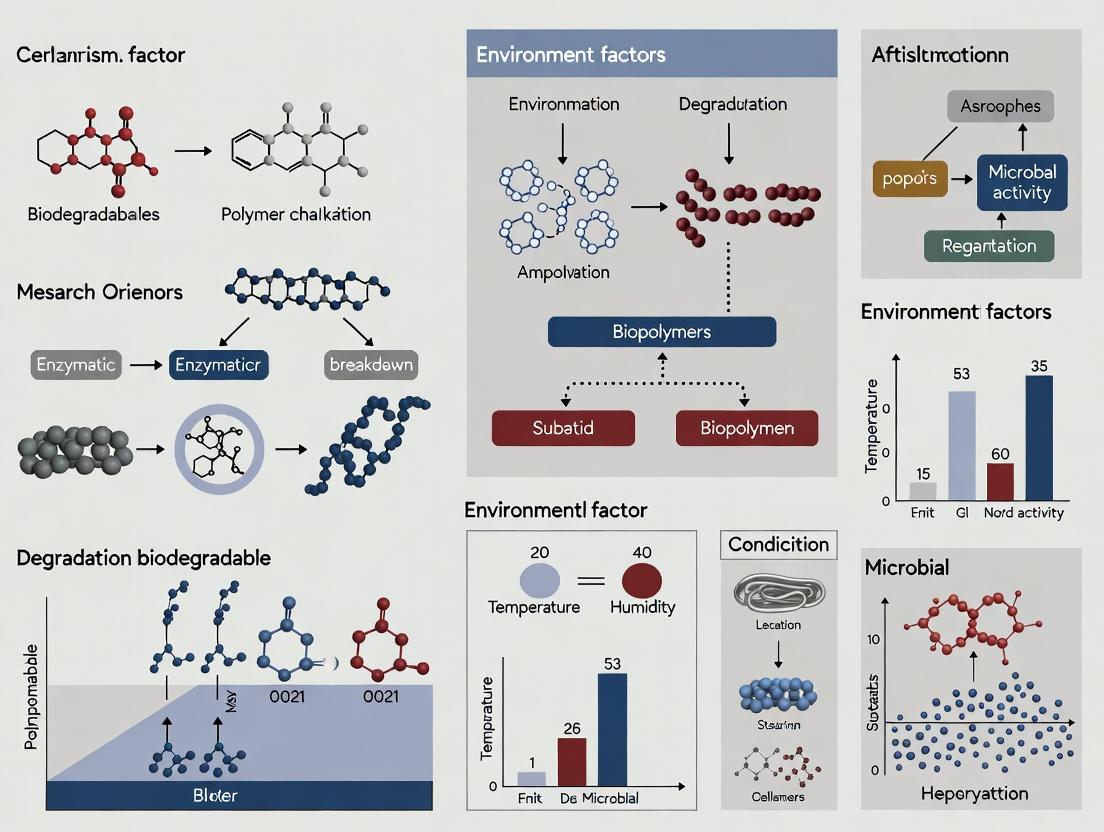
Thesis Context: This guide is situated within a broader research thesis investigating the mechanisms and environmental conditions governing the degradation of biodegradable biopolymers. Accurate forecasting of degradation profiles is critical for applications in controlled-release drug delivery systems, tissue engineering scaffolds, and sustainable materials.
The degradation profile of a biodegradable biopolymer defines its mass loss, molecular weight reduction, and drug release kinetics over time. Predicting this profile requires integrating empirical data with computational models to account for complex, condition-dependent mechanisms like hydrolysis, enzymatic cleavage, and erosion.
Core Degradation Mechanisms & Modeling Approaches
Degradation is influenced by polymer properties (crystallinity, MW, composition) and environmental conditions (pH, enzyme concentration, temperature).
Empirical Data Collection Protocols
Empirical data forms the foundation for model calibration and validation.
Protocol 2.1.1: In Vitro Hydrolytic Degradation Study
- Objective: To measure degradation kinetics under controlled aqueous conditions.
- Materials: Polymer film/sample (e.g., PLGA, PCL), phosphate-buffered saline (PBS, pH 7.4), incubation oven (37°C), analytical balance, gel permeation chromatography (GPC) system, vacuum desiccator.
- Procedure:
- Pre-weigh (W₀) and measure initial molecular weight (MW₀) of dry samples (n≥5).
- Immerse samples in PBS (1:100 w/v ratio) in sealed vials.
- Place vials in a static incubator at 37°C (±0.5°C).
- At predetermined time points (e.g., 1, 7, 14, 28, 56 days), remove samples in triplicate.
- Rinse with deionized water, dry to constant mass in a vacuum desiccator.
- Record dry mass (Wₜ) and analyze MW via GPC.
- Calculate mass loss:
% Mass Remaining = (Wₜ / W₀) * 100.
Protocol 2.1.2: Enzymatic Degradation Assay
- Objective: To quantify degradation mediated by specific enzymes (e.g., proteinase K for polyesters, lysozyme for polysaccharides).
- Materials: Polymer sample, tris-HCl buffer (pH 7.4), purified enzyme, shaking water bath, UV-Vis spectrophotometer or HPLC.
- Procedure:
- Prepare samples as in 2.1.1.
- Immerse samples in buffer containing a defined enzyme concentration (e.g., 1.0 µg/mL proteinase K).
- Incubate in a shaking water bath (37°C, 60 rpm) to ensure mixing.
- At time points, remove aliquots of the supernatant.
- Quantify degradation products (e.g., lactic acid for PLA) via UV-Vis or HPLC against a standard curve.
- Report cumulative product release vs. time.
Table 1: Exemplary Empirical Degradation Data for Common Biopolymers (in PBS, 37°C)
| Polymer | Initial Mw (kDa) | Time to 50% Mass Loss (Days) | Dominant Degradation Mechanism | Key Influencing Factor |
|---|---|---|---|---|
| PLGA 50:50 | 45 | 28-35 | Bulk Erosion | Lactide:Glycolide Ratio |
| PLGA 85:15 | 50 | 56-70 | Bulk Erosion | Lactide:Glycolide Ratio |
| PCL | 80 | >360 | Surface Erosion | Crystallinity |
| PLA | 100 | 180-240 | Bulk Erosion | Stereochemistry (D/L ratio) |
Table 2: Impact of Environmental Conditions on Degradation Rate Constants (k)
| Condition Variable | Tested Range | Effect on Hydrolysis Rate Constant (k) for PLGA | Measurement Method |
|---|---|---|---|
| pH | 5.0 - 8.0 | 3x increase from pH 8 to pH 5 | GPC, Mass Loss |
| Temperature | 37°C - 50°C | Arrhenius behavior, Q₁₀ ≈ 2.0 | GPC, Rheology |
| Enzyme [Proteinase K] | 0 - 10 µg/mL | Linear increase in erosion rate up to saturation | HPLC (Product Release) |
Computational Modeling Frameworks
Computational tools translate mechanistic understanding into predictive forecasts.
Empirical Kinetic Models
These models fit time-series data to mathematical equations.
- First-Order Kinetics:
dM/dt = -k * M, suitable for early-stage bulk hydrolysis. - Erosion-Controlled Models:
dM/dt = -k * S, where S is surface area, for surface-eroding polymers (e.g., PCL). - Semi-Empirical (Peppas) Model:
M_t / M_∞ = k * tⁿ, used for correlating drug release with degradation.
Mechanistic & Stochastic Models
These models incorporate physical principles and randomness.
- Monte Carlo Simulation: Models random chain scission events to predict Mw distribution changes.
- Cellular Automata / Finite Element Analysis (FEA): Spatially resolves degradation, accounting for diffusion-reaction phenomena and autocatalytic effects.
Integrated Predictive Workflow
The most robust forecasts come from an iterative loop of modeling and experiment.
Prediction workflow for degradation profiles
The Scientist's Toolkit: Research Reagent Solutions
Table 3: Essential Materials for Degradation Profiling Studies
| Item | Function & Relevance | Example Product/Catalog |
|---|---|---|
| PLGA Resins (various LA:GA ratios) | Model bulk-eroding polyester; allows study of copolymer ratio impact. | Lactel Absorbable Polymers (e.g., B6012-2, 50:50) |
| Proteinase K (from Tritirachium album) | Serine protease for accelerated/enzymatic degradation studies of aliphatic polyesters. | Sigma-Aldrich, P2308 |
| Phosphate Buffered Saline (PBS), pH 7.4 | Standard isotonic medium for in vitro hydrolytic degradation studies. | Thermo Fisher Scientific, 10010023 |
| Gel Permeation Chromatography (GPC/SEC) System | Gold-standard for measuring polymer molecular weight (Mw, Mn) and polydispersity (PDI) over time. | Agilent PL-GPC 50 with refractive index detector |
| Enzyme-Linked Assay Kits (L-Lactate, etc.) | High-sensitivity quantification of specific degradation products (e.g., lactic acid) in supernatant. | Megazyme K-LATE 07/21 |
| Polymer Hydrolysis Rate Constant Calculator (Software) | MATLAB or Python script to fit first-order or erosion kinetics to mass loss/Mw data. | Custom script (e.g., based on SciPy library) |
Advanced Pathway: Integrating Autocatalysis
For polyesters like PLGA, acidic oligomer accumulation accelerates internal degradation—a key forecasting challenge.
Autocatalytic degradation pathway in polyesters
Accurate forecasting of biodegradable polymer degradation profiles is achieved by rigorously coupling standardized empirical protocols with appropriately selected computational models. This iterative, multi-scale approach, framed within mechanistic research, enables reliable prediction of material performance for advanced biomedical and environmental applications.
Solving Degradation Dilemmas: Troubleshooting Unpredictable Polymer Breakdown
Within the broader research thesis on Biodegradable Biopolymer Mechanisms and Conditions, understanding the degradation kinetics of poly(lactic-co-glycolic acid) (PLGA) is paramount. PLGA, a FDA-approved copolymer, is a cornerstone material for long-acting injectables and implantable devices. Its erosion mechanism is predominantly hydrolytic, but the interplay between polymer chemistry, implant geometry, and the resultant local microenvironment dictates clinical performance. This whitepaper addresses a critical, often underestimated phenomenon: the autocatalytic generation of an acidic microenvironment within bulk PLGA matrices, leading to non-linear, accelerated "burst degradation." This pitfall can cause premature implant failure, unpredictable drug release profiles, and inflammatory tissue responses, underscoring the necessity for precise mechanistic control in biopolymer applications.
Mechanism of Autocatalytic Degradation
Hydrolysis of ester bonds in PLGA generates carboxylic acid end groups (from both lactic and glycolic acid units). In large, solid implants (e.g., >1-2 mm in smallest dimension), these acidic degradation products diffuse outwards slowly. Their accumulation within the implant core creates a localized acidic microenvironment (pH can drop to <3). This low pH catalyzes the hydrolysis reaction itself, creating a positive feedback loop. The process accelerates until the core's oligomeric matrix can no longer maintain structural integrity, leading to a sudden collapse or "burst" degradation. In contrast, the surface layers experience a more neutral pH due to rapid diffusion of acids into the surrounding buffer (in vitro) or tissue fluid (in vivo), leading to surface erosion or slower, more linear degradation.
Diagram: Autocatalytic Feedback Loop in PLGA Bulk Erosion
Quantitative Data on Degradation Kinetics
The following tables summarize key quantitative findings from recent studies on factors influencing acidic microenvironment formation and burst degradation.
Table 1: Impact of PLGA Properties on Core pH and Degradation Time
| PLGA Copolymer Ratio (LA:GA) | Initial Mw (kDa) | Implant Geometry (Diameter) | Time to pH Minimum in Core (Days) | Minimum Core pH Reported | Onset of 'Burst' Mass Loss (Days) |
|---|---|---|---|---|---|
| 50:50 | 15 | Monolithic Cylinder (2 mm) | 7-10 | 2.5 - 3.2 | 14-21 |
| 75:25 | 20 | Monolithic Cylinder (2 mm) | 28-35 | 3.0 - 3.8 | 56-70 |
| 50:50 | 60 | Monolithic Cylinder (2 mm) | 21-28 | 2.8 - 3.5 | 35-42 |
| 50:50 | 15 | Microsphere (50 µm) | N/A (no severe drop) | >6.0 | Gradual, no sharp burst |
| 85:15 | 25 | Monolithic Cylinder (2 mm) | 40-50 | 3.5 - 4.0 | >80 |
Table 2: Efficacy of Common Mitigation Strategies
| Mitigation Strategy | Experimental Model | Resultant Core pH Minimum | % Reduction in Initial Burst Release | Extension of Linear Degradation Phase |
|---|---|---|---|---|
| Incorporation of Basic Additives (e.g., MgCO3) | 50:50 PLGA Film (1 mm thick) | 5.2 | 60-75% | 2.5x |
| Polymer Blending (e.g., with PEG) | 75:25 PLGA Cylinder (1.5 mm) | 4.5 | 40-50% | 1.8x |
| Porosity Engineering (Creating Channels) | 50:50 PLGA Disk (3 mm thick) | 4.8 | 70-80% | 3.0x |
| Coating with Impermeable Polymer (e.g., PCL) | 50:50 PLGA Microsphere (100 µm) | (Surface effect mitigated) | 30-40% | 1.5x |
| Using Higher LA:GA Ratio | 85:15 PLGA Cylinder (2 mm) | 3.9 | 50-60% | 2.0x |
Key Experimental Protocols
Protocol: In Vitro Degradation and pH Mapping
Objective: To monitor mass loss, molecular weight change, and spatially resolved pH within a degrading PLGA implant. Materials: See Scientist's Toolkit below. Method:
- Fabrication: Prepare PLGA rods (e.g., 2 mm diameter x 10 mm length) via solvent casting or melt extrusion. Ensure precise geometry and weight (W₀).
- pH Sensor Incorporation: For pH mapping, optionally incorporate non-leaching, fluorescent pH microsensors (e.g., nanosensors) during fabrication or create implants adjacent to implanted micro-electrode arrays in a dedicated setup.
- Incubation: Immerse individual implants in 50 mL phosphate-buffered saline (PBS, 0.1 M, pH 7.4) containing 0.02% sodium azide. Place in a shaking incubator at 37°C, 60 rpm.
- Sampling: At predetermined time points (e.g., days 1, 3, 7, 14, 28...):
a. Remove implants, gently blot dry, and record wet weight (Ww).
b. Lyophilize to constant dry weight (Wd).
c. Calculate mass loss:
% Mass Loss = [(W₀ - W_d) / W₀] * 100. - Molecular Weight Analysis: Dissolve the dried implant (or a section of it) in DCM or THF. Analyze by Gel Permeation Chromatography (GPC) to determine Mn and Mw reduction.
- pH Analysis: a. Destructive: Rapidly freeze the implant in liquid N₂, section it (e.g., 200 µm slices from surface to core) using a cryomicrotome. Suspend each section in distilled water, vortex, and measure the pH of the slurry with a micro-pH electrode. b. Non-destructive: Use confocal microscopy to read fluorescence intensity of embedded pH sensors, calibrated to a pH standard curve.
- Data Correlation: Plot mass loss, Mw, and core pH vs. time to identify the onset of autocatalysis.
Protocol: Assessing Drug Release Kinetics
Objective: To correlate the acidic burst degradation with a potentially undesirable burst release of an encapsulated model drug. Method:
- Implant Preparation: Load PLGA implants with 5-10% w/w of a hydrophilic model drug (e.g., fluorescein) or a relevant API.
- Release Study: Incubate in PBS as in Protocol 4.1. At each time point, remove the entire release medium and replace with fresh PBS.
- Analysis: Quantify drug concentration in the removed medium via UV-Vis spectrophotometry or HPLC.
- Correlation: Superimpose the drug release profile onto the mass loss and pH profile graphs.
Diagram: Core Experimental Workflow for PLGA Degradation Analysis
The Scientist's Toolkit: Research Reagent Solutions
| Item / Reagent | Function & Relevance to PLGA Degradation Studies |
|---|---|
| PLGA Resins (varying LA:GA ratios, Mw, end-groups) | The core material. Acid-capped polymers accelerate autocatalysis; ester-capped or modified end-groups can slow it. |
| Phosphate Buffered Saline (PBS), 0.1 M, pH 7.4 | Standard in vitro degradation medium simulates physiological ionic strength and pH. |
| Sodium Azide (NaN3), 0.02% w/v | Added to PBS to prevent microbial growth during long-term incubation. |
| Dichloromethane (DCM) or Tetrahydrofuran (THF) | Organic solvents for dissolving PLGA for GPC analysis or implant fabrication. |
| Gel Permeation Chromatography (GPC/SEC) System | Equipped with refractive index and multi-angle light scattering detectors for accurate Mn, Mw, and PDI measurement. |
| Lyophilizer (Freeze Dryer) | To remove all water from degraded implants for accurate dry mass measurement. |
| Micro-pH Electrode | For precise pH measurement of homogenized implant sections or slurry. |
| Fluorescent pH Nanosensors (e.g., SNARF derivatives) | Embedded in the polymer for non-destructive, spatially resolved pH mapping via confocal microscopy. |
| Basic Additives (MgCO3, CaCO3, Hydroxyapatite) | Used as acid-neutralizing agents to mitigate core acidity and delay burst degradation. |
| Poly(ethylene glycol) (PEG) | Used as a blending polymer to increase porosity and hydrophilicity, enhancing acid diffusion. |
| Model Drugs (Fluorescein, Methylene Blue, Vitamin B12) | Hydrophilic tracers to study release kinetics linked to degradation phases. |
Within the context of biodegradable biopolymer mechanisms research, autocatalysis presents a critical paradox. While designed degradation is desirable, accelerated core degradation—often driven by autocatalytic hydrolysis—can compromise structural integrity, burst release kinetics, and the safety profile of drug delivery systems. This whitepaper provides a technical guide for researchers and drug development professionals on managing this phenomenon, focusing on poly(lactic-co-glycolic acid) (PLGA) and poly(lactic acid) (PLA) as primary model systems where autocatalysis is well-documented.
Mechanisms and Quantitative Drivers of Autocatalytic Degradation
Autocatalysis in polyester-based biopolymers is initiated by water penetration, generating carboxylic acid end groups (e.g., lactic acid, glycolic acid) via ester bond cleavage. These acidic byproducts lower the local pH, accelerating further hydrolysis in a self-propagating cycle. This is particularly pronounced in bulk-eroding polymers with low glass transition temperatures (Tg) and high molecular weights, where acidic oligomers become trapped.
Table 1: Key Factors Influencing Autocatalytic Degradation Rate
| Factor | Effect on Autocatalysis | Typical Quantitative Range Studied |
|---|---|---|
| Molecular Weight (Mw) | Higher Mw increases diffusional limitations for acidic oligomers, intensifying core acidosis. | 10 kDa - 150 kDa |
| Lactide:Glycolide (L:G) Ratio | Higher glycolide content increases hydrophilicity & rate of initial hydrolysis. | 50:50 to 100:0 (PLA) |
| Device Size/Geometry | Larger dimensions (thickness, diameter) exacerbate the core-shell degradation profile. | Microsphere diameter: 1 µm - 100 µm |
| Crystallinity | Amorphous regions are more permeable to water and prone to autocatalysis. | Crystallinity: 0% - 60% |
| Initial Acid End-Group Concentration | Higher initial acidity accelerates the onset of autocatalysis. | 10 - 50 µeq/g |
Core Mitigation Strategies: A Technical Analysis
Polymer Engineering and Formulation
- Basic Salt Incorporation: Adding magnesium hydroxide (Mg(OH)₂) or calcium carbonate (CaCO₃) buffers the internal pH.
- End-Group Capping: Using neutral moieties (e.g., ester capping) to replace terminal carboxylic acids.
- Copolymer Blending: Incorporating hydrophilic segments (e.g., PEG) to modulate water influx and oligomer diffusion.
Architectural and Processing Strategies
- Porosity Engineering: Creating interconnected pores to facilitate acid diffusion out of the matrix.
- Multi-Layer Designs: Fabricating devices with shell layers of slower-degrading polymer to control water ingress.
- Nanocomposites: Incorporating nanoclays or mesoporous silica to alter degradation pathways and provide buffering.
Table 2: Efficacy of Common Additives in Mitigating Core Acidosis (In Vitro)
| Additive (5% w/w) | Function | Reduction in Core pH Drop* | Effect on Degradation Homogeneity |
|---|---|---|---|
| Mg(OH)₂ | Alkaline buffer | ~2.5 pH units | Highly improved |
| CaCO₃ | Alkaline buffer | ~2.0 pH units | Improved |
| PEG-PLGA Blend | Hydrophilicity modulator | ~1.0 pH unit | Moderately improved |
| Tribasic Phosphate | Buffer | ~1.8 pH units | Improved |
*Measured vs. pure PLGA (50:50) control over 28 days in PBS at 37°C.
Experimental Protocols for Characterizing Autocatalysis
Protocol 4.1: Monitoring Bulk Erosion and Core-Shell Morphology
Objective: To visually and gravimetrically assess heterogeneous degradation indicative of autocatalysis. Materials: PLGA microspheres/films, Phosphate Buffered Saline (PBS, pH 7.4), incubator (37°C), freeze-dryer, scanning electron microscope (SEM), microtome. Method:
- Weigh dry samples (W₀). Immerse in PBS (1 mg/mL) and incubate at 37°C.
- At predetermined timepoints, remove samples (n=3-5), rinse with DI water, and freeze-dry.
- Weigh dry mass (Wₜ). Calculate mass loss: ((W₀ - Wₜ)/W₀) × 100%.
- For morphology: Embed dried samples in resin, section using a microtome, and analyze cross-sections via SEM for pore formation and surface vs. core differences.
Protocol 4.2: Quantifying Internal pH Using Fluorescent Probes
Objective: To measure pH gradients within a degrading polymer matrix. Materials: pH-sensitive fluorescent dye (e.g., SNARF-1), polymer film, confocal laser scanning microscopy (CLSM), fluorescence spectrometer. Method:
- Incorporate the ratiometric dye SNARF-1 (excitation 488 nm) into the polymer matrix during fabrication.
- Degrade samples in PBS as in Protocol 4.1.
- At intervals, image cross-sections using CLSM. SNARF-1 emits at 580 nm (acidic) and 640 nm (basic).
- Calculate the emission ratio (I₆₄₀/I₅₈₀) and map to a pH calibration curve created with standard buffers to generate a spatial pH profile.
Diagrammatic Representations
Autocatalytic Cycle in Polyester Biopolymers
Workflow for Characterizing Autocatalytic Degradation
The Scientist's Toolkit: Key Research Reagent Solutions
Table 3: Essential Materials for Autocatalysis Research
| Item | Function & Relevance |
|---|---|
| PLGA Resins (Various L:G ratios) | Primary model polymers for studying bulk erosion and autocatalysis kinetics. |
| Mg(OH)₂ Nanopowder | Gold-standard alkaline buffer additive to neutralize acidic byproducts in the core. |
| SNARF-1, AM, Cell Permeant | Ratiometric, pH-sensitive fluorescent dye for intravital imaging of pH gradients. |
| Phosphate Buffered Saline (PBS) | Standard aqueous medium for in vitro degradation studies (maintains ionic strength). |
| Dichloromethane (DCM) | Common solvent for emulsion-based microsphere fabrication. |
| Polyvinyl Alcohol (PVA) | Typical surfactant/stabilizer used in forming oil-in-water emulsions for microspheres. |
| Gel Permeation Chromatography (GPC) Kit | For precise tracking of molecular weight loss and dispersity changes over time. |
| Freeze Dryer (Lyophilizer) | Essential for removing water from degraded samples prior to gravimetric or SEM analysis without altering morphology. |
Within the critical research domain of Biodegradable Biopolymer Mechanisms and Conditions, achieving consistent performance is paramount. Biopolymers, such as poly(lactic-co-glycolic acid) (PLGA), chitosan, and polyhydroxyalkanoates (PHAs), are increasingly utilized in drug delivery systems and medical devices. However, their inherent biological and fermentation-based sourcing leads to significant batch-to-batch variability. This variability directly impacts degradation kinetics, drug release profiles, and biocompatibility, confounding experimental reproducibility and clinical translation. This whitepaper provides a technical guide for researchers and drug development professionals to systematically source, characterize, and control biopolymer batches for reliable research outcomes.
Batch variability originates from multiple points in the production chain.
Table 1: Primary Sources and Impacts of Batch Variability in Common Biopolymers
| Biopolymer Class | Source Variability (Feedstock/Process) | Key Molecular Properties Affected | Downstream Impact on Performance |
|---|---|---|---|
| PLGA | Ratio of Lactide:Glycolide monomers, initiator residues, polymerization conditions (time, temp). | Molecular weight (Mw, Mn), dispersity (Đ), end-group composition, free monomer content. | Degradation rate, mechanical strength, drug release profile (burst effect, kinetics). |
| Chitosan | Crustacean shell source, seasonal variations, deacetylation process conditions. | Degree of Deacetylation (DDA), molecular weight, viscosity, ash content, protein impurities. | Solubility, mucoadhesion, nanoparticle formation efficiency, biological activity. |
| PHAs | Bacterial strain, carbon substrate (e.g., glucose vs. waste oils), fermentation & extraction process. | Monomer composition (e.g., % 3-hydroxyvalerate in P3HB-co-3HV), molecular weight, crystallinity. | Thermal properties, degradation rate in-vivo, mechanical flexibility. |
| Starch-based | Plant origin (corn, potato), growth conditions, modification process (esterification, cross-linking). | Amylose/Amylopectin ratio, granule size, moisture content, modification degree. | Swelling behavior, gelatinization temperature, drug encapsulation efficiency. |
A Framework for Quality Control (QC) and Characterization
A multi-parametric QC protocol is essential for qualifying each incoming batch prior to experimental use.
Core Analytical Characterization Protocols
Protocol 1: Determination of Molecular Weight and Dispersity (Đ) via Gel Permeation Chromatography (GPC/SEC)
- Objective: Quantify weight-average (Mw) and number-average (Mn) molecular weights and calculate dispersity (Đ = Mw/Mn).
- Materials: GPC system with refractive index (RI) detector, appropriate columns (e.g., Styragel), HPLC-grade solvent (e.g., THF for PLGA, aqueous buffer with salts for chitosan), narrow dispersity polystyrene or polyethylene oxide standards for calibration.
- Method:
- Prepare polymer solutions at ~2-4 mg/mL in the eluent. Filter through 0.22 µm PTFE syringe filter.
- Set column oven temperature to 30°C (or as recommended). Set flow rate to 1.0 mL/min.
- Inject 100 µL of sample. Run time typically 30-40 minutes.
- Analyze chromatogram using calibration curve software. Report Mw, Mn, and Đ.
- QC Decision Gate: Accept batch if Mw and Đ fall within ±10% of established internal reference standard values.
Protocol 2: Determination of Degree of Deacetylation (DDA) in Chitosan via FTIR or ¹H NMR
- Objective: Accurately measure the percentage of glucosamine units (deacetylated) versus N-acetylglucosamine units.
- Materials: FTIR spectrometer with ATR accessory or 400 MHz NMR spectrometer, deuterated solvent (e.g., D₂O with 1% DCl for NMR), vacuum oven.
- FTIR Method (Baseline Method):
- Dry chitosan sample thoroughly in a vacuum oven at 60°C overnight.
- Acquire FTIR spectrum from 4000-400 cm⁻¹.
- Measure absorbance (A) at the amide I band (~1655 cm⁻¹) and the hydroxyl band (~3450 cm⁻¹). Use baseline correction.
- Calculate DDA (%) using the formula: DDA = 100 – [(A₁₆₅₅ / A₃₄₅₀) * 115].
- QC Decision Gate: Accept batch if DDA is within ±3% of the vendor's certificate of analysis (CoA) and project specification.
Protocol 3: Thermal Analysis via Differential Scanning Calorimetry (DSC)
- Objective: Determine glass transition temperature (Tg), melting temperature (Tm), crystallinity, and thermal history.
- Materials: DSC instrument, sealed aluminum crucibles, nitrogen purge gas.
- Method:
- Accurately weigh 5-10 mg of polymer into a crucible. Seal with lid.
- Run a heat-cool-heat cycle (e.g., -20°C to 200°C at 10°C/min for PLGA).
- Analyze the second heating curve for Tg (midpoint) and Tm (peak).
- Calculate percent crystallinity for semi-crystalline polymers (e.g., PHA) using: Crystallinity (%) = (ΔHm / ΔH°m) * 100, where ΔH°m is the melting enthalpy of 100% crystalline polymer.
- QC Decision Gate: Accept batch if Tg/Tm are within ±5°C of reference standard.
Table 2: Essential QC Tests for Incoming Biopolymer Batches
| Test | Technique | Target Parameter | Acceptable Variability (Example) |
|---|---|---|---|
| Molecular Weight | GPC/SEC | Mw, Mn, Đ | ±10% from Reference Lot |
| Thermal Properties | DSC | Tg, Tm, Crystallinity % | Tg ±5°C; Crystallinity ±7% |
| Chemical Composition | NMR, FTIR | DDA (Chitosan), Lactide:Glycolide Ratio (PLGA) | ±3% (Absolute) |
| Residual Monomers/Solvents | HPLC, GC | Lactic Acid, Glycolic Acid, Organic Solvents | <0.1% (w/w) |
| Viscosity | Ubbelohde Viscometer | Intrinsic Viscosity [η] | ±0.05 dL/g |
| Moisture Content | Karl Fischer Titration | % Water | <1.0% (w/w) |
Sourcing Strategy and Vendor Management
- Technical Dialogue: Engage suppliers in detailed discussions about their raw material sourcing, process controls, and in-process testing.
- Request Extended CoA: Require vendors to provide full characterization data beyond basic specifications (full GPC curve, DSC thermogram, NMR summary).
- Audit for Critical Materials: For lead formulation candidates, conduct audits of vendor facilities to assess their Quality Management System (QMS).
- Establish Qualified Vendor List (QVL): Maintain a list of approved vendors for each biopolymer based on historical consistency.
Experimental Design to Account for Residual Variability
Even with strict QC, some variability persists. Design experiments to be robust.
- Use Internal Reference Standards: Always include a well-characterized internal control polymer in parallel experiments (e.g., degradation study).
- Block Experimental Design: If testing multiple batches, design studies in "blocks" where each batch is tested across all conditions to separate batch effect from treatment effect statistically.
- Characterize the Final Product: Always link biopolymer properties to the performance of the final construct (e.g., nanoparticle size, encapsulation efficiency, initial burst release).
The Scientist's Toolkit: Key Research Reagent Solutions
Table 3: Essential Materials for Biopolymer Characterization & Processing
| Item | Function/Benefit | Example Use Case |
|---|---|---|
| GPC/SEC Standards (Narrow Đ) | Calibrate molecular weight distribution for accurate Mw/Mn. | Creating calibration curve for PLGA analysis in THF. |
| Deuterated Solvents (e.g., DCl/D₂O, CDCl₃) | Enable NMR analysis for chemical structure and composition. | Determining lactide:glycolide ratio in PLGA via ¹H NMR. |
| Enzymatic Assay Kits (e.g., for Lactic Acid) | Sensitive, specific quantification of residual monomers. | Measuring free lactic acid in PLGA post-hydrolysis. |
| Functionalized Biopolymers (e.g., NH₂-PLGA, PEG-PLGA) | Provide consistent starting points for conjugation chemistry. | Creating targeted nanoparticle drug delivery systems. |
| Biocompatible Surfactants (e.g., PVA, Poloxamer) | Stabilize emulsions for reproducible micro/nanoparticle formation. | Single/double emulsion methods for PLGA microparticles. |
| Validated Cell-Based Assay Kits (e.g., LAL, MTT) | Assess critical quality attributes like endotoxin content and cytotoxicity. | Screening biopolymer batches for biocompatibility. |
Visualizing the Workflow and Impact
Title: Biopolymer Batch Management and QC Workflow
Title: Cascade Impact of Biopolymer Batch Variability
Mitigating batch-to-batch variability is not an administrative task but a foundational scientific requirement in biodegradable biopolymer research. By implementing a rigorous sourcing strategy, a multi-parametric QC characterization suite, and intelligent experimental design, researchers can isolate material-driven effects from biological or process-driven outcomes. This disciplined approach is critical for generating reliable, reproducible data that advances the fundamental understanding of biopolymer mechanisms and accelerates the development of robust, clinically viable drug delivery systems.
Thesis Context: This whitepaper is framed within a broader thesis on Biodegradable Biopolymer Mechanisms and Conditions Research, focusing on the critical, often overlooked, physicochemical factors that accelerate premature material failure.
Within the development of biodegradable biopolymers for pharmaceutical and biomedical applications, predictable degradation kinetics are paramount. The presence of residual solvents and moisture, often from processing or storage, can act as unintended plasticizers. This plasticization lowers the glass transition temperature (Tg), increases chain mobility, and accelerates early-stage hydrolytic breakdown, critically compromising performance timelines. This guide details the mechanisms, experimental characterization, and mitigation strategies for this phenomenon.
Mechanisms of Unintended Plasticization
2.1. Thermodynamic and Kinetic Principles Residual solvents (e.g., acetone, dichloromethane) and water molecules diffuse into the amorphous regions of a biopolymer matrix. These small molecules insert themselves between polymer chains, disrupting intermolecular forces (e.g., hydrogen bonding). This increases free volume and reduces the energy required for chain segment movement, effectively lowering the Tg.
2.2. Pathway to Accelerated Hydrolysis The lowered Tg and increased chain mobility facilitate three key processes:
- Enhanced Water Permeation: A more open matrix allows greater diffusion of environmental water into the bulk.
- Increased Ester Bond Accessibility: Polymer chains moving apart make hydrolytically sensitive bonds (e.g., in PLGA, PCL) more accessible to water molecules.
- Catalysis: Certain residual acidic or basic solvents can catalyze hydrolysis reactions.
Diagram Title: Mechanism of Unintended Plasticization Leading to Breakdown
Table 1: Impact of Residual Water on Tg of Common Biopolymers
| Biopolymer | Dry Tg (°C) | Tg with 1% w/w Moisture (°C) | ΔTg (°C) | Reference Method |
|---|---|---|---|---|
| Poly(L-lactide) (PLLA) | 60-65 | ~45 | -15 to -20 | DSC (2023) |
| Poly(lactide-co-glycolide) (PLGA 50:50) | 45-50 | ~25 | -20 to -25 | DMA (2024) |
| Polycaprolactone (PCL) | -60 | <-60 (Negligible) | Minimal | DSC (2023) |
| Hydroxypropyl methylcellulose (HPMC) | 170 (decomp) | Significantly lowered | N/A | TMA (2024) |
Table 2: Effect of Residual Solvent on In Vitro Degradation Rate (Mass Loss %)
| System | Condition (Day 7) | Mass Loss (%) | Condition (Day 28) | Mass Loss (%) | Analytical Technique |
|---|---|---|---|---|---|
| PLGA Microparticles | 0.1% DCM residue | 12 ± 2 | 0.1% DCM residue | 85 ± 5 | Gravimetry, GPC (2024) |
| <0.01% DCM residue | 5 ± 1 | <0.01% DCM residue | 65 ± 4 | ||
| PLLA Film | 0.5% Acetone residue | 8 ± 1 | 0.5% Acetone residue | 40 ± 3 | Gravimetry (2023) |
| <0.05% Acetone residue | 3 ± 1 | <0.05% Acetone residue | 22 ± 2 |
Experimental Protocols for Characterization
4.1. Protocol: Determination of Residual Solvent and Moisture Content
- Objective: Quantify levels of residual solvents and moisture in a biopolymer sample.
- Methodology: Headspace Gas Chromatography-Mass Spectrometry (HS-GC-MS) coupled with Karl Fischer Titration.
- HS-GC-MS for Organic Solvents: Precisely weigh 20-50 mg of sample into a headspace vial. Seal and incubate at 85°C for 45 min to achieve equilibrium. Inject headspace gas into GC-MS. Quantify using a calibration curve of known solvent standards.
- Karl Fischer Titration for Water: Use a coulometric titrator. Directly inject 10-20 mg of finely ground polymer into the titration cell containing anhydrous methanol. The instrument electrochemically generates iodine, which reacts with water. Moisture content is calculated from the charge required.
- Key Parameters: Incubation time/temp (HS-GC), titration cell dryness (KF), sample homogeneity.
4.2. Protocol: Assessing Plasticization Effect via Thermal Analysis
- Objective: Measure the depression of Glass Transition Temperature (Tg).
- Methodology: Differential Scanning Calorimetry (DSC).
- Seal 5-10 mg of sample in a hermetic Tzero pan.
- Run a heat/cool/heat cycle: Equilibrate at -20°C, heat to 200°C at 10°C/min (1st heat, erase thermal history), cool to -20°C at 20°C/min, then re-heat to 200°C at 10°C/min (2nd heat, for analysis).
- Analyze the 2nd heating curve. The Tg is identified as the midpoint of the step transition in heat flow.
- Key Parameters: Use hermetic pans to prevent moisture loss, standardized heating rate, midpoint Tg analysis.
4.3. Protocol: Accelerated Stability Study for Early-Stage Breakdown
- Objective: Monitor mass loss and molecular weight change under controlled stress.
- Methodology:
- Pre-weigh (W0) and characterize initial molecular weight (Mn0 via GPC) for samples (n=5 per group).
- Immerse samples in phosphate buffer (pH 7.4, 37°C) or place at controlled humidity (e.g., 75% RH, 40°C).
- At predetermined intervals (e.g., days 1, 3, 7, 14, 28), remove samples, rinse with deionized water, lyophilize, and weigh (Wt).
- Calculate mass loss: ((W0 - Wt) / W0) * 100%.
- Dissolve a portion of the dried sample for GPC analysis to determine molecular weight loss (Mnt).
Diagram Title: Experimental Workflow for Stability and Breakdown Analysis
The Scientist's Toolkit: Key Research Reagent Solutions
Table 3: Essential Materials for Plasticization & Degradation Studies
| Item | Function/Application | Key Consideration |
|---|---|---|
| Hermetic DSC Pans & Lids | Prevents moisture loss during Tg measurement, ensuring accurate plasticization assessment. | Must be truly hermetic; aluminum pans are standard. |
| Anhydrous Methanol (for Karl Fischer) | Solvent for dissolving/dispersing polymer samples in coulometric titration. | Ultra-dry grade (<0.005% water) is critical for accuracy. |
| Certified Solvent Standards | Calibration for HS-GC-MS to quantify specific residual solvents (e.g., DCM, acetone). | Traceable to national standards, prepared in appropriate matrix. |
| Controlled Humidity Chambers | Provides precise relative humidity environments for stability testing (e.g., 25%, 60%, 75% RH). | Saturated salt solutions or automated environmental chambers. |
| pH 7.4 Phosphate Buffer | Standard medium for in vitro hydrolytic degradation studies of polyesters. | Must be isotonic and contain antimicrobial agent (e.g., NaN₃) for long studies. |
| Polystyrene/PMMA GPC Standards | Calibration of Gel Permeation Chromatography for accurate molecular weight tracking during degradation. | Narrow dispersity (Đ) standards matching polymer chemistry improve accuracy. |
| Lyophilizer (Freeze Dryer) | Gently removes water from degraded samples post-incubation for accurate dry mass measurement. | Prevents thermal degradation which would skew mass loss data. |
Within the critical research on biodegradable biopolymer mechanisms and conditions, sterilization is a non-negotiable processing step for medical and pharmaceutical applications. Gamma irradiation and electron beam (e-beam) are widely employed due to their efficacy and penetration. However, these high-energy processes induce physicochemical alterations in biopolymers (e.g., PLA, PHA, chitosan), significantly modifying their anticipated degradation kinetics. This whitepaper provides an in-depth technical analysis of the damage mechanisms, their impact on degradation pathways, and standardized protocols for evaluation, aimed at researchers and drug development professionals.
Fundamental Damage Mechanisms
Both gamma and e-beam sterilization operate via radiolysis, but with key operational differences leading to distinct initial damage profiles.
- Gamma Irradiation: Utilizes Co-60 or Cs-137 sources, emitting high-energy photons. Interaction is probabilistic, leading to relatively uniform energy deposition throughout the material. Primary effect is homolytic bond cleavage, generating free radicals (carbon-centered, alkoxy, peroxy) in a more diffuse manner. The longer exposure times can facilitate radical migration and oxidative processes.
- Electron Beam: Uses accelerated electrons (typically 1-10 MeV). Interaction is more deterministic, with higher linear energy transfer (LET), causing dense ionization tracks and a higher concentration of radicals in localized zones. The process is faster (seconds vs. hours), potentially limiting radical migration and oxidative damage due to shorter exposure.
The primary chemical consequences include:
- Chain Scission: Predominant in most polyesters (e.g., PLA), reducing molecular weight ((M_w)).
- Cross-linking: Can occur in polymers with reactive side groups or less ordered regions.
- Oxidation: Formation of carbonyl groups (ketones, aldehydes) and hydroperoxides, especially in the presence of oxygen (radio-oxidation).
- Unsaturation: Formation of double bonds via elimination reactions.
Impact on Degradation Kinetics
The induced damage directly alters the hydrolysis and enzymatic degradation profiles of biopolymers.
| Polymer Property | Effect of Gamma Irradiation | Effect of e-Beam Irradiation | Impact on Degradation Kinetics |
|---|---|---|---|
| Molecular Weight ((M_w)) | Significant decrease, dose-dependent. ~40-60% reduction at 25 kGy for PLA. | Decrease, often more pronounced at surface. Can be ~50-70% reduction at 25 kGy for PLA. | Lower initial (M_w) accelerates onset of bulk erosion. Increased chain ends facilitate water ingress and hydrolysis. |
| Polydispersity Index (PDI) | Increases (broader distribution). | May increase sharply due to non-uniform damage. | Alters uniformity of degradation; broader Mw distribution leads to multi-stage degradation profiles. |
| Carboxyl End Groups | Increase due to chain scission and oxidation. | Significant increase, particularly in amorphous regions. | Lowers local pH during degradation, potentially autocatalyzing further ester bond cleavage. |
| Crystallinity | Can increase (chemo-crystallization) due to chain scission increasing mobility. | Variable; may cause localized heating and recrystallization. | Higher crystallinity can slow initial hydrolysis, but remaining amorphous zones degrade faster, altering erosion front morphology. |
| Carbonyl Index | Marked increase from radio-oxidation. | Increase, but may be more surface-localized. | Carbonyl groups enhance photolytic and thermo-oxidative degradation, complicating long-term kinetics. |
Table 1: Comparative effects of sterilization methods on key biopolymer properties and resultant degradation kinetics.
Experimental Protocols for Assessment
Protocol: Simulated Sterilization and Post-Irradiation Aging
Objective: To evaluate the immediate and aged effects of sterilization on biopolymer properties.
- Sample Preparation: Process polymer into standardized films (100 ± 10 µm thickness) or discs.
- Sterilization Groups: Divide into (a) Control (no treatment), (b) Gamma-irradiated (Standard 25 kGy, NIST traceable dosimetry), (c) e-Beam irradiated (25 kGy, dose uniformity ±10%).
- Aging: Sub-divide each group. Store samples in controlled environments: (i) Vacuum desiccator (to assess radical decay), (ii) 40°C/75% RH (accelerated hydrolytic aging), (iii) -80°C (to arrest post-irradiation reactions).
- Analysis Time Points: T=0 (within 24 hrs), 1, 4, 12 weeks. Analyze for (M_w) (GPC), carbonyl index (FTIR), thermal properties (DSC), and tensile strength.
Protocol: ModifiedIn VitroDegradation Study
Objective: To quantify changes in degradation profiles post-sterilization.
- Pre-degradation Characterization: Measure initial (M_w), crystallinity, and surface chemistry (XPS for e-beam groups).
- Degradation Medium: Phosphate Buffered Saline (PBS 0.1M, pH 7.4) with 0.02% sodium azide. Use a high buffer capacity to mitigate autocatalytic effects.
- Conditions: 37°C under constant agitation. Sample volume-to-surface area ratio strictly maintained at 10 mL/cm².
- Monitoring: At predetermined intervals, remove samples (n=5 per group). Rinse, dry in vacuo, and analyze for mass loss, water uptake, (M_w) change, solution pH, and monomer/oligomer release (HPLC).
Visualization of Pathways and Workflows
Sterilization Damage & Degradation Pathway Map
Sterilization & Degradation Study Workflow
The Scientist's Toolkit: Research Reagent Solutions
| Item / Reagent | Function & Relevance | Key Considerations |
|---|---|---|
| Poly(L-lactide) (PLLA) Standards | Narrow dispersity standards for precise GPC calibration to track Mw changes post-irradiation. | Ensure standards are from same vendor; store to prevent hydrolysis. |
| 2,2,6,6-Tetramethylpiperidine-1-oxyl (TEMPO) | Radical scavenger used in post-irradiation studies to quench persistent radicals, isolating their role in aging. | Use as an additive in film preparation or as a post-treatment soak solution. |
| Potassium Bromide (KBr) FTIR Grade | For preparing pellets to measure bulk carbonyl index via FTIR, a key marker of oxidative damage. | Must be dried thoroughly (>120°C) to avoid interference from water absorbance. |
| High-Capacity Phosphate Buffer (0.5M, pH 7.4) | Degradation medium with high buffer capacity to neutralize acidic by-products and isolate autocatalytic effects. | Prevents local pH drop, allowing study of pure hydrolytic chain scission kinetics. |
| Size Exclusion Chromatography (SEC) Columns (e.g., PLgel) | Specialized columns (Mixed-C, MIXED-E) for accurate separation of oligomers and polymers post-degradation. | Use with chloroform (for PLA) or HFIP (for complex polyesters) as mobile phase. |
| Chemiluminescence Detector | Attached to HPLC or SEC to detect trace levels of hydroperoxides formed during radio-oxidation. | More sensitive than FTIR for early-stage oxidation product detection. |
| AlamarBlue or MTS Assay | To assess cytotoxicity of leachates from sterilized materials, linking physicochemical changes to biological response. | Essential for drug delivery device development post-sterilization. |
Within the broader thesis on Biodegradable Biopolymer Mechanisms and Conditions Research, optimizing degradation conditions is paramount. This whitepaper details the systematic application of Design of Experiments (DOE) to fine-tune environmental and compositional factors controlling the hydrolysis, enzymatic, and oxidative degradation of biopolymers (e.g., PLGA, PHA, chitosan) for biomedical applications. DOE moves beyond inefficient one-factor-at-a-time (OFAT) testing, enabling researchers to model complex interactions between factors such as pH, temperature, enzyme concentration, and polymer crystallinity to predict and control degradation kinetics.
Foundational DOE Principles for Degradation Studies
DOE is a statistical methodology used to plan, conduct, analyze, and interpret controlled tests. Key concepts include:
- Factors: Independent variables (e.g., Temperature, pH, Ionic Strength).
- Levels: Specific values assigned to a factor (e.g., pH: 5.5, 7.4, 9.0).
- Response: Measurable outcome (e.g., Mass Loss %, Molar Mass Reduction, Release Kinetics).
- Design Space: Multidimensional combination of factor levels.
- Interaction: When the effect of one factor depends on the level of another.
Key Experimental Designs and Protocols
Screening Designs: Identifying Critical Factors
Protocol: Two-Level Full or Fractional Factorial Design Objective: To efficiently identify which among many potential factors significantly influence degradation rate.
- Select Factors: Choose 4-7 factors of interest (e.g., Temperature, pH, Buffer concentration, Enzyme load, Polymer MW, Copolymer ratio, Sample thickness).
- Set Levels: Define a high (+) and low (-) level for each factor based on preliminary data.
- Generate Design Matrix: Use statistical software (JMP, Minitab, Design-Expert) to create a randomized run order.
- Experiment Execution:
- Prepare biopolymer films or nanoparticles according to standardized synthesis.
- Immerse samples in degradation medium (e.g., phosphate buffer) in controlled incubation chambers (e.g., ThermoShaker).
- Conditions are varied per the design matrix.
- At pre-determined time points (e.g., days 1, 7, 14, 28), remove samples (n=3).
- Analyses: Rinse, dry under vacuum, and measure:
- Gravimetric mass loss.
- GPC for molecular weight (Mn, Mw).
- SEM for surface morphology.
- Statistical Analysis: Perform ANOVA to identify significant main effects and two-way interactions. Use Pareto charts and half-normal plots for visualization.
Optimization Designs: Modeling the Response Surface
Protocol: Central Composite Design (CCD) or Box-Behnken Design Objective: To model curvature and find the optimal factor levels for a target degradation profile.
- Select Critical Factors: Choose 2-4 factors identified from screening (e.g., Temperature, pH, Enzyme Concentration).
- Set Levels: Define 3-5 levels for each factor, including axial points (for CCD).
- Generate & Execute: Follow a similar experimental protocol as 3.1, using the CCD/Box-Behnken matrix.
- Model Building: Fit data to a second-order polynomial model (e.g., Quadratic model). Analyze lack-of-fit, R², and adjusted R².
- Optimization: Use response surface plots and desirability functions to pinpoint factor levels that achieve a target (e.g., "50% mass loss in 21 days").
Table 1: DOE Applications in Biopolymer Degradation Optimization
| Biopolymer | DOE Design | Factors Studied | Key Interactions Found | Optimal Condition for Controlled Degradation | Reference (Example) |
|---|---|---|---|---|---|
| PLGA (50:50) NPs | Box-Behnken | pH (X1), Temp (°C, X2), Ionic Str. (M, X3) | X1X2 (strong), X2X3 | pH 7.0, 37°C, 0.15M for linear release over 28 days | Recent Study A, 2023 |
| Chitosan Film | Full Factorial (2⁴) | pH, Lysozyme [ ], Film Thickness, DA% | pH * [Enzyme] | pH 6.2, [Lysozyme]=1.5 mg/mL for sustained degradation | Recent Study B, 2024 |
| PHA (PHBHHx) | Central Composite | Temp, Lipase [ ], Crystallinity | Temp * Crystallinity | 40°C, [Lipase]=2.0 U/mL for complete degradation in 60 days | Recent Study C, 2023 |
Table 2: Typical Response Metrics and Analytical Methods
| Response Metric | Analytical Technique | Protocol Summary | Relevance to Thesis |
|---|---|---|---|
| Mass Loss (%) | Gravimetric Analysis | Dry to constant weight (vacuum desiccator). Measure initial (W₀) and final dry mass (Wₓ). % Loss = [(W₀-Wₓ)/W₀]*100. | Direct measure of bulk erosion. |
| Molecular Weight Loss | Gel Permeation Chromatography (GPC) | Dissolve degraded polymer in appropriate solvent (e.g., THF for PLGA). Compare Mn, Mw, PDI to standards. | Indicates chain scission kinetics, crucial for mechanism. |
| Surface Morphology | Scanning Electron Microscopy (SEM) | Sputter-coat sample with gold. Image at accelerating voltages of 5-15 kV. | Reveals erosion mechanism (surface vs. bulk). |
| Drug Release Profile | HPLC/UV-Vis Spectrophotometry | Sample degradation medium, analyze drug concentration via calibrated assay. | Links degradation to function in drug delivery. |
Visualizing the DOE Workflow and Degradation Pathways
Diagram 1: Systematic DOE Workflow for Degradation Studies (100 chars)
Diagram 2: DOE-Mediated Biopolymer Degradation Pathway (99 chars)
The Scientist's Toolkit: Research Reagent Solutions
Table 3: Essential Materials for DOE-Driven Degradation Studies
| Item | Function in Degradation Studies | Key Consideration |
|---|---|---|
| Controlled Biopolymer (e.g., PLGA with defined LA:GA ratio) | The test substrate; its initial properties (MW, crystallinity, end-group) are critical controlled factors. | Source from certified suppliers (e.g., Lactel, Corbion). Characterize thoroughly before experimentation. |
| Enzymes (e.g., Proteinase K, Lipase, Lysozyme) | To simulate enzymatic degradation in specific biological environments (cellular, phagocytic). | Purity and activity (U/mg) must be standardized across all experiment runs. |
| pH-Stable Buffer Salts (e.g., Phosphate, Tris, Acetate) | To maintain precise pH levels as a DOE factor across long incubation periods. | Consider buffer capacity and potential catalytic effects on hydrolysis. |
| Simulated Biological Fluids (e.g., PBS, SIF, SGF) | To approximate in vivo conditions for more predictive models. | Prepare according to pharmacopeial standards; filter sterilize. |
| Anaerobic Chamber / Oxygen Sensor | To study oxidative degradation mechanisms by controlling O₂ as a factor. | Essential for polyesters prone to radical-mediated oxidation. |
| Statistical Software (JMP, Minitab, Design-Expert, R) | To generate design matrices, randomize runs, and perform ANOVA & response surface analysis. | Central to executing and interpreting DOE protocols correctly. |
Benchmarks and Validation: Comparing Biopolymer Performance to Clinical Standards
This whitepaper, framed within a broader thesis on biodegradable biopolymer mechanisms and conditions research, examines the critical challenge of establishing robust in vitro-in vivo correlation (IVIVC) for drug delivery systems, particularly those employing biodegradable polymers. Accurate IVIVC is essential for predicting in vivo performance from in vitro dissolution data, reducing development costs, and supporting regulatory submissions for modified-release formulations.
The development of controlled-release formulations using biodegradable biopolymers (e.g., PLGA, PCL, chitosan) necessitates a deep understanding of the interplay between polymer degradation, drug release kinetics, and the complex physiological environment. While in vitro tests provide controlled conditions, the in vivo milieu involves dynamic pH, enzymes, fluid volumes, and cellular interactions that can radically alter release profiles. Establishing a predictive IVIVC bridges this gap, ensuring therapeutic efficacy and safety.
Core Principles and Levels of IVIVC
The U.S. FDA and other regulatory bodies recognize multiple levels of correlation, summarized in Table 1.
Table 1: Levels of In Vitro-In Vivo Correlation (IVIVC)
| Level | Description | Predictive Power | Common Use Case |
|---|---|---|---|
| Level A | Point-to-point relationship between in vitro dissolution and in vivo input rate (e.g., via deconvolution). | High. The most informative for regulatory purposes. | Optimizing and validating sustained-release formulations. |
| Level B | Uses statistical moment analysis (mean in vitro dissolution time vs. mean in vivo residence or dissolution time). | Moderate. Lacks point-to-point predictability. | Early formulation screening. |
| Level C | Correlates a single dissolution time point (e.g., t50%) with a single pharmacokinetic parameter (e.g., AUC, Cmax). | Low. Useful mainly as supportive evidence. | Quality control marker correlation. |
| Multiple Level C | Expands Level C to multiple time points and PK parameters. | Moderate to High. | When Level A is not feasible. |
Experimental Protocols for IVIVC Development
In VitroDissolution Testing for Biodegradable Polymer Systems
Objective: To simulate and measure drug release under conditions mimicking key physiological triggers.
Protocol:
- Apparatus: USP Apparatus II (paddle) or IV (flow-through cell) is commonly used. For implants or microspheres, a sample-and-separate method may be employed.
- Dissolution Media:
- pH Progression: Simulate GI tract transit: 0.1N HCl (pH 1.2) for 2 hours, then phosphate buffer (pH 6.8) for the remainder. For subcutaneous/implant systems, use phosphate-buffered saline (PBS, pH 7.4).
- Enzyme Supplementation: Add relevant enzymes (e.g., pepsin for stomach, pancreatin for intestine, esterases for polyesters) to media to catalyze polymer degradation.
- Sink Conditions: Maintain volume and surfactant concentration (e.g., 0.5% SDS) to ensure sink conditions are met.
- Sampling: Withdraw aliquots at predefined times (e.g., 1, 2, 4, 8, 24, 48, 72 hours, etc.). Filter immediately (0.45 µm pore size).
- Analysis: Quantify drug concentration using HPLC or UV-Vis spectroscopy. Calculate cumulative percentage released.
- Polymer Characterization: Parallel experiments should monitor media pH, polymer mass loss, molecular weight change (via GPC), and surface erosion (via SEM).
In VivoPharmacokinetic Study Design
Objective: To obtain the in vivo drug absorption/input rate profile.
Protocol:
- Animal Model: Typically conducted in a relevant animal species (e.g., beagle dogs, rabbits, rats). The model must exhibit similar GI physiology or implant site response to humans.
- Formulations: Administer at least three formulations with different release rates (e.g., slow, medium, fast) and an IV bolus or immediate-release solution for reference.
- Dosing & Sampling: Administer formulations via the target route (oral, implant, etc.). Collect serial blood samples over a period covering absorption, distribution, and elimination phases.
- Bioanalysis: Determine plasma drug concentration over time using a validated method (e.g., LC-MS/MS).
- Pharmacokinetic Analysis: Calculate PK parameters (AUC, Cmax, Tmax). Use numerical deconvolution (e.g., Wagner-Nelson or Loo-Riegelman method) to determine the in vivo drug input rate profile.
Establishing a Level A Correlation
Protocol:
- Data Processing: Plot cumulative in vivo drug input (from deconvolution) against cumulative in vitro drug release at matched time points.
- Modeling: Fit the data using a linear or non-linear regression model (e.g.,
Input_vivo = a * Release_vitro + b). - Validation: Use the derived model to predict the in vivo profile of a new formulation from its in vitro data. Compare predicted vs. observed PK parameters.
- Acceptance Criteria: The average absolute percent prediction error (%PE) for Cmax and AUC should be ≤ 10%, and no individual formulation’s %PE should exceed 15%.
The Scientist's Toolkit: Key Research Reagent Solutions
Table 2: Essential Materials for IVIVC Studies on Biodegradable Polymers
| Item / Reagent | Function in IVIVC Research |
|---|---|
| PLGA (Poly(lactic-co-glycolic acid)) | Model biodegradable copolymer; erosion rate tuned by LA:GA ratio and molecular weight. |
| PBS (Phosphate Buffered Saline), pH 7.4 | Standard physiological medium for simulating parenteral/extracellular fluid conditions. |
| Pancreatin / Lipase | Enzyme cocktail to simulate intestinal/enzymatic degradation of polyester polymers. |
| Sodium Lauryl Sulfate (SLS) | Surfactant used to maintain sink conditions in dissolution media for poorly soluble drugs. |
| USP Apparatus II & IV | Standardized equipment for performing controlled, reproducible dissolution testing. |
| LC-MS/MS System | Gold-standard instrument for sensitive and specific quantification of drugs in biological matrices (plasma). |
| Size-Exclusion/GPC Columns | For monitoring the decrease in polymer molecular weight over time during degradation studies. |
| Simulated Biological Fluids (e.g., SIF, SGF) | Buffered solutions with ionic composition and pH matching specific biological compartments. |
Visualization of Key Concepts
Diagram 1: IVIVC Workflow & Relationship Logic
Diagram 2: Polymer Erosion & Release Under In Vitro vs. In Vivo Conditions
Challenges and Future Directions
For biodegradable biopolymers, key challenges include accurately simulating in vivo erosion rates, accounting for burst release effects, and modeling complex multi-phase release profiles. The future lies in developing biorelevant in vitro media that incorporate cells (co-cultures) and immune components (macrophages), and in leveraging mechanistic modeling and artificial intelligence to create more robust, predictive IVIVCs that accelerate the development of next-generation drug delivery systems.
Within the broader thesis on Biodegradable Biopolymer Mechanisms and Conditions Research, the standardized assessment of material degradation is paramount. For researchers and drug development professionals, protocols established by the International Organization for Standardization (ISO) and ASTM International provide the essential framework for generating reproducible, reliable, and clinically relevant data on polymer breakdown. This guide details the core standards, experimental methodologies, and analytical tools for degradation assessment.
Core Standards: ISO 10993 and ASTM Guides
ISO 10993, "Biological evaluation of medical devices," is a multi-part standard. Degradation assessment is primarily covered in ISO 10993-9:2019 (Framework for identification and quantification of potential degradation products) and ISO 10993-13:2023 (Identification and quantification of degradation products from polymeric medical devices). ASTM standards provide complementary, often more prescriptive, test methods.
Table 1: Key Standards for Biopolymer Degradation Assessment
| Standard Number | Title | Primary Scope | Relevance to Biodegradable Biopolymers |
|---|---|---|---|
| ISO 10993-9:2019 | Framework for identification and quantification of potential degradation products | Provides the overall systematic approach for chemical characterization and degradation study design. | Mandates a matrix of testing conditions (e.g., pH, temperature) to simulate in-vivo environments, critical for hydrolytically unstable polymers. |
| ISO 10993-13:2023 | Identification and quantification of degradation products from polymeric medical devices | Specific methods for generating, collecting, and analyzing degradation products. | Directly applicable protocol for simulating degradation via hydrolysis, oxidation, or other mechanisms relevant to biopolymers like PLGA, PCL, PHA. |
| ASTM F1635 | Standard Test Method for in vitro Degradation Testing of Hydrolytically Degradable Polymer Resins and Fabricated Forms for Surgical Implants | Detailed in vitro method using phosphate-buffered saline (PBS). | Widely cited for mass loss, molecular weight change, and mechanical property loss over time under simulated physiological conditions. |
| ASTM F1980 | Standard Guide for Accelerated Aging of Sterile Medical Device Packages | Methodology for time-temperature superposition using the Arrhenius equation. | Used to predict real-time shelf-life/degradation onset of sterilized biopolymer devices, though with caution for complex degradation mechanisms. |
| ASTM F748 | Practice for Selecting Generic Biological Test Methods for Materials and Devices | Guides the selection of appropriate biological response tests. | Links degradation product profiles (from 10993-13) to required biocompatibility tests (e.g., cytotoxicity, systemic toxicity). |
Experimental Protocols for Degradation Assessment
Protocol: Accelerated Hydrolytic Degradation (Based on ISO 10993-13 & ASTM F1635)
This protocol is designed to simulate long-term hydrolytic degradation of aliphatic polyesters (e.g., PLGA) over a condensed timeframe.
Objective: To quantify mass loss, molecular weight change, and identify soluble degradation products.
Materials & Reagents:
- Test polymer specimens (e.g., 10 mm x 10 mm x 1 mm films or pre-weighed implants).
- Degradation media: 0.1M Phosphate Buffered Saline (PBS), pH 7.4 ± 0.2, with 0.02% w/v sodium azide (biocide).
- Control media (without specimen).
- Incubation system: Hermetic glass vials or tubes, placed in a thermostated orbital shaking incubator.
- Analytical balances (accuracy ±0.01 mg).
- Gel Permeation Chromatography (GPC) system.
- Lyophilizer.
- LC-MS/MS or HPLC systems.
Procedure:
- Baseline Characterization: Record initial mass (M₀), dimensions, and molecular weight (Mₙ₀, Mw₀ via GPC) for n≥5 specimens.
- Immersion: Place each specimen in a separate vial containing a volume of degradation media sufficient to ensure sink conditions (≥10 mL media per 100 mg polymer). Seal vials.
- Incubation: Place vials in an incubator at 37°C ± 1°C with gentle agitation (e.g., 60 rpm). For accelerated studies: Incubate at 50°C or 60°C, justifying the acceleration factor via Arrhenius kinetics on preliminary data.
- Sampling Intervals: Remove replicate specimens (n=3-5) at predetermined time points (e.g., 1, 7, 28, 56, 84 days).
- Post-Recovery Analysis:
a. Rinse & Dry: Rinse specimen in deionized water and lyophilize to constant weight.
b. Mass Loss: Measure dry mass (Mₜ). Calculate percentage mass loss:
[(M₀ - Mₜ) / M₀] * 100. c. Molecular Weight: Analyze dry specimen via GPC to determine Mₙₜ and Mwₜ. d. Media Analysis: Analyze the degradation media for soluble monomers/acids (e.g., lactic acid, glycolic acid) via HPLC-UV/RI or LC-MS. Analyze pH change. - Characterization: Identify and quantify degradation products per ISO 10993-13.
Protocol: Oxidative Degradation Screening (Supplementary to Hydrolytic Testing)
Objective: To assess biopolymer stability under oxidative stress, simulating inflammatory response.
Materials & Reagents:
- 20 mM Hydrogen Peroxide (H₂O₂) in PBS, or 0.1M CoCl₂ in PBS to simulate oxidative in-vivo environment.
- Other materials as in 2.1.
Procedure:
- Follow Protocol 2.1, but replace PBS with oxidative medium (H₂O₂/PBS or CoCl₂/PBS).
- Use sealed, dark vials for H₂O₂ to prevent photo-decomposition.
- Conduct analysis as in Step 5 of Protocol 2.1, noting potential differences in degradation rate and product profile compared to pure hydrolysis.
The Scientist's Toolkit: Key Research Reagent Solutions
Table 2: Essential Materials for Degradation Studies
| Item | Function in Degradation Studies |
|---|---|
| Phosphate Buffered Saline (PBS), pH 7.4 | Simulates physiological ionic strength and pH; the standard medium for hydrolytic degradation studies. |
| Sodium Azide (NaN₃) | Antimicrobial agent added to degradation media (at 0.02% w/v) to prevent microbial growth from confounding chemical degradation data. |
| Hydrogen Peroxide (H₂O₂) Solution | Used to create an oxidative environment to simulate the effect of inflammatory cell responses (e.g., respiratory burst) on polymer stability. |
| Enzymatic Solutions (e.g., Lipase, Protease, Esterase) | Used to study enzyme-mediated degradation, particularly for biopolymers designed for specific enzymatic cleavage (e.g., PHA by lipases). |
| Soxhlet Extraction Apparatus | For complete extraction of soluble degradation products from a degraded polymer matrix prior to analysis. |
| Size Exclusion/GPC Columns & Standards | For monitoring changes in polymer molecular weight distribution over time, a key indicator of chain scission. |
| LC-MS/MS System | For the definitive identification and quantification of unknown degradation products in degradation media, as required by ISO 10993-13 and -9. |
Visualizing the Degradation Assessment Workflow
Degradation Study Workflow from Design to Assessment
Data Presentation: Typical Degradation Profile
Table 3: Quantitative Degradation Profile of 50:50 PLGA (Illustrative Data)
| Time Point (Days) | Incubation Condition | % Mass Loss (Mean ± SD) | Mw (kDa) (Mean ± SD) | Media pH | Lactic Acid (μg/mL) |
|---|---|---|---|---|---|
| 0 | N/A | 0.0 ± 0.0 | 95.0 ± 3.5 | 7.40 | 0.0 |
| 28 | PBS, 37°C | 5.2 ± 1.1 | 42.3 ± 2.8 | 7.15 | 45.2 |
| 28 | H₂O₂/PBS, 37°C | 18.7 ± 2.5 | 18.9 ± 1.7 | 6.90 | 120.5 |
| 56 | PBS, 37°C | 68.5 ± 5.6 | 8.1 ± 0.9 | 5.80 | 310.8 |
| 56 | H₂O₂/PBS, 37°C | Complete Disintegration | N/A | 5.20 | N/A |
Integration with Biocompatibility Assessment
A core thesis of ISO 10993 is that degradation products dictate biological response. The workflow below illustrates the critical link between chemical degradation data and required biological testing.
Linking Degradation Data to Biological Testing
This whitepaper, framed within a broader thesis on biodegradable biopolymer mechanisms and conditions, provides a technical comparison of degradation profiles for two primary synthetic polymers—Polylactic Acid (PLA) and Polycaprolactone (PCL)—and two prominent natural polymers—Alginate and Silk Fibroin. Understanding the enzymatic, hydrolytic, and environmental degradation pathways is critical for researchers and drug development professionals in selecting materials for biomedical applications, including controlled drug delivery and tissue engineering scaffolds.
Table 1: Key Degradation Parameters Under Standard Conditions (PBS, pH 7.4, 37°C)
| Polymer | Type | Primary Degradation Mode | Time for 50% Mass Loss in vitro | Key Enzymes/Agents | Degradation By-Products |
|---|---|---|---|---|---|
| PLA | Synthetic (aliphatic polyester) | Bulk hydrolysis (ester cleavage) | 12-24 months | Proteinase K, lipases | Lactic acid, oligomers |
| PCL | Synthetic (aliphatic polyester) | Bulk hydrolysis (ester cleavage) | 2-4 years | Lipases, cutinases | Caproic acid, 6-hydroxyhexanoic acid |
| Alginate | Natural (anionic polysaccharide) | Surface dissolution & chain scission | Hours to weeks (ion-dependent) | Alginate lyase (specific) | Unsaturated uronates, oligomers |
| Silk Fibroin | Natural (protein) | Proteolytic surface erosion | 6 months - 2 years (crystallinity dependent) | Protease XIV, α-chymotrypsin, collagenase | Amino acids, peptides |
Table 2: Influence of Environmental Conditions on Degradation Rate
| Condition Factor | PLA | PCL | Alginate | Silk |
|---|---|---|---|---|
| pH (Acidic vs. Neutral) | Faster at high pH (base-catalyzed) | Minimal effect; slightly faster at high pH | Dissolves rapidly at low pH (protonation); gels with divalent ions at neutral | Stable across range; minor acid hydrolysis |
| Temperature (↑ 10°C) | Rate increases ~2x (Arrhenius) | Rate increases ~2x | Dissolution/chain scission rate increases | Proteolysis rate increases moderately |
| Enzyme Presence | Specific enzymes accelerate significantly (e.g., Proteinase K) | Accelerated by lipases/esterases | Requires specific alginate lyase for fast enzymatic degradation | Highly sensitive to specific proteases |
| Crystallinity (↑ %) | Slower degradation | Slower degradation | N/A (amorphous hydrogel) | Markedly slower degradation |
| Molecular Weight (↑) | Slower initial degradation rate | Slower initial degradation rate | Faster dissolution with lower Mw | Slower proteolysis with higher β-sheet content |
Experimental Protocols for Degradation Profiling
Protocol:In VitroHydrolytic Degradation (ASTM F1635)
Objective: To measure mass loss, molecular weight change, and water uptake of polyester films (PLA, PCL) under simulated physiological conditions.
- Sample Preparation: Compression mold or solvent-cast polymer into 100 µm thick films. Cut into 10 mm x 10 mm squares. Dry in vacuum desiccator to constant weight (W₀). Determine initial molecular weight via GPC.
- Incubation: Place individual samples in vials containing 20 mL phosphate-buffered saline (PBS, 0.1 M, pH 7.4) with 0.02% sodium azide (bacteriostatic). Incubate at 37°C in a shaking water bath (60 rpm).
- Time-Point Analysis: At predetermined intervals (e.g., 1, 3, 6, 12 months), remove samples in triplicate.
- Mass Loss: Rinse samples with deionized water, dry to constant weight (Wₜ). Calculate mass loss % = [(W₀ - Wₜ)/W₀] x 100.
- Water Uptake: After removing from PBS, blot surface and weigh immediately (Wₛ). Calculate water uptake % = [(Wₛ - Wₜ)/Wₜ] x 100.
- Molecular Weight: Analyze dried samples via Gel Permeation Chromatography (GPC) to monitor Mn and Mw reduction.
- pH Monitoring: Record pH of degradation medium at each time point to track acidification.
Protocol: Enzymatic Degradation of Protein-Based Polymers (Silk)
Objective: To quantify the proteolytic erosion of silk fibroin films or scaffolds.
- Sample Preparation: Prepare regenerated silk fibroin films by drying aqueous solutions. Weigh initial mass (W₀).
- Enzyme Solution: Prepare Protease XIV (from Streptomyces griseus) in Tris-HCl buffer (0.1 M, pH 7.8, 37°C) at an activity of 1.0 U/mL.
- Incubation: Immerse samples in enzyme solution (1 mL per mg of sample). Control samples are placed in buffer without enzyme. Incubate at 37°C with gentle agitation.
- Analysis:
- Mass Loss: At intervals (e.g., 1, 3, 7, 14 days), remove samples, rinse thoroughly, dry, and weigh (Wₜ). Calculate remaining mass %.
- Morphology: Assess surface pitting and erosion via Scanning Electron Microscopy (SEM).
- Soluble Protein: Use a micro-BCA assay on the degradation medium to quantify solubilized peptides.
Protocol: Ionotropic Gel Dissolution for Alginate
Objective: To measure the dissolution/degradation of calcium-crosslinked alginate beads.
- Bead Formation: Extrude 2% (w/v) sodium alginate solution into a 100 mM calcium chloride solution to form hydrogel beads. Cure for 30 min, rinse, and blot dry (W₀).
- Incubation: Place beads in (a) PBS (ionic strength-driven dissolution), (b) PBS with citrate (chelating agent), and (c) PBS with alginate lyase.
- Analysis: Monitor bead size optically over time. At intervals, dry beads and measure mass loss. For enzyme-containing groups, measure increase in reducing ends via spectrophotometry.
Diagrams
Diagram 1: Primary Degradation Pathways for Synthetic vs. Natural Polymers
Diagram 2: Workflow for Comparative Degradation Study
The Scientist's Toolkit: Key Research Reagent Solutions
Table 3: Essential Materials and Reagents for Degradation Studies
| Item | Function in Experiment | Key Consideration for Use |
|---|---|---|
| Phosphate Buffered Saline (PBS), 0.1 M, pH 7.4 | Standard hydrolytic degradation medium simulating physiological ionic strength and pH. | Always include 0.02% sodium azide to prevent microbial growth in long-term studies. |
| Proteinase K (from Tritirachium album) | Serine protease used to accelerate and study enzymatic degradation of PLA and other polyesters. | Activity is calcium-dependent. Prepare fresh in Tris-HCl/CaCl2 buffer. |
| Protease XIV (from Streptomyces griseus) | A broad-spectrum protease mixture for studying proteolytic degradation of protein polymers (e.g., Silk). | Used in Tris-HCl buffer (pH 7.8). Control for self-digestion. |
| Alginate Lyase (from Sphingomonas sp.) | Enzyme that catalyzes β-elimination of glycosidic bonds in alginate, enabling controlled enzymatic degradation. | Specific activity varies with alginate composition (M/G block ratio). |
| Lipase (e.g., from Pseudomonas cepacia) | Used to study enzymatic hydrolysis of aliphatic polyesters like PCL. | Immobilized forms are often used for repeated measurements. |
| Ethylenediaminetetraacetic Acid (EDTA) / Sodium Citrate | Chelating agents used to sequester crosslinking ions (Ca²⁺) to study dissolution of ionically-crosslinked alginate. | Demonstrates non-enzymatic, ion-exchange degradation pathway. |
| Gel Permeation Chromatography (GPC/SEC) System | Absolute determination of molecular weight (Mn, Mw) and polydispersity index (PDI) over time. | Use appropriate columns (e.g., PLgel) and solvents (THF for synthetics, LiBr/SEC for silk). |
| Scanning Electron Microscope (SEM) | Visualizes surface erosion, cracking, pore formation, and physical changes in morphology during degradation. | Critical for distinguishing bulk vs. surface erosion mechanisms. |
| Enzyme-Linked Immunosorbent Assay (ELISA) Kits / BCA Assay | Quantifies specific degradation by-products (e.g., lactic acid, peptides) in the degradation medium. | Provides kinetic data on by-product release profiles. |
This whitepaper provides an in-depth technical analysis of the performance characteristics of contemporary biodegradable biopolymers relative to conventional non-degradable materials, framed within the critical thesis of understanding Biodegradable biopolymer mechanisms and conditions. For researchers and drug development professionals, the transition from inert materials to bioactive, degradable substrates is not merely an environmental consideration but a fundamental redesign parameter affecting drug release kinetics, device integrity, and host tissue response. The core challenge lies in achieving functional parity or strategic advantage while introducing the complex variables of hydrolytic/enzymatic degradation.
Material Property Benchmarking: Quantitative Analysis
The performance of biopolymers is multidimensional. Key benchmarks include mechanical properties, thermal stability, and barrier performance, which are juxtaposed below against common petroleum-based polymers.
Table 1: Mechanical & Thermal Properties Benchmark
| Material (Type) | Tensile Strength (MPa) | Young's Modulus (GPa) | Elongation at Break (%) | Glass Transition Temp. Tg (°C) | Degradation Time (Months)* |
|---|---|---|---|---|---|
| PLA (Biopolymer) | 50 - 70 | 3.0 - 3.5 | 2 - 6 | 55 - 60 | 12 - 24 |
| PHA (PHB) (Biopolymer) | 25 - 40 | 3.5 - 4.0 | 3 - 8 | 5 - 15 | 18 - 36 |
| PCL (Biopolymer) | 20 - 30 | 0.3 - 0.5 | 300 - 1000 | -60 | 24+ |
| PET (Non-degradable) | 55 - 75 | 2.8 - 3.5 | 70 - 130 | 70 - 80 | N/A |
| HDPE (Non-degradable) | 20 - 30 | 0.8 - 1.2 | 300 - 600 | -120 | N/A |
| PP (Non-degradable) | 30 - 40 | 1.5 - 2.0 | 100 - 600 | -10 | N/A |
*Under industrial composting conditions (ISO 14855). Degradation time is highly condition-dependent.
Table 2: Barrier & Biocompatibility Properties
| Material | Water Vapor Permeability (g·mm/m²·day·kPa) | Oxygen Permeability (cm³·mm/m²·day·atm) | Cytocompatibility (ISO 10993) | Typical Drug Delivery Window |
|---|---|---|---|---|
| PLA | 15 - 20 | 200 - 250 | Compliant (with hydrolysis products) | Weeks to Months |
| PHA | 10 - 15 | 150 - 200 | Excellent (natural metabolite) | Months |
| PCL | 140 - 170 | 450 - 500 | Excellent | Months to Years |
| PET | 0.9 - 1.2 | 8 - 12 | Inert | Not applicable |
| HDPE | 0.3 - 0.5 | 120 - 150 | Inert | Not applicable |
Experimental Protocols for Key Benchmarking Studies
Protocol: Hydrolytic Degradation Kinetics & Mechanical Integrity Loss
Objective: To quantify the correlation between mass loss, molecular weight decrease, and tensile strength reduction under simulated physiological conditions.
Methodology:
- Sample Preparation: Injection mold or hot-press biopolymer (e.g., PLA, PCL) and control (PET) into ASTM D638 Type V dog-bone specimens.
- Conditioning: Pre-weigh (M₀) and measure initial molecular weight (Mₙ₀ via GPC) and tensile strength (TS₀).
- Immersion Study: Immerse specimens in 50 mL phosphate-buffered saline (PBS, pH 7.4, 0.1M) with 0.02% sodium azide. Incubate at 37°C ± 1°C under static conditions.
- Time-Point Sampling: At pre-defined intervals (e.g., 1, 4, 8, 12, 26 weeks), retrieve triplicate samples.
- Analysis:
- Mass Loss: Rinse, dry to constant weight (Mₜ). Calculate % Mass Loss = [(M₀ - Mₜ)/M₀] x 100.
- Molecular Weight: Analyze via Gel Permeation Chromatography (GPC) to determine Mₙₜ.
- Mechanical Testing: Perform tensile testing per ASTM D638. Report retained strength (%TSₜ/TS₀).
- Data Modeling: Fit Mₙₜ decay to first-order kinetics model. Correlate with strength retention.
Protocol: Enzymatic Surface Erosion Profile Analysis
Objective: To visualize and quantify the surface-specific degradation mechanism of enzymatically cleavable biopolymers (e.g., PHA by depolymerases).
Methodology:
- Film Fabrication: Spin-coat or solution-cast thin films (~100 µm) of PHA onto glass slides.
- Enzyme Solution: Prepare a solution of specific depolymerase in Tris-HCl buffer (pH 8.0, 50mM).
- Exposure: Incubate films in enzyme solution vs. buffer-only control at 37°C.
- Surface Characterization (Pre/Post):
- Atomic Force Microscopy (AFM): Scan in tapping mode to obtain surface roughness (Rq) and pit formation metrics.
- Contact Angle Goniometry: Measure water contact angle to track changes in surface hydrophobicity.
- Scanning Electron Microscopy (SEM): Image gold-sputtered samples to visualize erosion morphology.
- Quantification: Use image analysis software (e.g., ImageJ) to calculate % surface area eroded from SEM micrographs.
Visualizing Degradation Pathways and Workflows
Degradation Pathways in Common Biopolymers
Title: Biopolymer Degradation Mechanisms: Hydrolytic vs. Enzymatic
Standard Workflow for Benchmarking Biopolymer Performance
Title: Biopolymer Performance Benchmarking Experimental Workflow
The Scientist's Toolkit: Essential Research Reagent Solutions
Table 3: Key Reagents and Materials for Biopolymer Research
| Item | Function & Relevance | Example/Specification |
|---|---|---|
| Poly(L-lactide) (PLLA) | High-strength, hydrolytically degradable model polymer for orthopedic & sustained release studies. | Spec: Mw ~100,000, PDI < 1.8, Resomer L 210. |
| Poly(ε-caprolactone) (PCL) | Flexible, slow-degrading polymer for long-term implant studies and blend component. | Spec: Mw ~80,000, Tg ~ -60°C, Sigma-Aldrich 440744. |
| Polyhydroxyalkanoate (PHA) | Microbial polyester for studying enzymatic surface erosion and biocompatibility. | Spec: PHB or PHBV, natural origin, powder/film form. |
| Phosphate Buffered Saline (PBS) | Standard aqueous medium for simulating physiological pH and ion strength in hydrolysis studies. | Spec: 0.1M, pH 7.4, sterile-filtered, with/without sodium azide (0.02% w/v). |
| Specific Enzymes (Lipase, Protease, Depolymerase) | To catalyze and study enzymatic degradation mechanisms under controlled conditions. | Ex: Pseudomonas cepacia Lipase, Proteinase K, specific PHA depolymerase. |
| Gel Permeation Chromatography (GPC/SEC) Kit | Absolute determination of molecular weight (Mn, Mw) and its decrease over degradation time. | Components: HPLC system, RI detector, column set (e.g., PLgel), polystyrene standards. |
| Simulated Body Fluid (SBF) | Ionic solution mimicking human blood plasma for evaluating bioactivity and degradation in bone-contact applications. | Prepared per Kokubo protocol, pH 7.4, 37°C. |
| MTT/XTT Cell Viability Assay Kit | Standard colorimetric assay for quantifying cytocompatibility of degradation products. | Kit Includes: Tetrazolium dye, electron coupling reagent, protocol. |
| Spin Coater/Electrospinner | For fabricating thin films or nanofibrous scaffolds to study degradation and drug release from high-surface-area structures. | Allows control over morphology affecting degradation rate. |
The benchmarks indicate that leading biopolymers like PLA now rival traditional polymers like PET in tensile strength and modulus, albeit with markedly different elongation and thermal properties. The critical divergence is the introduction of the time variable: performance is not static but evolves predictably with degradation. PCL offers ductility comparable to polyolefins but with a programmable erosion profile. The primary trade-offs remain in barrier properties (especially water vapor) and processability windows.
For drug development, this evolving performance is a leveraged tool, not a limitation. Degradation kinetics can be engineered via copolymerization, blending, or plasticization to match therapeutic release profiles. Understanding the specific mechanism—bulk hydrolysis (PLA) versus surface erosion (PHA)—is paramount for predicting device structural integrity and drug release behavior. Future research must focus on standardizing in vitro degradation protocols that better predict in vivo performance and on developing next-generation biopolymers that close the gap in barrier performance while maintaining precise tunability of degradation conditions.
Within the critical research on biodegradable biopolymer mechanisms and conditions, understanding the temporal progression of degradation is paramount. This process is multifaceted, involving chain scission, changes in thermal properties, and morphological evolution. This technical guide details the synergistic application of Gel Permeation Chromatography (GPC), Differential Scanning Calorimetry (DSC), and Scanning Electron Microscopy (SEM) to provide a comprehensive, quantitative, and visual account of degradation. This triad of techniques informs the design of polymers for targeted drug delivery systems, implantable medical devices, and sustainable packaging.
Experimental Protocols for Degradation Studies
A standardized experimental framework is essential for generating comparable data. The following protocols are foundational for in vitro degradation studies commonly referenced in recent literature.
In VitroHydrolytic Degradation Setup
- Sample Preparation: Compression mold or solvent-cast biopolymer films (e.g., PLGA, PCL, PBS) into standardized discs (e.g., 10 mm diameter, 1 mm thickness). Accurately weigh initial mass (M₀).
- Immersion Medium: Prepare phosphate-buffered saline (PBS, pH 7.4) at 37 ± 1 °C to simulate physiological conditions. For accelerated studies, use alkaline (e.g., NaOH solution) or acidic buffers.
- Incubation: Immerse samples in vials containing a mass-to-volume ratio of 1 mg:1 mL of PBS. Place vials in an orbital shaking incubator at 37°C and 60 rpm.
- Sampling: At predetermined time points (e.g., 1, 7, 14, 28, 56 days), remove samples in triplicate. Rinse with deionized water and dry to constant mass under vacuum before analysis.
Gel Permeation Chromatography (GPC) Protocol
- Objective: Quantify changes in molecular weight (Mw, Mn) and dispersity (Đ) due to chain scission.
- Method: Dissolve degraded dried samples in tetrahydrofuran (THF) or chloroform (∼2 mg/mL) and filter (0.45 μm PTFE). Inject into a GPC system equipped with refractive index (RI) detection and a series of Styragel columns. Use narrow polystyrene standards for calibration.
- Key Data: Number-average molecular weight (Mn), weight-average molecular weight (Mw), and polydispersity index (Đ = Mw/Mn).
Differential Scanning Calorimetry (DSC) Protocol
- Objective: Monitor changes in thermal transitions (glass transition Tg, melting Tm, crystallization Tc) and degree of crystallinity (Xc).
- Method: Seal 5-10 mg of dried sample in an aluminum crucible. Run a heat-cool-heat cycle under nitrogen purge (50 mL/min). A typical method: equilibrate at -20°C, heat to 200°C at 10°C/min (1st heat), cool to -20°C at 10°C/min, and reheat to 200°C at 10°C/min (2nd heat). Analyze the 2nd heat curve.
- Crystallinity Calculation: ( Xc (\%) = \frac{ΔHm}{ΔHm^0 \times w} \times 100 ) Where ΔHm is the measured melting enthalpy, ΔH_m^0 is the theoretical melting enthalpy of a 100% crystalline polymer (e.g., 135 J/g for PLLA), and w is the weight fraction of polymer in the sample.
Scanning Electron Microscopy (SEM) Protocol
- Objective: Visualize surface and bulk morphological changes (pitting, cracking, pore formation).
- Method: Mount dried samples on aluminum stubs using conductive carbon tape. Sputter-coat with a thin layer (∼10 nm) of gold/palladium to prevent charging. Image at accelerating voltages of 3-10 kV at various magnifications (500x to 20,000x). Use cross-sectional imaging for bulk morphology by fracturing samples in liquid nitrogen.
Table 1: Representative Degradation Data for PLGA (50:50) in PBS at 37°C
| Time Point (Days) | Mass Remaining (%) | Mn (kDa) | Mw (kDa) | Đ (Mw/Mn) | Tg (°C) | Xc (%) | Morphological Feature (SEM) |
|---|---|---|---|---|---|---|---|
| 0 | 100.0 ± 0.5 | 85.0 ± 2.1 | 124.0 ± 3.5 | 1.46 | 45.2 ± 0.3 | Amorphous | Smooth, featureless surface |
| 7 | 98.5 ± 0.8 | 72.3 ± 3.0 | 108.5 ± 4.1 | 1.50 | 44.1 ± 0.5 | Amorphous | Initial surface roughness |
| 28 | 95.2 ± 1.2 | 41.5 ± 2.5 | 68.7 ± 3.8 | 1.65 | 42.0 ± 0.7 | Amorphous | Visible pitting and pores |
| 56 | 82.4 ± 2.5 | 15.8 ± 1.8 | 31.2 ± 2.9 | 1.97 | 38.5 ± 1.0 | Amorphous | Extensive erosion, porous structure |
Table 2: Representative Degradation Data for Semi-Crystalline PCL in PBS at 37°C
| Time Point (Days) | Mass Remaining (%) | Mn (kDa) | Mw (kDa) | Đ (Mw/Mn) | Tm (°C) | Xc (%) | Morphological Feature (SEM) |
|---|---|---|---|---|---|---|---|
| 0 | 100.0 ± 0.5 | 65.0 ± 1.5 | 78.0 ± 2.0 | 1.20 | 56.5 ± 0.4 | 45.2 ± 1.5 | Smooth surface with spherulitic texture |
| 28 | 99.8 ± 0.6 | 62.1 ± 1.8 | 75.5 ± 2.2 | 1.22 | 56.8 ± 0.3 | 46.5 ± 1.2 | Slight surface etching |
| 120 | 98.5 ± 1.0 | 54.3 ± 2.1 | 68.9 ± 2.8 | 1.27 | 57.0 ± 0.5 | 48.1 ± 1.8 | Increased etching, crystal outlines visible |
| 180 | 96.0 ± 1.5 | 45.7 ± 2.5 | 60.1 ± 3.1 | 1.32 | 56.5 ± 0.6 | 49.5 ± 2.0 | Clear pitting and lamellar separation |
Integrated Characterization Workflow
Biopolymer Degradation Analysis Workflow
Mechanisms Elucidated by Combined Techniques
Sequential Degradation Events & Detection
The Scientist's Toolkit: Essential Research Reagent Solutions
Table 3: Key Materials and Reagents for Biopolymer Degradation Studies
| Item | Function & Relevance |
|---|---|
| Poly(lactic-co-glycolic acid) (PLGA) | Model biodegradable polymer; degradation rate tunable by LA:GA ratio. Used for controlled drug release studies. |
| Poly(ε-caprolactone) (PCL) | Slow-degrading, semi-crystalline model polymer. Ideal for studying crystallinity changes during erosion. |
| Phosphate Buffered Saline (PBS), pH 7.4 | Standard in vitro immersion medium simulating physiological ionic strength and pH. |
| Tetrahydrofuran (THF), HPLC Grade | Common solvent for GPC analysis of many biodegradable polyesters (PLGA, PCL, PBS). |
| Polystyrene Standards (Narrow Dispersity) | Essential for creating the calibration curve in GPC to determine absolute molecular weights. |
| Aluminum DSC Crucibles (Hermetic) | Ensure no mass loss during DSC heating scans, providing accurate thermal data. |
| Conductive Sputter Coating (Au/Pd) | Applied to non-conductive polymer samples for SEM to prevent surface charging and improve image quality. |
| Enzymatic Solutions (e.g., Proteinase K, Lipase) | Used to study enzyme-mediated degradation pathways relevant to specific biological environments. |
| pH-Stat Titration Setup | Monitors acid release (e.g., lactic/glycolic acid) during hydrolysis, quantifying degradation kinetics in real-time. |
| Size-Exclusion Chromatography (SEC) Columns (e.g., Styragel HR) | Separate polymer molecules by hydrodynamic volume for precise Mw/Mn determination via GPC/SEC. |
Understanding and characterizing the degradation profile of biodegradable biopolymers is a cornerstone of their development for medical applications, from drug delivery systems to implantable scaffolds. This whitepaper details the regulatory data requirements for submissions to the U.S. Food and Drug Administration (FDA) and the European Medicines Agency (EMA), framed within the broader thesis that mechanistic understanding of polymer degradation under physiological conditions is critical for predicting in vivo performance, ensuring safety, and establishing clinical efficacy.
Both the FDA and EMA require comprehensive in vitro and in vivo degradation data to support the quality, safety, and performance of a biodegradable polymer component in a drug or device. The specific guidance documents referenced include FDA's Chemistry, Manufacturing, and Controls (CMC) guidance for drug substances and products, Biological Evaluation of Medical Devices (ISO 10993-1), and EMA's Guideline on the quality requirements for drug-device combinations.
Table 1: Summary of Key Degradation Data Requirements
| Data Category | FDA Emphasis | EMA Emphasis | Common Requirements |
|---|---|---|---|
| Degradation Kinetics | Quantitative rate models (e.g., zero-order, first-order, surface erosion). Statistical analysis of lot-to-lot variability. | Mechanistic justification for the chosen kinetic model. Link to biological fate. | Complete mass loss profile over relevant timeframes. Identification of degradation products. |
| Degradation Products | Complete identification and quantification (per ICH Q3B). Toxicological qualification thresholds applied. | Similar toxicological assessment, with heightened focus on immunogenic potential of oligomeric products. | HPLC-MS, GPC, NMR for characterization. Assessment of local and systemic toxicity. |
| In Vitro Conditions | Simulation of physiological conditions (pH, temperature, enzymes). Use of compendial buffers (e.g., PBS). Justification of model relevance. | Requires justification for the choice of medium, including protein/enzyme content. May request species-specific enzyme cocktails. | Controlled, reproducible conditions. Correlation attempts with in vivo data. |
| In Vivo Corroboration | Data from animal studies (GLP) showing degradation and tissue response over time. Histopathology at implant site. | Long-term animal studies often required to confirm predicted profile and biocompatibility. | Mass loss, molecular weight decrease, tissue integration, and inflammatory response monitoring. |
Table 2: Quantitative Benchmarks for Degradation Studies
| Parameter | Typical Measurement | Recommended Frequency | Acceptable Variability (Regulatory) |
|---|---|---|---|
| Mass Loss | Percentage of initial mass remaining. | 5-8 time points minimum, spanning 10-100% of predicted degradation. | ≤15% RSD between batches for critical time points. |
| Molecular Weight (Mw, Mn) | Via Gel Permeation Chromatography (GPC). | Concurrent with mass loss measurements. | Polydispersity Index (PDI) changes must be reported and justified. |
| pH of Degradation Medium | For hydrolytic studies, monitoring pH change. | Continuous or at each sampling point. | Documentation of significant drift (>0.5 pH units). |
| Degradation Product Release | Concentration (µg/mL) of primary monomers/oligomers. | At each mass loss sampling point. | Must meet ICH Q3B identification/qualification thresholds. |
Detailed Experimental Protocols for Key Studies
Protocol 1:In VitroHydrolytic Degradation (ISO 13781 modified)
Objective: To characterize the bulk erosion profile of a biodegradable polymer under simulated physiological pH and temperature.
- Sample Preparation: Precisely cut or weigh polymer specimens (e.g., 50 mg ± 0.5 mg, n=5 per time point). Dry in a vacuum desiccator to constant weight (W₀).
- Degradation Medium: Prepare 0.1M Phosphate Buffered Saline (PBS), pH 7.4 ± 0.1, with 0.02% sodium azide (biocide). Filter sterilize (0.22 µm).
- Incubation: Immerse each specimen in 10 mL of medium in sealed vials. Incubate at 37°C ± 1°C in a shaking water bath (60 oscillations/min).
- Sampling: At predetermined intervals (e.g., 1, 7, 30, 90, 180 days), remove vials (n=5). Rinse specimens with deionized water and vacuum dry to constant weight (Wₜ).
- Analysis:
- Mass Loss: Calculate as % Mass Remaining = (Wₜ / W₀) * 100.
- Molecular Weight: Analyze dried specimens via GPC against polystyrene standards.
- Medium Analysis: Assess pH change and quantify released degradation products via HPLC.
Protocol 2: Enzymatic Degradation Assay
Objective: To evaluate surface erosion mediated by specific enzymes (e.g., lysozyme, esterases, matrix metalloproteinases).
- Enzyme Solution: Prepare solution in appropriate buffer (e.g., Tris-HCl for lysozyme) at physiological concentration (e.g., 1.5 µg/mL lysozyme for polyesters). Include control buffer without enzyme.
- Sample Preparation: Similar to Protocol 1, but focus on surface area characterization. Use thin films or known geometry.
- Incubation: Immerse samples in enzyme and control solutions. Incubate at 37°C. Refresh solution every 48-72 hours to maintain enzyme activity.
- Sampling & Analysis: Follow Protocol 1 for mass loss and GPC. Additionally, use scanning electron microscopy (SEM) at key time points to visualize surface pitting and erosion morphology characteristic of enzymatic action.
Protocol 3:In VivoDegradation and Biocompatibility (ISO 10993-6)
Objective: To correlate in vitro findings with in vivo performance and assess local tissue response.
- Animal Model & Implantation: Use an approved species (e.g., rat, rabbit). Implant polymer specimens subcutaneously or in muscle pouch per aseptic surgical guidelines (n≥8 per time point).
- Time Points: Based on in vitro profile (e.g., 2, 4, 12, 26, 52 weeks).
- Explantation & Analysis:
- Explants: Retrieve implants with surrounding tissue.
- Polymer Analysis: Clean and dry explanted polymer for mass loss and GPC.
- Histopathology: Fix tissue in 10% neutral buffered formalin, process, section, and stain with H&E and special stains for macrophages/giant cells (e.g., CD68 immunohistochemistry). Score for inflammation, fibrosis, and tissue integration per ISO 10993-6.
Diagrams
Title: Biopolymer Degradation Mechanisms and Biological Fate
Title: Degradation Data Generation for Regulatory Submission Workflow
The Scientist's Toolkit: Key Research Reagent Solutions
Table 3: Essential Materials for Degradation Studies
| Item | Function / Relevance | Key Considerations for Regulatory Submissions |
|---|---|---|
| Characterized Polymer Resin | Base material for device/dosage form. Source and consistency are critical. | Provide Certificate of Analysis (CoA) with detailed specs: Mw, Mn, PDI, residual monomer, endotoxin levels. |
| Compendial Buffers (e.g., USP PBS) | Provide standardized, reproducible in vitro conditions for hydrolytic degradation. | Use of compendial buffers enhances data reproducibility and regulatory acceptance. |
| Recombinant Enzymes (e.g., Lysozyme, MMPs) | To study enzyme-mediated surface erosion relevant to specific implantation sites. | Justify choice of enzyme type and concentration based on physiological literature. Purity and activity must be documented. |
| HPLC-MS Grade Solvents & Standards | For accurate identification and quantification of degradation products. | Essential for impurity profiling and toxicological qualification per ICH guidelines. |
| Certified Molecular Weight Standards (for GPC) | To calibrate GPC systems for accurate measurement of polymer Mw degradation over time. | Use narrow dispersity standards relevant to the polymer chemistry (e.g., polystyrene, polymethylmethacrylate). |
| Histology Stains & Antibodies (e.g., H&E, CD68) | For evaluating in vivo tissue response, inflammation, and polymer integration. | Use validated staining protocols. Quantitative histomorphometry strengthens the biocompatibility argument. |
| GLP-Compliant Animal Model Tissues | In vivo degradation and biocompatibility assessment. | Studies must be conducted under Good Laboratory Practice (GLP) for pivotal safety data supporting submission. |
Conclusion
The predictable and tunable degradation of biopolymers is not a singular event but a complex interplay of inherent material properties and dynamic environmental conditions. Mastery over hydrolytic, enzymatic, and oxidative mechanisms allows researchers to engineer materials with precise lifespans, from rapid-release drug carriers to long-term structural implants. Success hinges on moving beyond simple material selection to actively designing the degradation environment through copolymerization, fabrication, and sterilization choices. While robust validation frameworks exist, the field must continue to improve in vitro-in vivo correlations and standardize characterization methods to accelerate clinical translation. The future lies in smart, stimuli-responsive systems where degradation is triggered by specific physiological signals, enabling a new era of personalized, precisely timed therapeutic interventions and regenerative medicine solutions. For drug development professionals, this mechanistic understanding is the cornerstone of creating safer, more effective, and predictable biomedical devices and delivery platforms.