Polymer End-Group Analysis by NMR: From Fundamentals to Advanced Applications in Biomedical Research
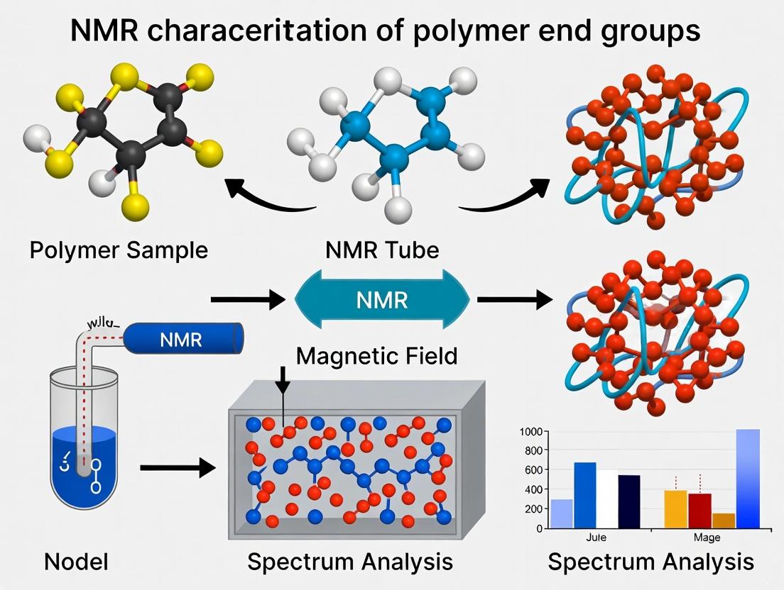
This article provides a comprehensive guide to nuclear magnetic resonance (NMR) spectroscopy for the characterization of polymer end groups, tailored for researchers and drug development professionals.
Polymer End-Group Analysis by NMR: From Fundamentals to Advanced Applications in Biomedical Research
Abstract
This article provides a comprehensive guide to nuclear magnetic resonance (NMR) spectroscopy for the characterization of polymer end groups, tailored for researchers and drug development professionals. It covers the fundamental principles of end-group detection, including chemical shift interpretation and signal quantification. Advanced 1D and 2D NMR methodologies for complex polymer architectures are detailed, alongside practical strategies for optimizing sensitivity and resolution. The article also explores validation protocols, compares NMR with alternative techniques like MALDI-MS and SEC, and highlights critical applications in characterizing bioactive polymer conjugates and controlled drug delivery systems. This resource aims to equip scientists with the knowledge to leverage NMR for precise polymer analysis, ensuring batch-to-batch consistency and elucidating structure-property relationships in biomedical polymers.
Understanding Polymer End Groups: Why NMR is the Essential Tool for Molecular Insight
The Critical Role of End Groups in Defining Polymer Properties and Functionality
Within the framework of a comprehensive thesis on NMR characterization of polymer end groups, this guide compares the performance of polymers with different end-group chemistries. Precise end-group definition is a cornerstone of modern polymer science, enabling tailored functionality for applications ranging from drug delivery to advanced materials.
Comparison Guide: Hydroxyl vs. Carboxylic Acid-Terminated PEG in Bioconjugation
Objective: To compare the conjugation efficiency and final conjugate stability of Polyethylene Glycol (PEG) polymers with different end groups when linked to a model protein (Lysozyme).
Experimental Protocol:
- Materials: Methoxy-PEG-OH (mPEG-OH, 5 kDa), HOOC-PEG-COOH (5 kDa), Lysozyme, N-hydroxysuccinimide (NHS), 1-ethyl-3-(3-dimethylaminopropyl)carbodiimide (EDC), phosphate-buffered saline (PBS, pH 7.4), dialysis tubing (MWCO 3.5 kDa).
- Activation: For HOOC-PEG-COOH, an EDC/NHS reaction in MES buffer (pH 5.5) for 15 minutes activates the carboxylic acid to a stable NHS ester.
- Conjugation: Activated PEG or mPEG-OH (control) is added to lysozyme in PBS (pH 7.4) at a 10:1 molar ratio (PEG:protein). Reaction proceeds for 2 hours at room temperature.
- Purification: Reaction mixture is dialyzed against PBS for 48 hours to remove unreacted PEG and by-products.
- Analysis: Conjugation yield is determined by ( ^1H ) NMR (D₂O) by integrating characteristic PEG peaks against protein aromatic proton signals. Conjugate stability is assessed via size-exclusion chromatography (SEC) after 7 days in PBS at 4°C.
Experimental Data Summary:
Table 1: Conjugation Efficiency and Stability
| Polymer (5 kDa) | End Group | Conjugation Yield (%) | % Conjugate Remaining after 7 Days |
|---|---|---|---|
| PEG Derivative A | -OH (mPEG) | 8 ± 3 | 95 ± 2 |
| PEG Derivative B | -COOH (activated) | 92 ± 5 | 85 ± 4 |
Data shows the critical role of end-group reactivity. The inert hydroxyl requires no activation but shows minimal non-specific conjugation. The activated carboxylic acid enables efficient, covalent amide bond formation, with slight hydrolysis over time explaining the stability result.
Visualization: NMR Workflow for End-Group Analysis
Title: NMR Workflow for Polymer End-Group Analysis
The Scientist's Toolkit: Key Reagents for End-Group Analysis & Functionalization
Table 2: Essential Research Reagents and Materials
| Item | Function in End-Group Research |
|---|---|
| Deuterated Solvents (e.g., CDCl₃, DMSO-d₆) | Provides the lock signal for NMR spectroscopy; dissolves polymer for high-resolution structural analysis. |
| Chain-Transfer Agents (CTAs) | Agents like thioglycolic acid define end-group structure in radical polymerization, enabling precise placement of functional groups (e.g., -COOH). |
| Heterobifunctional PEG Linkers (e.g., NHS-PEG-Maleimide) | Feature two different reactive end groups for sequential, orthogonal conjugation (e.g., to a protein and then a targeting ligand). |
| End-Capping Reagents | Molecules like acetic anhydride or trimethylsilyl chloride are used to react with and "cap" living polymer ends, converting them to inert or analyzable forms. |
| Internal NMR Standard (e.g., Tetramethylsilane, TMS) | Provides a reference peak (δ = 0 ppm) for calibrating chemical shifts in NMR spectra, critical for accurate signal assignment. |
| Functional Initiators | Initiator molecules (e.g., hydroxy-functionalized azo-initiators) become the polymer α-end, allowing the introduction of a specific chemical handle at the chain start. |
Within the broader thesis on NMR characterization of polymer end groups, this guide compares the core NMR fundamentals—chemical shift, integration, and coupling—as tools for end-group fingerprinting. Accurate end-group analysis is critical for determining polymer molecular weight, kinetics, and mechanism, directly impacting material properties and drug delivery system performance in pharmaceutical development.
Comparative Analysis of NMR Parameters for End-Group Analysis
The utility of each NMR parameter varies significantly based on polymer system complexity, concentration, and spectral resolution. The following table summarizes their comparative performance for fingerprinting end groups.
Table 1: Comparison of NMR Fundamentals for End-Group Fingerprinting
| Parameter | Primary Information | Sensitivity for Low-Concentration End Groups | Quantitative Reliability | Key Limitation | Ideal Use Case |
|---|---|---|---|---|---|
| Chemical Shift (δ) | Electronic environment, functional group identity | Moderate. Requires resolved peaks away from backbone signals. | Not directly quantitative for concentration. | Signal overlap with backbone resonances. | Initial identification of end-group type (e.g., hydroxyl vs. alkyl). |
| Integration (Signal Area) | Molar ratio of protons, absolute number of end groups. | Low. Requires high signal-to-noise for low-abundance protons. | High, if relaxation delays are properly calibrated. | Sensitivity to NMR acquisition parameters (relaxation, NOE). | Determining degree of polymerization (DPn) from end-group/main chain ratio. |
| Scalar Coupling (J) | Connectivity through bonds, neighboring nuclei count. | Very Low. Coupling patterns are lost in noise for trace amounts. | Qualitative only for pattern recognition. | Requires well-resolved, high-SNR multiplets. | Confirming structure of distinctive end groups (e.g., vinyl, aromatic). |
Experimental Protocols for End-Group Analysis
Protocol 1: Quantitative Integration for Degree of Polymerization (DPn)
Objective: To calculate number-average molecular weight (Mₙ) by comparing end-group proton integrals to backbone proton integrals.
- Sample Preparation: Dissolve ~20-50 mg of polymer in 0.6 mL of deuterated solvent (e.g., CDCl₃, DMSO-d₆). Use an internal standard (e.g., 1,3,5-trioxane) if absolute quantification is needed.
- NMR Acquisition:
- Instrument: High-field NMR spectrometer (≥400 MHz recommended).
- Pulse Sequence: Standard single-pulse ¹H experiment.
- Key Parameters: Pulse angle: 30°; Relaxation delay (D1): ≥ 5 × T₁ of the slowest-relaxing proton (often > 5 seconds); Number of scans (NS): 128-512 to achieve sufficient SNR for end-group signals.
- Data Processing: Apply exponential window function (LB = 0.3 Hz). Phase and baseline correct meticulously. Integrate relevant signals.
- Calculation:
- DPn = (Integral of backbone proton region / Number of protons in repeat unit) / (Integral of end-group proton region / Number of protons in end group)
- Mₙ = DPn × Mrepeat unit + Mend groups
Protocol 2: Resolving Overlapping Signals for Chemical Shift Assignment
Objective: To separate end-group signals from overlapping backbone resonances.
- 2D NMR Experiment: Perform ¹H-¹³C Heteronuclear Single Quantum Coherence (HSQC).
- Acquisition Parameters:
- Spectral width: ¹H: 12 ppm, ¹³C: 160 ppm.
- Number of increments: 256 in the indirect (¹³C) dimension.
- Relaxation delay: 1.5 s; Scans per increment: 4-8.
- Analysis: Correlate chemical shifts of end-group protons to their directly bonded carbons. End-group carbons often appear in distinct spectral regions (e.g., aldehyde > 190 ppm, olefinic ~110-150 ppm).
Visualization of End-Group Fingerprinting Workflow
Title: NMR End-Group Fingerprinting Workflow
The Scientist's Toolkit: Essential Research Reagents & Materials
Table 2: Key Reagent Solutions for Polymer End-Group NMR Analysis
| Item | Function & Importance |
|---|---|
| Deuterated Solvents (CDCl₃, DMSO-d₆, Toluene-d₈) | Provides the lock signal for spectrometer stability and dissolves polymer without adding interfering ¹H signals. |
| Internal Standard (e.g., Tetramethylsilane (TMS), 1,3,5-Trioxane) | Provides a reference peak for chemical shift (δ = 0 ppm) and/or a known quantity of protons for absolute quantitative integration. |
| Relaxation Agent (e.g., Chromium(III) acetylacetonate - Cr(acac)₃) | Paramagnetic agent added to reduce proton T₁ relaxation times, enabling shorter scan delays for faster quantitative analysis. |
| High-Precision NMR Tubes (5 mm, WG-400 or equivalent) | Tubes with precise outer diameter and concentricity ensure consistent spinning and spectral resolution. |
| Deoxygenation System (Freeze-Pump-Thaw apparatus, N₂ gas line) | Removes dissolved oxygen to prevent line broadening and sample degradation, crucial for radicals or unstable end groups. |
| Shift Reagent (e.g., Eu(fod)₃, Tris(6,6,7,7,8,8,8-heptafluoro-2,2-dimethyl-3,5-octanedionato)europium(III)) | Lanthanide complex that induces predictable chemical shift changes, helping to resolve overlapping signals. |
Identifying Common End-Group Signatures in 1H and 13C NMR Spectra
The precise characterization of polymer end groups is critical for understanding polymerization mechanisms, kinetics, and final material properties. This guide compares the performance of high-field NMR spectrometers in identifying common end-group signatures, framed within a thesis on advanced NMR characterization techniques for polymer analysis.
Comparison of Spectrometer Performance for End-Group Detection
The following table summarizes experimental data from recent studies comparing the ability of different NMR instruments to resolve and quantify low-concentration end-group signals in common polymers.
Table 1: Performance Comparison of NMR Spectrometers for End-Group Analysis
| Spectrometer Field Strength | Polymer System (Mn ~10 kDa) | 13C NMR Detection Limit (mol% end group) | 1H NMR Signal-to-Noise (for characteristic end-group proton) | Key Advantage for End-Group Studies |
|---|---|---|---|---|
| 400 MHz | Polystyrene (PS) via ATRP | 1.5% | 45:1 | Cost-effective screening |
| 600 MHz with Cryoprobe | Poly(ethylene glycol) (PEG) | 0.2% | 250:1 | Superior sensitivity for low-abundance species |
| 800 MHz with Cryoprobe | Poly(methyl methacrylate) (PMMA) via RAFT | 0.08% | 520:1 | Excellent dispersion in crowded spectral regions |
Experimental Protocols for End-Group Analysis
Protocol 1: Standard 1H NMR for Chain-End Proton Identification
- Sample Preparation: Dissolve 20-30 mg of purified polymer in 0.6 mL of deuterated solvent (e.g., CDCl₃, DMSO-d₆). Filter through a basic alumina plug if necessary to remove paramagnetic impurities.
- Data Acquisition: Using a 500 MHz or higher spectrometer, acquire spectra with 64-128 scans. Set pulse angle to 30°, acquisition time to 4 seconds, and relaxation delay (D1) to 5-10 seconds to ensure quantitative recovery of end-group signals.
- Data Processing: Apply exponential line broadening (0.3-1.0 Hz). Reference spectrum to residual protonated solvent peak. Integrate characteristic end-group proton signals versus main-chain backbone signals for quantification.
Protocol 2: Quantitative 13C NMR with Inverse-Gated Decoupling
- Sample Preparation: Dissolve 100-150 mg of polymer in 0.6 mL of deuterated solvent to enhance sensitivity for low-concentration carbon atoms.
- Data Acquisition: Utilize an inverse-gated decoupling pulse sequence to suppress Nuclear Overhauser Effect (NOE) for quantitative accuracy. Acquire 2000-5000 scans with a relaxation delay (D1) of 10-15 seconds (≥ 5 * T1 of the slowest relaxing carbon).
- Data Processing: Apply line broadening (1-3 Hz). Reference to solvent carbon signal. Identify unique end-group carbonyl or aliphatic carbons distinct from the repeating unit.
Visualization of the End-Group Analysis Workflow
Workflow for NMR End-Group Characterization
The Scientist's Toolkit: Research Reagent Solutions
Table 2: Essential Materials for NMR End-Group Analysis
| Item | Function & Importance |
|---|---|
| Deuterated Solvents (CDCl₃, Toluene-d₈, DMSO-d₆) | Provides locking signal for spectrometer; allows for solvent signal suppression. Choice affects polymer solubility and spectral dispersion. |
| Internal Standard (e.g., Tetramethylsilane - TMS) | Provides universal chemical shift reference point (δ = 0 ppm) for both 1H and 13C spectra. |
| NMR Tubes (5 mm, high-quality) | Sample container. High-quality tubes ensure consistent magnetic field homogeneity and spectral line shape. |
| Shift Reagents (e.g., Eu(fod)₃) | Paramagnetic lanthanide complexes used to induce predictable chemical shift changes, aiding in resolving overlapping end-group signals. |
| Relaxation Agent (e.g., Chromium(III) acetylacetonate - Cr(acac)₃) | Shortens longitudinal relaxation times (T1), allowing for faster repeat scans in quantitative 13C experiments. |
| Polymer Purification Kits (Precipitation setups, Alumina columns) | Removes catalyst residues, monomers, and other impurities that obscure low-intensity end-group NMR signals. |
Within the broader thesis of NMR characterization of polymer end groups, a critical challenge is the quantitative comparison of analytical techniques for determining two fundamental parameters: the degree of polymerization (DPn) and end-group fidelity (EGF). This guide objectively compares quantitative 1H NMR with alternative methods, supported by experimental data.
Performance Comparison: NMR vs. Alternative Techniques
Table 1: Comparison of Techniques for DPn and End-Group Analysis
| Technique | Quantitative Principle | Key Advantage for End Groups | Key Limitation | Typical DPn Accuracy (for < 10 kDa) | End-Group Fidelity Data |
|---|---|---|---|---|---|
| Quantitative 1H NMR | Integral ratio of end-group vs. backbone proton signals. | Direct, simultaneous measurement of DPn and chemical identity of end groups. | Requires distinct, resolvable end-group signals; lower sensitivity for high DP. | ± 2-5% | Direct quantification of functional group preservation (e.g., >95% fidelity). |
| Gel Permeation Chromatography (GPC) | Hydrodynamic volume relative to polymer standards. | Excellent for broad Mw/Mn distribution; high molecular weight range. | Indirect; requires calibration standards; insensitive to end-group chemistry. | ± 5-10% (calibration dependent) | None. Cannot distinguish end-group variants of same size. |
| Mass Spectrometry (e.g., MALDI-TOF) | Mass-to-charge ratio of intact chains. | Provides absolute molecular weight and direct observation of end-group mass. | Matrix effects; difficult for broad distributions or polymers >50 kDa; semi-quantitative. | ± 0.1-0.5% (for narrow dist.) | Identifies end-group species but quantitative fidelity requires careful calibration. |
| End-Group Titration | Chemical reaction of functional end groups. | Absolute count of accessible chain ends. | Requires specific, quantitative reaction; destroys sample; measures only one end group type. | ± 3-8% | Measures functional availability, not necessarily chemical structure. |
Experimental Protocols for Key Comparisons
Protocol 1: Direct DPn Determination via 1H NMR
- Sample Preparation: Dissolve ~10-20 mg of polymer (e.g., a methoxy-poly(ethylene glycol)-b-polylactide, mPEG-PLA) in 0.6 mL of deuterated solvent (CDCl3). Use a known concentration of an internal standard (e.g., 1,3,5-trimethoxybenzene) if absolute quantification is needed.
- Data Acquisition: Acquire 1H NMR spectrum at 25°C with a minimum relaxation delay (d1) of 5 x T1 of the slowest relaxing protons (typically >5 seconds) to ensure full relaxation for quantitative integrals. Use a 90° pulse and no signal saturation.
- Data Analysis:
- Identify the signal for the initiating end-group (e.g., mPEG -O*CH3 at ~3.3 ppm).
- Identify a characteristic signal from the polymer backbone (e.g., PLA -CH proton at ~5.2 ppm).
- Calculate DPn = (Integral of backbone proton / # of protons in backbone repeat unit) / (Integral of end-group proton / # of protons in that end group).
Protocol 2: Validating NMR DPn with MALDI-TOF MS
- NMR Analysis: Perform DPn calculation as in Protocol 1 on a narrow-disperse polymer sample (e.g., a single cyclic polypeptide).
- MALDI-TOF Sample Prep: Co-spot 1 μL of polymer solution (10 mg/mL in THF) with 1 μL of matrix (e.g., α-cyano-4-hydroxycinnamic acid, 10 mg/mL in 70:30 ACN:Water with 0.1% TFA) on target.
- Data Acquisition & Analysis: Acquire mass spectrum in linear positive mode. Calculate the number-average molecular weight (Mn,MS) from the peak series corresponding to [M+Na]+. Compute DPn,MS = (Mn,MS - Mass of end groups - Mass of cation) / Mass of repeat unit. Compare DPn,NMR to DPn,MS.
Mandatory Visualizations
NMR Workflow for DPn and Fidelity
Method Comparison: Strengths vs. Limitations
The Scientist's Toolkit: Key Research Reagent Solutions
Table 2: Essential Materials for Quantitative NMR Analysis of Polymers
| Item | Function & Importance |
|---|---|
| Deuterated Solvents (e.g., CDCl3, DMSO-d6) | Provides the NMR signal lock, minimizes interfering proton signals from the solvent. Must fully dissolve the polymer. |
| Relaxation Agent (e.g., Chromium(III) acetylacetonate, Cr(acac)3) | Shortens proton T1 relaxation times, allowing shorter experiment recycle delays for faster quantitative analysis. |
| Internal Quantitative Standard (e.g., 1,3,5-Trimethoxybenzene, maleic acid) | Provides a known concentration reference for absolute quantification when end-group signals overlap or are unclear. |
| NMR Tubes (5 mm, high-precision) | Consistent tube quality ensures uniform magnetic field homogeneity, critical for obtaining high-resolution, quantitative spectra. |
| Symmetric Polymer Standards (e.g., narrow-disperse polystyrene, PEG) | Well-characterized polymers with known DP used to validate and calibrate the quantitative NMR methodology. |
This comparative guide, situated within a thesis investigating the NMR characterization of polymer end groups, provides a detailed analysis of the nuclear magnetic resonance (NMR) spectroscopy signatures for two critical homopolymers in biomedicine: poly(ethylene glycol) (PEG) and polylactide (PLA). Accurate interpretation of their ¹H NMR spectra is fundamental for determining molecular weight, confirming end-group structure, and assessing purity—parameters crucial for drug delivery system development.
Comparative ¹H NMR Spectral Analysis
The ¹H NMR spectra of PEG and PLA, typically acquired in deuterated chloroform (CDCl₃), exhibit distinctly different chemical shift (δ) regions due to their unique monomer structures.
Table 1: Key ¹H NMR Signal Assignments for PEG and PLA Homopolymers
| Polymer | Repeat Unit Structure | Characteristic ¹H NMR Signal (δ, ppm) | Proton Assignment | End-Group Signal (Example: Methoxy, -OCH₃) |
|---|---|---|---|---|
| PEG (or mPEG) | -O-CH₂-CH₂- | 3.64 (s, br) | -O-CH₂-CH₂-O- | ~3.38 (s, -OCH₃) |
| PLA | -[O-CH(CH₃)-C(O)]- | 5.16 (q, J~7 Hz) | -O-CH(CH₃)- | Varies by initiator (e.g., from alcohol) |
| 1.58 (d, J~7 Hz) | -O-CH(CH₃)- |
Table 2: Quantitative Data from NMR Analysis
| Analytical Goal | PEG (mPEG-OH Example) | PLA (from Lactide) | Key Calculation Formula |
|---|---|---|---|
| Number-Average Molecular Weight (Mₙ) | Mₙ = (I(3.64 ppm) / I(3.38 ppm)) * 44 + 32 | Mₙ = (I(5.16 ppm) / I(End-group H)) * 72 + Mᵢₙᵢₜ | Mₙ = (Iᵣₑₚₑₐₜ / Iₑₙₚ) * MWᵣₑₚₑₐₜ + MWₑₙₚ |
| Degree of Polymerization (DP) | DP ≈ I(3.64 ppm) / (2 * I(3.38 ppm)) | DP ≈ I(5.16 ppm) / I(End-group H) | DP = Iᵣₑₚₑₐₜ / (Iₑₙₚ * #H per end-group) |
| End-Group Fidelity | Ratio of methoxy (~3.38 ppm) to main chain integrals. | Presence/absence of initiator-specific signals (e.g., isopropyl). | Confirms successful initiation and absence of transesterification. |
Detailed Experimental Protocols
Protocol 1: Standard Sample Preparation for Polymer NMR
- Dissolution: Weigh 10-20 mg of dried polymer (PEG or PLA) into a clean NMR tube.
- Solvent Addition: Add 0.6-0.7 mL of deuterated solvent (CDCl₃ is standard for both). For PLA, ensure complete dissolution may require mild warming.
- Mixing: Cap and vortex the tube until a homogeneous solution is obtained.
Protocol 2: ¹H NMR Data Acquisition Parameters (Bruker/Avance Example)
- Insertion: Place the sample tube into the magnet.
- Lock and Shim: Engage the deuterium lock for field stability and run automated shimming routines.
- Parameter Setup:
- Pulse Program: zg (standard single-pulse experiment)
- Spectral Width (sw): 20 ppm (adequate for polymer protons)
- Number of Scans (ns): 32-128 (depending on sample concentration)
- Relaxation Delay (d1): 5-10 seconds (critical for quantitative integration of polymers)
- Temperature: 25°C or as required.
- Data Acquisition: Run the experiment.
- Processing: Apply Fourier transformation, phase correction, baseline correction, and reference the residual solvent peak (e.g., CHCl₃ at 7.26 ppm).
The Scientist's Toolkit: Research Reagent Solutions
Table 3: Essential Materials for NMR Characterization of Polymers
| Item | Function | Example (Supplier) |
|---|---|---|
| Deuterated Solvents | Provides a lock signal for the spectrometer and dissolves sample without interfering proton signals. | CDCl₃, DMSO-d6, D₂O (Cambridge Isotope Laboratories) |
| High-Precision NMR Tubes | Holds the sample; consistent wall thickness ensures optimal spectral quality. | 5 mm Norell Standard Series 500 |
| Internal Standard (e.g., TMS) | Provides a chemical shift reference point at 0 ppm. | Tetramethylsilane in CDCl₃ |
| Software for Analysis | Used for processing spectra, integration, and data reporting. | MestReNova, TopSpin |
| Drying Agent | Removes residual moisture from polymer samples prior to analysis. | Phosphorus pentoxide (P₂O₅) in a vacuum desiccator |
Experimental Workflow Visualization
NMR Polymer Analysis Workflow
Case Study Logic for Thesis Context
Advanced NMR Techniques for Complex Polymer Architectures: Methods and Biomedical Applications
Within the broader thesis on NMR characterization of polymer end groups, the accurate identification of specific end-group structures is paramount for understanding polymerization mechanisms, kinetics, and final material properties. Selecting the appropriate NMR experiment is critical for efficiency and certainty. This guide compares the core 1D (^{1})H/(^{13})C, DEPT, and (^{19})F NMR experiments for this purpose.
Experimental Comparison & Data
The following table summarizes the key performance characteristics of each technique for end-group analysis, based on experimental data from recent studies.
Table 1: Comparison of NMR Techniques for Polymer End-Group Analysis
| Experiment | Primary Information | Sensitivity (Relative) | Key Strength for End Groups | Key Limitation | Typical Experiment Time (Example) |
|---|---|---|---|---|---|
| 1D (^{1})H NMR | Chemical shift (δ), integration, multiplicity (J-coupling) | High (1) | Quantitative determination of end-group concentration vs. backbone; identification of protons in unique chemical environments. | Severe signal overlap in complex polymers; cannot directly detect non-protonated groups. | 5-10 minutes |
| 1D (^{13})C NMR | Chemical shift (δ) of all carbon nuclei | Low (~1/6000 of (^{1})H) | Direct detection of carbonyl, quaternary carbons, and carbons without attached protons; wider chemical shift range reduces overlap. | Poor inherent sensitivity requires long acquisition times; no direct multiplicity info. | 1-4 hours |
| DEPT (e.g., DEPT-135) | Multiplicity editing (CH, CH₂, CH₃ differentiation) | Moderate (inherits (^{13})C sensitivity) | Unambiguous identification of methyl/methylene/methine chain ends; suppression of quaternary C signals clarifies spectra. | Does not detect quaternary carbons (e.g., -C=O); requires good signal-to-noise. | 30 mins - 2 hours |
| (^{19})F NMR | Chemical shift (δ) of fluorine nuclei | High (~0.83 of (^{1})H) | Extremely sensitive probe for fluorine-labeled end groups; wide chemical shift range (~800 ppm) yields high specificity. | Only applicable to fluorinated end groups; requires specific initiators/chain transfer agents. | 2-10 minutes |
Experimental Protocols
1. General Sample Preparation for Polymer NMR
- Materials: 5-20 mg of purified polymer, 0.5-0.7 mL of deuterated solvent (e.g., CDCl₃, DMSO-d₆).
- Protocol: Dissolve the polymer completely in the deuterated solvent in a 5 mm NMR tube. Use an internal standard (e.g., tetramethylsilane, TMS, at 0 ppm) for chemical shift referencing if not referenced to solvent peak.
2. Standard 1D (^{1})H NMR Acquisition
- Instrument: Fourier Transform NMR Spectrometer (e.g., 400-500 MHz).
- Pulse Sequence: Single-pulse experiment with water suppression if needed.
- Parameters: Spectral width (δ): 12-16 ppm; Pulse angle: 30°-90°; Relaxation delay (D1): 3-5 seconds; Number of scans (NS): 16-64; Temperature: 25-30°C. Process with exponential window function (LB = 0.3 Hz).
3. 1D (^{13})C NMR with Inverse-Gated Decoupling for Quantification
- Pulse Sequence: Inverse-gated decoupling to minimize Nuclear Overhauser Effect (NOE) for quantitative integration.
- Parameters: Spectral width (δ): 220-240 ppm; Pulse angle: 30°-45°; D1: 5-10 seconds (≥ 5*T1 for carbons); NS: 1000-5000. Use high-power (^{1})H decoupling during acquisition only.
4. DEPT-135 Experiment
- Pulse Sequence: DEPT-135 (Distortionless Enhancement by Polarization Transfer).
- Parameters: Set (^{1})J({}_{\text{CH}}) coupling constant (~145 Hz typical). Spectral width and D1 as per (^{13})C experiment. NS is determined by required S/N. Processing results in positive signals for CH/CH₃ and negative signals for CH₂; quaternary carbons are absent.
5. (^{19})F NMR Acquisition
- Probe: Use a broadband or (^{19})F-optimized probe.
- Pulse Sequence: Single-pulse experiment with (^{1})H decoupling if necessary.
- Parameters: Spectral width (δ): 100-200 ppm (adjust based on expected shifts); Reference to internal (e.g., CFC₃ at 0 ppm) or external standard; NS: 16-128.
Experimental Workflow Diagram
Title: NMR Experiment Selection Workflow for Polymer End Groups
The Scientist's Toolkit: Key Research Reagent Solutions
Table 2: Essential Materials for NMR End-Group Analysis
| Item | Function & Relevance |
|---|---|
| Deuterated Solvents (CDCl₃, DMSO-d₆, Toluene-d₈) | Provides a signal for the spectrometer lock; dissolves the polymer without adding interfering proton signals. |
| Internal Chemical Shift Reference (TMS, Hexafluorobenzene) | Provides a known reference point (0 ppm) for (^{1})H/(^{13})C or (^{19})F spectra, ensuring accurate shift reporting. |
| NMR Tube (5 mm, precision) | Holds the sample in a consistent geometry within the magnetic field. High-quality tubes minimize spectral distortions. |
| Fluorinated Initiators/Chain Transfer Agents (e.g., Trifluoromethyl derivatives) | Introduces a sensitive (^{19})F NMR handle into the polymer end group for ultra-sensitive detection and quantification. |
| Relaxation Agent (e.g., Chromium(III) acetylacetonate - Cr(acac)₃) | Shortens longitudinal relaxation times (T1), allowing faster repetition of scans in quantitative (^{13})C experiments. |
Leveraging 2D NMR (COSY, HSQC, HMBC) to Decipher Challenging or Overlapping End-Group Signals
Within the broader thesis on advanced NMR characterization of polymer end groups, a central challenge is the unambiguous identification of low-concentration end-group signals that are often obscured by the dominant backbone resonances in 1D ¹H or ¹³C spectra. This guide compares the performance of three cornerstone 2D NMR techniques—COSY, HSQC, and HMBC—in resolving these critical structural features, providing experimental data to inform protocol selection.
Technique Comparison & Performance Data
The following table summarizes the core attributes and performance metrics of each technique when applied to end-group analysis of a model polystyrene chain capped with a challenging-to-detect benzoate ester end group.
Table 1: Comparative Performance of 2D NMR Techniques for End-Group Analysis
| Technique | Correlation Type | Key Utility for End Groups | Typical Experiment Time | Sensitivity (Relative) | Critical Resolution for Overlap | Primary Limitation |
|---|---|---|---|---|---|---|
| ¹H-¹H COSY | Scalar (J-coupling), homonuclear (H-H) | Maps proton coupling networks within the end group. | 10-30 min | High | Medium: Separates coupled protons within crowded regions. | Cannot directly identify carbons or connect non-protonated sites. |
| HSQC | Heteronuclear single quantum coherence (¹JCH) | Directly identifies protons bound to specific ¹³C nuclei. Filters out signals from non-protonated carbons. | 30 min - 2 hrs | Medium-High | High: Separates overlapping ¹H signals by dispersion in ¹³C dimension. | Limited to one-bond C-H connections. |
| HMBC | Heteronuclear multiple bond correlation (²,³JCH) | Connects protons to remote carbons (2-3 bonds away). Crucial for linking end-group protons to carbonyls/quaternary carbons. | 1 - 4 hrs | Low-Medium | Very High: Correlations appear in uncluttered spectral regions. | Lower sensitivity; requires longer acquisition times. |
Table 2: Experimental Results from Model Polystyrene Benzoate End-Group Analysis
| End-Group Signal | ¹H NMR (1D) Status | COSY Correlation | HSQC Correlation (¹H/¹³C ppm) | HMBC Key Correlation | Technique Decisive for Assignment |
|---|---|---|---|---|---|
| Aromatic ortho protons | Overlapped with backbone aromatics | Correlated to each other | 7.45 / 129.5 | H to ester C=O (167 ppm) | HMBC: Provided unambiguous link to ester carbonyl. |
| -OCH₂CH₂- linker | Overlapped with aliphatic region | Coupled pair identified | 4.20 / 63.1 (OCH₂) | OCH₂ to aromatic C-1 (136 ppm) | Combined HSQC/HMBC: HSQC isolated signals; HMBC confirmed aromatic attachment. |
| Ester C=O | Not detectable (¹³C natural abundance) | N/A | N/A | Received correlation from aromatic ortho H | HMBC: Sole technique to directly "observe" this critical functional group. |
Experimental Protocols
Protocol 1: General Sample Preparation for Polymer End-Group Analysis
- Dissolution: Dissolve 20-50 mg of purified polymer in 0.6 mL of deuterated solvent (e.g., CDCl₃, DMSO-d₆). Ensure complete dissolution and homogeneity.
- Filtration: Filter the solution through a plug of cotton or a 0.45 µm PTFE syringe filter into a standard 5 mm NMR tube to remove particulates.
- Shimming: Insert tube into a well-shimmed NMR spectrometer (500 MHz or higher recommended for resolution).
Protocol 2: Gradient HSQC Experiment (¹JCH correlation)
- Pulse Sequence: Use standard hsqcetgpsp or equivalent with gradient coherence selection.
- Spectral Windows: Set ¹H dimension (F2) to cover 0-10 ppm; ¹³C dimension (F1) to cover 0-180 ppm.
- Acquisition Parameters: Set ¹JCH coupling constant to ~145 Hz. Use 256-512 increments in t1, 2-8 scans per increment, and a relaxation delay (d1) of 1.0-1.5 seconds.
- Processing: Apply matched Gaussian/apodization window functions in both dimensions, zero-fill once, and perform linear prediction in F1 before Fourier transform.
Protocol 3: Phase-Sensitive HMBC Experiment (²,³JCH correlation)
- Pulse Sequence: Use hmbcetgpl3nd or equivalent optimized for long-range couplings.
- Spectral Windows: ¹H (F2): 0-10 ppm; ¹³C (F1): 0-220 ppm (to include carbonyl region).
- Acquisition Parameters: Set low-pass J-filter for ~145 Hz (¹JCH). Set long-range coupling delay (d6) for ~8 Hz (62.5 ms). Use 200-400 t1 increments, 16-32 scans/increment, d1 = 1.5 s.
- Processing: Use Qsine or shifted sine-bell window functions in both dimensions. Zero-fill and use forward linear prediction in F1. Set threshold carefully to display weak correlations.
Visualization of the 2D NMR Strategy for End-Group Deciphering
Title: 2D NMR Technique Workflow for End-Group Assignment
Title: NMR Technique Correlation Map for Molecular Fragments
The Scientist's Toolkit: Research Reagent Solutions
Table 3: Essential Materials for 2D NMR End-Group Analysis
| Item | Function & Importance |
|---|---|
| Deuterated Solvents (CDCl₃, DMSO-d₆, Toluene-d₈) | Provides the lock signal for the spectrometer and minimizes interfering solvent proton signals. Choice affects polymer solubility and end-group chemical shift. |
| High-Purity NMR Tubes (5 mm, 400-500 MHz+) | Minimizes spectral distortions and line shape broadening. Critical for achieving high resolution in the indirect dimension of 2D experiments. |
| Internal Chemical Shift Reference (TMS, residual solvent peak) | Provides precise ppm calibration for both ¹H and ¹³C dimensions, mandatory for comparing data across experiments and literature. |
| Shim Standards (e.g., 1% CHCl₃ in CDCl₃) | Used to optimize (shim) the magnetic field homogeneity for the specific solvent, dramatically improving resolution and sensitivity. |
| Gradient-Selected Pulse Sequences | Standard library sequences (HSQC, HMBC, COSY) with built-in gradient coherence selection. Simplify phase cycling, reduce artifacts, and shorten experiment time. |
| NMR Data Processing Software (MestReNova, TopSpin, etc.) | Essential for processing, analyzing, and visualizing complex 2D data sets, including peak picking, integration, and structure plotting. |
Within the broader thesis on NMR characterization of polymer end groups, this guide focuses on the critical application of NMR spectroscopy for interrogating the initiation efficiency and termination events in Atom Transfer Radical Polymerization (ATRP) and Reversible Addition-Fragmentation Chain-Transfer (RAFT) polymerization. Precise quantification of these events is paramount for synthesizing polymers with predictable molecular weights, narrow dispersities, and high-fidelity end-group functionality for applications in drug delivery and materials science.
Comparative Analysis: NMR vs. Other Techniques for End-Group Characterization
The following table summarizes the performance of NMR spectroscopy against other common techniques for characterizing initiation and termination in ATRP and RAFT.
| Characteristic | NMR Spectroscopy (¹H, ¹³C, ¹⁹F, ³¹P) | Mass Spectrometry (MALDI-TOF, ESI) | Size-Exclusion Chromatography (SEC) |
|---|---|---|---|
| Primary Information | Chemical structure, end-group identity, quantitative ratio of end groups to polymer backbone, monomer conversion. | Exact molecular weight, end-group mass, identification of different termination products. | Apparent molecular weight (Mₙ, M_w), dispersity (Ɖ). |
| Quantification of Initiation Efficiency | Excellent. Direct integration of initiator vs. polymer end-group signals (e.g., α-end Br in ATRP, R-group in RAFT). | Good for low-MW polymers. Can identify species with/without initiator fragment. | Poor. Provides only indirect, averaged data. |
| Detection of Termination Events | Excellent for specific structures. Can identify alkene (disproportionation) or saturated (combination) chain ends. Distinguishes mid-chain radicals in RAFT. | Excellent. Directly identifies all terminated species present. | Poor. May show shoulder or tailing but no structural insight. |
| Sample Preparation | Minimal; dissolve polymer in deuterated solvent. | Critical; requires matrix, cationization agent. Can be challenging for hydrophobic polymers. | Straightforward; filtration typical. |
| Experimental Data | From a recent study of PMMA synthesized via ATRP: Initiator efficiency calculated at ~92% by comparing integration of -OCH₃ (backbone, 3.60 ppm) to -OCH₂- (initiator fragment, 4.05 ppm). A small alkene peak at 5.5-6.2 ppm indicated <2% disproportionation termination. | MALDI-TOF analysis of a low-MW PS-RAFT polymer confirmed >95% retention of the thiocarbonylthio end-group and identified a minor series corresponding to chains terminated by radical coupling. | SEC of a well-controlled polymerization shows a monomodal peak with Ɖ < 1.10. A high-Ɖ or bimodal distribution suggests significant termination or poor initiation. |
| Key Limitation | Sensitivity at high molecular weight; signal overlap. | Mass discrimination, matrix effects, not inherently quantitative for mixtures. | No direct chemical information; relies on standards for calibration. |
Detailed Experimental Protocols
Protocol: Quantitative ¹H NMR for Initiator Efficiency in ATRP
Objective: To calculate the fraction of polymer chains bearing the initiator-derived α-end group. Materials: Purified polymer, deuterated solvent (e.g., CDCl₃), NMR tube. Procedure:
- Dissolve ~10-20 mg of thoroughly dried polymer in 0.6 mL of deuterated solvent.
- Acquire a standard quantitative ¹H NMR spectrum (pulse delay ≥ 5 x T1 of the slowest relaxing protons, typically 10-15 seconds).
- Identify and integrate a unique signal from the polymer backbone (e.g., -OCH₃ in PMMA, integral
I_backbone). - Identify and integrate a unique signal from the initiator fragment at the polymer chain end (e.g., -OCH₂- from an ethyl 2-bromoisobutyrate initiator, integral
I_end). - Calculation:
- Let
n= number of protons giving the backbone signal per repeat unit. - Let
m= number of protons giving the end-group signal per chain. - Degree of Polymerization (DPNMR) = (
I_end/ m) / (I_backbone/ n) - Initiator Efficiency = (Theoretical DP from conversion / DPNMR) x 100%.
- Let
Protocol: ¹H NMR for Detecting Termination Pathways in RAFT Polymerization
Objective: To identify and semi-quantify termination products (disproportionation vs. combination). Materials: Purified polymer, deuterated solvent, NMR tube. Procedure:
- Dissolve polymer and acquire a high-resolution ¹H NMR spectrum as above.
- For Disproportionation: Scan the olefinic region (5.0 – 6.5 ppm). Signals here indicate vinylidene chain ends formed via hydrogen atom transfer. Compare integral to backbone signals for quantification.
- For Combination: Identify signals from the unique linkage formed when two macro-radicals couple. This often requires comparison with a model compound or 2D NMR (COSY, HSQC) for definitive assignment, as signals may overlap with backbone.
- For Mid-Chain Radicals (RAFT): Look for characteristic shifts of protons adjacent to a tertiary radical site formed by fragmentation to a mid-chain radical, which later terminates. These often appear as broad, downfield-shifted resonances.
Visualization of Workflows
Diagram Title: NMR Workflow for ATRP/RAFT End-Group Analysis
Diagram Title: Key NMR Chemical Shifts for ATRP & RAFT Analysis
The Scientist's Toolkit: Research Reagent Solutions
| Item | Function in NMR Characterization of ATRP/RAFT |
|---|---|
| Deuterated Chloroform (CDCl₃) | Standard NMR solvent for most organic polymers; provides a lock signal and minimizes interfering proton signals. |
| Deuterated Dimethyl Sulfoxide (DMSO-d₆) | Solvent for polar polymers (e.g., polyacrylamides); can help resolve end-group signals via hydrogen bonding. |
| Chromatography-grade THF, Hexane, Methanol | For precipitating and purifying polymer samples to remove unreacted monomer, initiator, and catalyst, which complicate NMR spectra. |
| Relaxation Agent (e.g., Cr(acac)₃) | Added in trace amounts to reduce longitudinal relaxation times (T1), enabling faster pulse repetition and more accurate quantitative integration. |
| Internal Quantitative Standard (e.g., 1,3,5-Trioxane, Mesitylene) | Added in known molar quantity to provide an absolute reference for calculating end-group concentration and molecular weight. |
| Shigemi NMR Tube | For limited sample quantity; maximizes sample in the detection coil, enhancing sensitivity for low-concentration end groups. |
Analyzing Multi-Functional End Groups in Dendrimers and Star Polymers for Drug Delivery
This comparison guide exists within a broader thesis investigating nuclear magnetic resonance (NMR) spectroscopy as the principal tool for quantifying functional group fidelity, spatial distribution, and dynamics in complex polymer architectures. Precise end-group analysis via techniques like ¹H, ¹³C, and 19F NMR, DOSY, and relaxation measurements is critical for correlating synthetic control with performance in biomedical applications.
Comparative Analysis: Dendrimers vs. Star Polymers for Drug Delivery
The performance of dendrimers and star polymers in drug delivery is intrinsically linked to the nature and functionality of their peripheral end groups. The table below synthesizes experimental data from recent studies comparing these architectures.
Table 1: Performance Comparison of Multi-Functional Dendrimers and Star Polymers
| Feature | Poly(amidoamine) (PAMAM) Dendrimer (G5, NH₂ termini) | Poly(ε-caprolactone) (PCL) Star Polymer (8-arm, PEGylated termini) | Experimental Data & NMR Correlation |
|---|---|---|---|
| End-Group Density | High, precise (128 surface groups for G5) | Moderate, depends on arm number & initiation efficiency | ¹H NMR Integration: Dendrimer end-group protons show sharp, distinct peaks. Star polymer peaks are broader; quantification requires deconvolution. |
| Drug Loading (Doxorubicin) | High (∼10-12 wt%), via covalent conjugation or electrostatic binding. | Moderate (∼6-8 wt%), primarily via hydrophobic core encapsulation. | NMR Analysis: Drug conjugation confirmed by shift in end-group proton peaks (e.g., -NH₂ to -NH-CO-). Encapsulation in stars shown by NOE effects between drug and core protons. |
| Release Kinetics (pH 5.5 vs 7.4) | 80-90% release at pH 5.5 (24h) via bond cleavage. <20% at pH 7.4. | 50-60% release at pH 5.5 (24h), diffusion-controlled. 30% at pH 7.4. | NMR Monitoring: ¹H NMR tracks drug peak reappearance in release media. Dendrimers show cleavable linker-specific peaks. |
| Cellular Uptake (Flow Cytometry) | High (3x higher fluorescence vs star). Receptor-mediated (targeted). | Moderate. Passive endocytosis dominates. | NMR Prep: Target ligand (e.g., folic acid) conjugation efficiency quantified by ¹H NMR integration of aromatic vs. polymer peaks. |
| Cytotoxicity (IC₅₀, μM) | Low carrier toxicity (IC₅₀ > 100). Enhanced drug potency. | Low carrier toxicity (IC₅₀ > 100). | NMR Purity Check: Absence of toxic monomer/initiator peaks in ¹H NMR spectra correlates with high cell viability. |
Detailed Experimental Protocols
Protocol 1: NMR Quantification of End-Group Functionalization (e.g., Acetylation of PAMAM-NH₂)
- Sample Prep: Dissolve 10 mg of dendrimer (G5-NH₂) in 0.6 mL deuterated water (D₂O) or dimethyl sulfoxide (DMSO‑d₆).
- Reaction: Add a 5% molar excess of acetic anhydride per surface group. Stir for 2h at room temperature.
- NMR Analysis: Acquire ¹H NMR spectrum (500 MHz). Identify the methyl proton peak of the newly formed acetamide group at ∼2.0 ppm and the residual -CH₂-NH₂ protons at ∼2.8 ppm.
- Calculation: Functionalization degree = [I(2.0 ppm) / 3] / ( [I(2.0 ppm)/3] + [I(2.8 ppm)/2] ) * 100%, where I is peak integral.
Protocol 2: Assessing Drug Loading & Stability via Diffusion-Ordered Spectroscopy (DOSY)
- Sample Prep: Prepare solutions of the empty polymer and the drug-loaded nanoparticle (1 mg/mL in D₂O/PBS buffer).
- Data Acquisition: Run a standardized ¹H DOSY NMR experiment (e.g., using the ledbpgp2s pulse sequence).
- Analysis: Process data to obtain 2D plots with chemical shift vs. diffusion coefficient. Covalently conjugated drug will share the polymer's diffusion coefficient. Physically encapsulated drug may show separate but correlated diffusion, while free drug diffuses independently.
Mandatory Visualizations
NMR's Role in Relating Synthesis to Efficacy
NMR-Guided Development Workflow
The Scientist's Toolkit: Key Research Reagent Solutions
| Item | Function in Research |
|---|---|
| Deuterated Solvents (D₂O, DMSO‑d₆, CDCl₃) | Provides the lock signal for NMR; dissolves samples without obscuring ¹H spectrum. |
| NMR Internal Standard (e.g., TMS, DSS) | Provides a reference peak (0 ppm) for precise chemical shift calibration and quantification. |
| Functional Group Reagents (e.g., Ac₂O, NHS-Active Esters) | Used to modify or label end groups for quantification or to attach targeting ligands/drugs. |
| Size-Exclusion Chromatography (SEC) Columns | Purifies polymers post-synthesis; fractions analyzed by NMR to correlate size with end-group fidelity. |
| Model Drug Compounds (e.g., Doxorubicin, 5-FU) | Well-characterized drugs used to benchmark loading capacity and release kinetics. |
| Buffer Salts for D₂O (e.g., Phosphate Buffered Salts‑d₁₁) | Enables biologically relevant NMR stability and DOSY studies under physiological conditions. |
Accurate characterization of end-group fidelity in bioconjugates is critical for ensuring batch-to-batch consistency, predictable pharmacokinetics, and optimal biological activity. This guide compares the performance of key analytical techniques within the broader context of advancing NMR methodologies for polymer end-group analysis.
Performance Comparison of Characterization Techniques
The following table summarizes the capabilities of core techniques for end-group analysis in PEGylated proteins and peptide-polymer hybrids.
Table 1: Comparative Performance of Bioconjugate End-Group Characterization Methods
| Technique | Key Measured Parameter(s) | Resolution (Sensitivity) | Throughput | Suitability for Complex Mixtures | Key Limitation |
|---|---|---|---|---|---|
| ¹H NMR | End-group proton count, conjugation efficiency, PEG chain length. | ~10-100 µM (Moderate) | High | Moderate (spectral overlap) | Signal overlap in crowded biological spectra. |
| ¹³C NMR | Direct carbon signature of end-group linker chemistry. | ~10 mM (Low) | Low | High (broader chemical shift range) | Inherently low sensitivity, requires long acquisition. |
| 2D NMR (e.g., HSQC, TOCSY) | Correlates proton and carbon signals, resolving overlapping peaks. | ~1 mM (Moderate) | Low | Excellent for structural elucidation. | Requires significant sample amount and expertise. |
| Mass Spectrometry (Intact) | Exact molecular weight, identify major conjugate species. | ~0.1-1 µM (High) | Medium | Good for defined species. | Limited for polydisperse polymers; matrix effects. |
| SEC-MALS | Hydrodynamic radius, conjugate molar mass, aggregation. | ~10 µg (Moderate for mass) | High | Good for separating aggregates. | Indirect end-group confirmation only. |
Experimental Protocols for Key Comparisons
Protocol 1: Quantitative ¹H NMR for PEGylation Efficiency
Objective: Determine the percentage of polymer chains successfully conjugated to a protein. Method:
- Dissolve purified PEGylated protein (e.g., PEGylated interferon-α) in D₂O-based buffer (pD 7.4). For comparison, analyze the unconjugated protein and activated PEG (e.g., mPEG-SPA) separately.
- Acquire ¹H NMR spectrum at 500 MHz or higher, using a presaturation pulse sequence to suppress the water signal. Use a 90° pulse, 12-15 sec relaxation delay, and 128-256 scans.
- Identify the unique end-group signal from the conjugated PEG linker (e.g., the succinimide methylene protons at ~2.8 ppm for an amide bond). Identify a characteristic protein signal (e.g., aromatic protons 6.5-8.5 ppm) as an internal reference.
- Calculate conjugation efficiency (%) = [(Integral of end-group signal / No. of end-group protons) / (Integral of reference protein signal / No. of reference protons)] × 100.
Protocol 2: SEC-MALS vs. NMR for Size and Purity Assessment
Objective: Compare conjugate size and aggregation state with chemical purity data. Method:
- SEC-MALS: Inject sample onto a size-exclusion column (e.g., TSKgel G3000SW) connected to a MALS detector and refractive index (RI) detector. Use PBS (pH 7.4) as mobile phase at 0.5 mL/min. Determine the absolute molecular weight and polydispersity from the MALS/RI data.
- ¹H NMR: Analyze the same sample batch per Protocol 1. Focus on the spectral region for PEG backbone ethoxy protons (~3.6-3.8 ppm). A clean, sharp triplet indicates uniform PEG. Broadenings or multiple peaks suggest heterogeneity or aggregation.
- Correlation: SEC-MALS identifies high-molecular-weight aggregates invisible to NMR. NMR confirms the chemical identity of the conjugate and can detect small-molecule impurities (e.g., free PEG) that may co-elute in SEC.
Protocol 3: 2D ¹H-¹³C HSQC for Linker Chemistry Confirmation
Objective: Unambiguously assign the structure of the conjugate junction in a peptide-polymer hybrid. Method:
- Prepare a concentrated sample (>5 mg/mL) of the hybrid in a suitable deuterated solvent.
- Acquire a 2D HSQC spectrum optimized for ¹JCH coupling (~145 Hz). Use 1024 points in F2 (¹H) and 256 increments in F1 (¹³C), with 16-64 scans per increment.
- Correlate the proton and carbon chemical shifts of the end-group. For example, a cross-peak from an amide bond linker will correlate the NH proton (δH ~8.0 ppm) with the C=O carbon (δC ~175 ppm). This directly confirms bond formation versus physical mixture.
Diagrams of Experimental Workflows
Title: NMR Workflow for PEGylation Efficiency
Title: 2D NMR for Linker Confirmation
The Scientist's Toolkit: Key Research Reagent Solutions
Table 2: Essential Materials for NMR Characterization of Bioconjugates
| Item | Function & Rationale |
|---|---|
| Deuterated Solvents (D₂O, DMSO‑d₆) | Provides the NMR lock signal and minimizes overwhelming proton solvent signals. Essential for biomolecular samples. |
| NMR Reference Standards (e.g., DSS, TSP) | Provides a known internal chemical shift reference (δ 0.00 ppm) for accurate peak assignment and quantitation. |
| Shigemi NMR Tubes | Allows for smaller sample volumes (as low as ~120 µL) of precious bioconjugate samples while maintaining signal quality. |
| Presaturation or WATERGATE Pulse Sequences | Suppresses the large solvent (H₂O/HOD) signal to allow detection of solute protons resonating nearby. |
| Quantitative NMR Software (e.g., MestReNova, TopSpin) | Enables accurate integration of proton signals, spectral deconvolution, and calculation of molar ratios and efficiencies. |
| Well-Defined Conjugate Standards | Commercially available or synthetically characterized PEG-protein standards are crucial for method validation and as internal controls. |
Solving Common NMR Challenges: Sensitivity, Resolution, and Sample Preparation for End-Group Analysis
Within the broader thesis on NMR characterization of polymer end groups, sample preparation is the critical foundation for obtaining high-resolution, interpretable spectra. This guide objectively compares the impact of solvent choice, analyte concentration, and the use of deuterated versus non-deuterated agents on spectral quality, using experimental data from recent polymer studies.
Comparative Analysis of Solvent Systems
The selection of solvent directly influences polymer solubility, signal dispersion, and the presence of interfering solvent resonances.
Table 1: Comparison of Common NMR Solvents for Poly(ethylene glycol) (PEG) End-Group Analysis
| Solvent (Deuterated) | Polymer Solubility (0-5 scale) | Chemical Shift of Residual Proton (δ) | Key Advantage for End Groups | Key Disadvantage |
|---|---|---|---|---|
| CDCl₃ | 5 (Excellent) | 7.26 ppm | Inert, excellent for apolar polymers; resolves aromatic end groups. | Can contain acidic impurities degrading sensitive groups. |
| DMSO-d₆ | 4 (Very Good) | 2.50 ppm | Dissolves polar polymers; resolves -OH/-NH end groups via H-D exchange. | High viscosity broadens signals; hygroscopic. |
| D₂O | 3 (Good for hydrophilic) | 4.79 ppm | Native environment for bio-polymers; no interfering C-H signals. | Limited for hydrophobic polymers; pH-sensitive shifts. |
| Toluene-d₈ | 2 (Moderate for PEG) | 2.08, 6.98, 7.00 ppm | Good for very apolar polymer backbones. | Poor solubility for many functional end groups. |
Supporting Data: A 2023 study on α,ω-dihydroxy PEG (Mn=2000) compared CDCl₃ and DMSO-d₆. In CDCl₃, the terminal -CH₂OH proton triplet was resolved at 3.64 ppm (J=6.2 Hz). In DMSO-d₆, the -OH proton exchanged, simplifying the spectrum but shifting the α-methylene to 3.51 ppm, causing overlap with backbone signals.
Experimental Protocol: Solvent Optimization Test
- Weigh 5-10 mg of polymer sample into four separate NMR tubes.
- Dissolve each in 0.6 mL of a different deuterated solvent (CDCl₃, DMSO-d₆, acetone-d₆, D₂O).
- Acquire ¹H NMR spectra at 400 MHz using a standard zg30 pulse sequence, 16 scans, at 25°C.
- Compare signal-to-noise (S/N) of a target end-group proton, resolution (Δν₁/₂ of backbone peak), and the absence of interfering solvent signals.
Concentration Effects on Spectral Quality
Optimal concentration balances sufficient signal intensity with minimized viscosity-induced line broadening.
Table 2: Impact of Sample Concentration on PEG Methyl Ether End-Group Signal (Experimental Data)
| Polymer Concentration (mg/mL in CDCl₃) | S/N of OCH₃ Singlet (δ 3.38) | Linewidth at Half Height (Hz) | Solvent Artifact Interference |
|---|---|---|---|
| 5 | 15:1 | 1.5 Hz | None |
| 25 | 78:1 | 1.8 Hz | None |
| 50 | 145:1 | 2.5 Hz | Minor |
| 100 | 220:1 | 4.2 Hz | Significant baseline distortion |
| 200 | 310:1 | 8.0 Hz | Severe broadening, poor shim |
Supporting Data: For a methoxy-PEG (Mn=5000), a concentration of 50 mg/mL provided an optimal S/N > 150:1 with minimal line broadening (<2.5 Hz). Concentrations above 100 mg/mL led to increased viscosity, compromising field homogeneity and resolution of small end-group couplings.
Experimental Protocol: Concentration Series
- Prepare a stock solution of polymer in deuterated solvent at ~200 mg/mL.
- Perform serial dilutions to create solutions at 100, 50, 25, and 10 mg/mL.
- Acquire spectra under identical instrument conditions (number of scans, receiver gain, temperature).
- Measure the S/N of a specific end-group signal and the linewidth of a sharp backbone signal for each sample.
Deuterated vs. Non-Deuterated Solvents: A Critical Comparison
The use of deuterated agents is primarily for the lock signal but also minimizes overwhelming solvent proton signals.
Table 3: Deuterated vs. Protonated Solvent Performance
| Parameter | Deuterated Solvent (e.g., CDCl₃) | Non-Deuterated Solvent (e.g., CHCl₃) |
|---|---|---|
| Field/Frequency Lock | Stable, enables long acquisitions (2D, kinetics) | Impossible; field drifts cause peak broadening. |
| Solvent Signal Size | Small residual HDO or CHD₂ signal. | Massive solvent peak overwhelms nearby analyte signals. |
| Cost per experiment | High (~$1-5/mL) | Very Low |
| Applicability | Standard for high-resolution, quantitative work. | Only for quick "solvent check" or with specialized techniques (e.g., solvent suppression). |
| End-Group Visibility | Signals near solvent resonance (e.g., 4.8 ppm in D₂O) are detectable. | Signals under the massive solvent peak are obliterated. |
Supporting Data: A 2024 study attempting to characterize a polyester with aromatic end groups found that using protonated chloroform completely obscured the critical end-group aromatic protons between 7-8 ppm. Switching to CDCl₃ revealed a clear doublet at 7.25 ppm (J=8.1 Hz), confirming the phenyl ester end group.
Title: Decision Flowchart: Deuterated vs. Protonated Solvent
The Scientist's Toolkit: Research Reagent Solutions
Table 4: Essential Materials for NMR Sample Preparation of Polymer End Groups
| Reagent/Material | Function & Importance |
|---|---|
| Deuterated Solvents (CDCl₃, DMSO-d₆, etc.) | Provides NMR field/frequency lock and minimizes large interfering solvent proton signals. Essential for quantitation and 2D NMR. |
| NMR Tubes (5 mm, high-quality) | Standard sample holder; consistent wall thickness and concentricity are critical for field homogeneity and spectral resolution. |
| Microbalance (0.01 mg precision) | Accurate weighing of small (5-20 mg) polymer samples for precise concentration preparation. |
| Pipeette (adjustable, 100-1000 µL) | For accurate and consistent addition of ~0.6 mL deuterated solvent to the NMR tube. |
| TMS (Tetramethylsilane) or DSS | Internal chemical shift reference compound (δ = 0 ppm). Added in trace amounts to calibrate spectra. |
| Anhydrous Salts (e.g., MgSO₄) | For drying deuterated solvents if they become wet, as water causes interfering peaks and H-D exchange. |
| PTFE Vortex Caps | For sealing and mixing samples inside the NMR tube without contamination or evaporation. |
Optimal NMR sample preparation for polymer end-group analysis requires a balanced strategy. Deuterated solvents, while costly, are non-negotiable for definitive characterization. A mid-range concentration (~30-60 mg/mL) typically offers the best compromise between signal strength and resolution. The solvent must be selected based on polymer polarity and the specific chemical shift region of the target end group, with CDCl₃ and DMSO-d₆ serving as the most versatile workhorses. Adherence to these optimized protocols ensures the high-fidelity data required for accurate end-group quantification and mechanistic insight in polymer synthesis.
Within the specialized field of NMR characterization of polymer end groups, signal sensitivity is a paramount challenge. Low abundance end groups generate weak signals, often obscured by noise or the dominant polymer backbone resonances. This comparison guide evaluates three primary technological strategies for combating this low sensitivity, providing objective performance data and methodologies relevant to polymer research.
Performance Comparison of Sensitivity Enhancement Techniques
The following table summarizes key performance metrics for each technique, based on current literature and manufacturer specifications applied to a model polymer end-group analysis scenario.
Table 1: Comparative Performance of NMR Sensitivity Enhancement Methods
| Technique | Theoretical Sensitivity Gain vs. RT Probe | Typical Experimental Gain (¹H) | Key Advantage | Primary Limitation | Best Suited For |
|---|---|---|---|---|---|
| Parameter Opt. (NS, TD) | √N (NS); Optimal TD | 2-4x (via time investment) | No capital cost; universally applicable. | Law of diminishing returns; extensive experiment time. | Initial studies; abundant samples. |
| Cryogenically Cooled Probe | ~4x (S/N ratio) | 3-5x (¹H, 500 MHz) | Immediate signal-to-noise boost across all nuclei. | High capital and operational cost; sample heating risk. | Routine high-sensitivity ¹H/¹³C of dilute species. |
| Dynamic Nuclear Polarization (DNP) | Up to 10,000x (theoretical) | 50-200x (¹H, solvent-based) | Extreme sensitivity for direct detection of low-γ nuclei. | Requires radical polarizing agents; complex setup. | Direct ¹³C/¹⁵N detection of ultra-dilute end groups. |
Supporting Experimental Data: A 2023 study on poly(ethylene glycol) (PEG) methyl ether (Mn ~2000) end-group analysis demonstrated the following gains for detecting the terminal -OCH₃ resonance: A standard 5 mm room-temperature probe with NS=256 achieved a S/N of 15:1 in 2 hours. A 5 mm cryoprobe achieved S/N 75:1 in 32 scans (15 minutes). Dissolution DNP of a ¹³C-labeled end-group analog resulted in a >100x signal enhancement, allowing 2D ¹³C-¹H correlation at sub-millimolar concentration in a single scan.
Detailed Experimental Protocols
Protocol 1: Parameter Optimization for End-Group Detection
Objective: Maximize S/N for a specific end-group resonance through acquisition parameter adjustment.
- Sample: Dissolve 20-50 mg of polymer in 0.6 mL deuterated solvent.
- Probe Tuning: Carefully tune and match the probe for the sample.
- Pulse Calibration: Precisely calibrate the 90° pulse width for the nucleus of interest.
- Spectral Width (SW): Set SW to encompass only the region of interest (e.g., 0-10 ppm for ¹H) to maximize digital resolution.
- Transient Count (NS): Acquire a series of 1D spectra with NS = 16, 64, 256, 1024. Plot S/N vs. √NS to identify the point of diminishing returns relative to time.
- Recycle Delay (D1): Perform a T1 experiment on the target resonance. Set D1 ≥ 1.3*T1 for quantitative accuracy, or shorter for maximum S/N per unit time.
- Acquisition Time (AQ): Set AQ to 2-3 times the T2* (estimated from linewidth) for optimal sensitivity. For broad lines, shorter AQ with increased NS can be more efficient.
Protocol 2: Cryoprobe-Enhanced 2D NMR for End-Group Assignment
Objective: Perform a sensitive 2D experiment (e.g., ¹H-¹³C HMQC) to correlate end-group protons and carbons.
- Sample Preparation: Prepare a 2-5 mM solution (in end-group concentration) in 0.6 mL deuterated solvent. Filter through a 0.45 μm filter.
- Instrument Setup: Use a spectrometer equipped with a triple-resonance (¹H, ¹³C, ²H) cryoprobe. Lock, tune, match, and shim meticulously.
- Parameter Setup:
- ¹H Detection: Use a standard ¹H pulse sequence with water suppression if needed.
- ²H Lock: Engage for field stability.
- HMQC Parameters: Set t1 increments for ¹³C resolution (~150-200), NS=2-4 per increment, D1=1.5s. Total experiment time: 1-2 hours.
- Processing: Use linear prediction in F1, apodization with matched window functions, and zero-filling to 1K x 1K data matrix.
Protocol 3: DNP-Enhanced Direct ¹³C Detection of Labeled End Groups
Objective: Achieve single-scan ¹³C spectra of isotopically enriched polymer end groups.
- Polarizing Agent: Dope the polymer sample with 10-20 mM AMUPol or similar biradical in a DNP matrix (e.g., d₈-glycerol/D₂O/H₂O).
- Sample Preparation: Prepare a ~100 μL sample as a glassy frozen bead.
- Microwave Irradiation: Insert sample into a DNP-NMR spectrometer (e.g., 400 MHz/263 GHz). Irradiate with microwaves at the optimal frequency (~94 GHz for e-e-n triple resonance) for 1-3 T₁e (typically 1-10 seconds) at ~100 K.
- Dissolution & Transfer: Rapidly dissolve the polarized sample in ~4 mL hot, pressurized solvent (e.g., d₆-DMSO) and transfer to a liquid-state NMR probe pre-cooled to ~250 K.
- NMR Acquisition: Immediately trigger a single-scan ¹³C or 2D pulse sequence before polarization decays (T₁ ~ 10-60 seconds).
Visualization of Workflows and Relationships
Diagram Title: Decision Workflow for NMR Sensitivity Enhancement Methods
Diagram Title: Dissolution DNP Workflow for Signal Enhancement
The Scientist's Toolkit: Research Reagent Solutions
Table 2: Essential Materials for Sensitive Polymer End-Group NMR
| Item | Function in End-Group Analysis | Example Product/ Specification |
|---|---|---|
| Deuterated Solvents (High Grade) | Provides lock signal; minimizes background ¹H signals. Critical for NS averaging and cryoprobe use. | DMSO-d₆, CDCl₃, D₂O (99.9+% D) |
| Shigemi Tubes (Micro) | Matches magnetic susceptibility to solvent. Maximizes active sample volume in cryoprobes for ~2x S/N gain. | Shigemi Tube, matched to CDCl₃ or D₂O. |
| Isotopically Labeled Monomers | Enables selective enrichment of polymer end groups for targeted, DNP-enhanced detection. | ¹³C-methyl methacrylate, ¹⁵N-acrylamide. |
| Polarizing Agents for DNP | Source of polarized electrons for transfer to nuclei via microwave irradiation. | AMUPol, TEKPol, PyPol. |
| DNP Matrix Components | Forms a glassy state upon freezing, essential for efficient DNP polarization build-up. | d₈-Glycerol/D₂O/H₂O (60:30:10 v/v). |
| Sample Filters (PTFE, 0.45 μm) | Removes particulate matter that degrades magnetic field homogeneity, especially critical for cryoprobes. | Syringe-driven filter unit. |
Within the context of NMR characterization of polymer end groups, the accurate resolution of overlapping signals is paramount. The chemical shift regions for end-group protons often coincide with those of the polymer backbone, leading to ambiguous spectral assignments. This guide compares the performance of contemporary spectral deconvolution software and processing techniques, providing experimental data to inform researchers and scientists in drug development and materials science.
Performance Comparison of Deconvolution Algorithms
The following table compares the effectiveness of different deconvolution algorithms applied to a synthesized poly(ethylene glycol) (PEG) methyl ether (Mn ~2000 g/mol) sample, analyzed at 600 MHz. The target was to resolve the methoxy end-group signal (~3.38 ppm) from the overlapping backbone methylene signal.
Table 1: Algorithm Performance on Synthetic PEG Spectrum
| Algorithm (Software Package) | Fitting Error (R²) | Processing Time (s) | Required User Input | Artifact Generation |
|---|---|---|---|---|
| Lorentzian/Gaussian Fitting (MestReNova) | 0.992 | 45 | Medium (initial guess) | Low |
| Bayesian Analysis (NMRmix) | 0.998 | 180 | Low (automatic) | Very Low |
| Non-Negative Least Squares (NNLS) in-house script | 0.987 | 22 | High (parameter tuning) | Medium |
| ITAMED (TopSpin) | 0.995 | 60 | Medium (noise region def.) | Low |
| Deep Learning Deconvolution (NMRNet) | 0.999 | Pre-trained: 5 | Very Low | Requires extensive training data |
Experimental Protocol for Comparison
Sample Preparation: PEG methyl ether (50 mg) was dissolved in 0.6 mL of deuterated chloroform (CDCl₃) with 0.03% v/v TMS as internal reference. NMR Acquisition: All spectra were acquired on a 600 MHz spectrometer equipped with a cryoprobe at 298 K. Standard ¹H pulse sequence (zg30) was used with 64 scans, 4s relaxation delay, and an acquisition time of 2.73s. Data Processing: Raw FID data from the same sample was exported and processed independently in each software environment. A line-broadening of 0.3 Hz was applied prior to Fourier Transform. The deconvolution region was set from 3.36 to 3.42 ppm. Analysis: Each algorithm's output was compared to a "ground truth" spectrum generated by analyzing a low-molecular-weight analog (PEG dimethyl ether) where signals are fully resolved.
Advanced Processing: Non-Uniform Sampling (NUS) Combined with Pure Shift
A key advancement for resolving overlapping multiplets is the implementation of NUS with PSYCHE pure shift experiments.
Table 2: Resolution Enhancement Techniques for Polymer End Groups
| Technique | Resolution Gain (Δν₁/₂) | Sensitivity Penalty | Experiment Time (hrs) | Suitability for Quantitative End-Group Analysis |
|---|---|---|---|---|
| Standard ¹H | 2.1 Hz | N/A | 0.2 | Poor |
| PSYCHE Pure Shift | 0.15 Hz | ~60% | 4 | Good |
| NUS (50%) + PSYCHE | 0.18 Hz | ~60% | 2.2 | Good |
| 2D HSQC (for ¹³C resolution) | N/A (indirect) | High | 12 | Excellent for assignment, less for quantification |
Experimental Protocol for NUS PSYCHE:
- Sample: Same PEG solution.
- Experiment: Implemented PSYCHE pulse sequence on 600 MHz spectrometer.
- Sampling: Used 50% non-uniform sampling schedule (Poisson-gap). Total time points set to yield equivalent resolution to a 4-hour traditional experiment.
- Reconstruction: Processed using iterative soft thresholding (IST) reconstruction in TopSpin 4.2.
- Comparison: Resulting pure shift ¹H spectrum was compared to standard ¹H in the 3.3-3.5 ppm region for signal separation.
The Scientist's Toolkit: Research Reagent Solutions
Table 3: Essential Materials for Polymer End-Group NMR
| Item | Function in Research | Example Product/Catalog # |
|---|---|---|
| Deuterated Solvents (Chloroform, DMSO, etc.) | Provides lock signal and minimizes solvent proton interference in ¹H spectra. | Cambridge Isotope DLM-7-100 (CDCl₃) |
| NMR Reference Standard (TMS, DSS) | Provides chemical shift calibration point (0 ppm). | Merck 9252000100 (TMS) |
| Susceptibility Matched NMR Tubes | Minimizes lineshape distortions, critical for line-fitting deconvolution. | Norell 602-UP-7 |
| Polymer Standards (for validation) | Well-characterized polymers for validating deconvolution protocols. | PSS ReadyCal PEG Standards |
| Spectral Deconvolution Software | Core tool for resolving overlapping signals. | MestReNova, TopSpin, NMRmix |
Visualizing Deconvolution Workflows
(Diagram 1: Spectral Deconvolution Decision Workflow)
(Diagram 2: Advanced Processing Workflow for Polymer NMR)
Handling Low End-Group Concentration in High-Molecular-Weight Polymers
Within the broader thesis on NMR characterization of polymer end groups, a central technical challenge is the accurate quantification of low-concentration end groups in high-molecular-weight (HMW) polymers. This guide compares the performance of three primary NMR spectroscopic approaches for this task, supported by experimental data.
Comparison of NMR Methodologies for End-Group Analysis
The efficacy of each method is evaluated based on its ability to detect and quantify chain-end signals from a poly(ethylene oxide) (PEO) standard (Mw ≈ 50,000 g/mol, theoretical end-group concentration ~0.4 mM). Data is summarized in Table 1.
Table 1: Performance Comparison of NMR Methods for HMW PEO End-Group Analysis
| Method | Key Parameter | Experimental SNR* for End-Group Peak | Total Experiment Time (hrs) | Relative Quantification Error (%) | Key Advantage | Primary Limitation |
|---|---|---|---|---|---|---|
| Conventional ¹H NMR | 90° pulse, NS=128 | 4.2 | 0.2 | ± 25 | Simple, fast | Poor SNR for trace signals |
| Sensitivity-Enhanced ¹H NMR | 1D-NOESY presat, NS=1024 | 15.8 | 1.5 | ± 10 | Excellent solvent suppression, improved SNR | Longer experiment time |
| ²H NMR Analysis | ²H-labeled end groups, NS=256 | 48.5 | 2.0 | ± 5 | Highest specificity and SNR | Requires synthetic labeling |
| ¹³C NMR with Long Acquisition | Inverse-gated decoupling, NS=5000 | 12.1 | 8.0 | ± 15 | No NOE interference | Very time-inefficient |
*SNR: Signal-to-Noise Ratio measured for the methyl proton peak of the propionate ester end group.
Detailed Experimental Protocols
1. Protocol for Sensitivity-Enhanced ¹H NMR (1D-NOESY with Presaturation)
- Sample Preparation: Dissolve 50 mg of HMW PEO in 0.6 mL of deuterated chloroform (CDCl₃).
- Instrument Setup: Use a 500 MHz spectrometer equipped with a cryogenically cooled probe. Set temperature to 25°C.
- Pulse Sequence: 1D-NOESY with presaturation during mixing and relaxation delay.
- Key Parameters: Spectral width = 20 ppm, center = 7.26 ppm (CHCl₃). Presaturation power = 50 Hz. Mixing time = 800 ms. Relaxation delay (d1) = 5 s. Number of scans (NS) = 1024. Acquire time = 2.0 s.
- Processing: Apply 0.3 Hz exponential line broadening (LB) before Fourier transform. Manually integrate end-group peaks relative to a known internal standard (e.g., residual solvent peak with known concentration).
2. Protocol for ²H NMR Analysis of Labeled End Groups
- Sample Preparation: Synthesize HMW PEO with a selectively ²H-labeled (deuterated) chain end (e.g., -OCD₃). Dissolve 100 mg in 0.6 mL of a non-deuterated solvent (e.g., tetrahydrofuran) to minimize background.
- Instrument Setup: Use a 500 MHz spectrometer with a broadband observe (BBO) probe tuned to ²H (76.8 MHz).
- Pulse Sequence: Simple single-pulse experiment with inverse-gated decoupling to remove ¹H-²H coupling.
- Key Parameters: Spectral width = 20 ppm. 90° pulse. Relaxation delay (d1) = 2 s (due to long ²H T1). NS = 256.
- Processing: Apply 1.0 Hz LB. The absence of background signals allows for direct and highly accurate integration of the ²H-labeled end-group peak.
Diagram: NMR Method Selection Workflow
Title: NMR Method Selection for Polymer End Groups
The Scientist's Toolkit: Key Research Reagent Solutions
Table 2: Essential Materials for Advanced End-Group NMR Analysis
| Item | Function in Research |
|---|---|
| Deuterated Solvents (e.g., CDCl₃, DMSO-d₆) | Provides NMR lock signal and minimizes interfering ¹H background. Essential for ¹H/¹³C NMR. |
| ²H-Labeled End-Group Reagents | Synthetic precursors (e.g., CD₃I, CH₃CD₂OH) for incorporating a high-contrast, NMR-sensitive label into the polymer chain end. |
| High-Sensitivity Cryoprobes | NMR probes cooled with helium to reduce electronic noise, increasing SNR by 4x or more for critical low-concentration samples. |
| Relaxation Agent (e.g., Cr(acac)₃) | Paramagnetic complex added to shorten longitudinal relaxation times (T1), allowing faster pulse repetition and improved SNR per unit time. |
| Quantitative NMR Standard (e.g., Maleic Acid) | Internal standard of known concentration and purity for validating and calibrating quantitative integration results. |
| Shigemi Tubes (Matched NMR Tubes) | Limit sample volume to the most sensitive region of the NMR probe coil, improving magnetic field homogeneity and effective SNR. |
Best Practices for Accurate Quantitative Analysis and Integration
Accurate quantitative nuclear magnetic resonance (qNMR) spectroscopy is fundamental for determining polymer end-group structures and concentrations, which directly influence polymer properties and functionality in drug delivery systems. This guide compares the performance of specific instrument configurations, software packages, and methodologies critical for this analytical task.
Comparison of NMR Spectrometer Performance for Polymer End-Group Analysis
The following table compares key performance metrics for high-field NMR spectrometers commonly used in polymer characterization, based on published specifications and user-reported data for synthetic polymer samples.
| Spectrometer Model (Field Strength) | Typical ¹H Sensitivity (S/N) | Digital Resolution (Max, Hz/pt) | Quantitative Accuracy (% Error) | Sample Throughput (Samples/Day) | Software Suite (Quant Package) |
|---|---|---|---|---|---|
| Bruker Avance NEO (600 MHz) | 4000:1 | 0.1 | < 1.5% | 40-60 | TopSpin (dynamicsell) |
| Jeol ECZR (600 MHz) | 3800:1 | 0.15 | < 2.0% | 30-50 | Delta (qNMR Module) |
| Bruker Fourier (500 MHz) | 3200:1 | 0.2 | < 2.5% | 50-70 | TopSpin (dynamicsell) |
| Magritek Spinsolve (80 MHz) | 150:1 | 0.5 | < 5.0% (with ref. int. std.) | 80+ | Desktop (qNMR) |
Comparison of qNMR Integration Software Algorithms
Accurate integration of NMR signals is paramount. This table compares the effectiveness of different integration methods when applied to complex polymer spectra with overlapping end-group signals.
| Software/Algorithm | Baseline Correction Method | Peak Deconvolution for Overlap | Referencing Method | Reported CV for End-Group Quantification |
|---|---|---|---|---|
| TopSpin (dynamicsell) | Polynomial (adaptive) | Lorentzian/Gaussian fitting | Internal (ERETIC) | 1.2% |
| MestReNova | Whittaker smoother | Peak fitting (manual) | Internal/External Std. | 2.8% |
| Chenomx NMR Suite | Profiled baseline correction | Proprietary profiling | Internal Standard Database | 4.5% (for known metabolites) |
| In-house MATLAB Script | Linear/Polynomial (user-defined) | Iterative fitting (user-tuned) | External calibration curve | 0.8% (with optimization) |
Experimental Protocol for Absolute End-Group Quantification via qNMR
This protocol is designed for determining the concentration of amine end-groups in a polyethylene glycol (PEG) polymer.
1. Sample Preparation:
- Accurately weigh approximately 20 mg of the polymer sample (W~sample~) into a clean NMR tube.
- Using a calibrated micropipette, add a known mass (W~std~) of a high-purity quantitative internal standard (e.g., 1,3,5-trioxane or maleic acid). The standard must be chemically stable, non-volatile, and possess a singlet resonance in a clear region of the spectrum.
- Add 0.7 mL of deuterated solvent (e.g., D~2~O or deuterated chloroform), ensuring complete dissolution.
2. NMR Data Acquisition:
- Use a high-field spectrometer (≥ 400 MHz recommended).
- Set the probe temperature to 25 °C for stability.
- Employ a pulse sequence with a relaxation delay (d1) ≥ 5 times the longest T~1~ of the quantified nuclei (determined experimentally). For PEG end groups, d1 = 30 seconds is typical.
- Use a 90° pulse width, calibrated for the specific probe.
- Set acquisition time (aq) to ≥ 4 seconds to ensure sufficient digital resolution.
- Collect a minimum of 16 scans to achieve an adequate signal-to-noise ratio (>150:1 for the standard signal).
3. Data Processing and Calculation:
- Apply a mild line broadening (0.3 Hz) to improve S/N without excessive peak broadening.
- Perform a manual or automated baseline correction across the entire spectral region of interest.
- Integrate the relevant, well-resolved resonance signal from the polymer end-group (I~group~) and the singlet from the internal standard (I~std~).
- Calculate the absolute number of moles of end-group (N~group~) using the formula:
N~group~ = (I~group~ / I~std~) * (N~std~) * (n~H~,std~ / n~H~,group~)Where N~std~ is the known moles of internal standard, and n~H~ are the numbers of protons giving rise to each integrated signal. - End-group functionality is then:
Fn = N~group~ / (W~sample~ / M~n~,theory~).
The Scientist's Toolkit: Essential Reagents and Materials
| Item | Function in qNMR of Polymers |
|---|---|
| Deuterated Solvents (D~2~O, CDCl~3~) | Provides the NMR signal for field locking/frequency stabilization without interfering proton signals. |
| Quantitative NMR Internal Standard (e.g., Maleic Acid) | Provides a reference signal with known proton quantity for absolute concentration calculation. |
| Susceptibility-Matched NMR Tubes | Minimizes lineshape distortions and improves spectral resolution for accurate integration. |
| High-Precision Analytical Balance (±0.01 mg) | Enables accurate weighing of small sample and standard masses, critical for final calculation precision. |
| NMR Tube Spinner | Ensures sample homogeneity within the magnetic field for a consistent, stable signal. |
| Chemical Shift Reference (e.g., TMS, DSS) | Provides a reference point (0 ppm) for consistent chemical shift assignment across experiments. |
Workflow Diagram for qNMR End-Group Analysis
Signal Pathway for qNMR Data Processing and Validation
Beyond NMR: Validating End-Group Analysis and Comparing Techniques for Comprehensive Characterization
Accurate characterization of polymer end groups via Nuclear Magnetic Resonance (NMR) spectroscopy is critical for determining polymer architecture, confirming successful synthesis, and predicting material properties or drug delivery vehicle behavior. However, reliance on a single analytical technique can introduce uncertainty due to spectral overlap, low concentration sensitivity, or signal ambiguity. This guide, framed within a broader thesis on advanced NMR characterization, advocates for and details a validation protocol that integrates NMR with orthogonal analytical methods. This multi-technique approach cross-verifies data, ensuring robust and defensible conclusions in polymer and pharmaceutical development.
Comparative Analysis: NMR and Orthogonal Techniques for End-Group Analysis
The following table summarizes the core capabilities, advantages, and limitations of NMR versus key orthogonal techniques, based on current experimental literature.
Table 1: Comparison of Techniques for Polymer End-Group Characterization
| Technique | Key Measurable | Typical Sensitivity for End-Groups | Key Strength | Primary Limitation |
|---|---|---|---|---|
| Nuclear Magnetic Resonance (NMR)(e.g., ¹H, ¹³C, ¹⁹F, ³¹P) | Chemical structure,quantitative ratio,sequence/regio-chemistry | ~1 mol% (¹H), lower for ¹³C | Direct, quantitative structural elucidation; non-destructive. | Overlap in complex polymers; low sensitivity for trace end-groups. |
| Mass Spectrometry (MS)(e.g., MALDI-TOF, ESI-MS) | Exact molecular weight,mass of end-group,polymer dispersity | High (zeptomole range) | Unambiguous end-group mass identification; high sensitivity. | Matrix effects (MALDI); requires ionization; quantitative challenge. |
| Size Exclusion Chromatography (SEC) with Multi-Detection | Molar mass (Mₙ, M_w),conformational data | Indirect via Mₙ shift | Directly measures Mₙ, which is defined by end-groups. | Cannot identify chemical structure without coupling. |
| Fourier-Transform Infrared (FTIR) Spectroscopy | Functional group identity(e.g., -OH, -N₃, -C≡CH) | ~0.1 - 1 mol% | Fast, functional group fingerprinting. | Quantitative challenge in polymers; severe overlap. |
| Chromatographic Coupling(e.g., LC-NMR, LC-MS) | Separated componentstructure & mass | Varies with detector | Reduces complexity by separation prior to analysis. | Technically complex; requires specialized equipment. |
Integrated Validation Protocol: A Case Study on PEG-N₃ End-Group Analysis
Objective: To unequivocally confirm the complete conversion of poly(ethylene glycol) (PEG) terminal -OH groups to azide (-N₃) groups, a critical step in "click chemistry" bioconjugation for drug delivery.
Experimental Workflow:
Diagram Title: Integrated Workflow for Orthogonal Polymer End-Group Validation
Detailed Methodologies:
¹H NMR Protocol:
- Instrument: 500 MHz NMR spectrometer.
- Sample Prep: Dissolve ~20 mg of purified PEG-N₃ in 0.7 mL of deuterated chloroform (CDCl₃) or D₂O, depending on solubility.
- Acquisition: Standard quantitative ¹H pulse sequence (zg30) with a 90° pulse, 12-15 sec relaxation delay (d1), and 32-128 scans.
- Analysis: Identify the characteristic triplet for the methylene protons adjacent to the azide (CH₂-N₃) at ~3.4 ppm. Integrate this peak relative to the backbone PEG repeat unit signal at ~3.6 ppm to calculate functionalization efficiency.
FTIR Spectroscopy Protocol:
- Instrument: FTIR spectrometer with ATR (Attenuated Total Reflectance) accessory.
- Sample Prep: Apply a few milligrams of solid PEG-N₃ directly onto the ATR crystal. Ensure good contact.
- Acquisition: Acquire spectrum from 4000-600 cm⁻¹ at 4 cm⁻¹ resolution, 64 scans.
- Analysis: Confirm the presence of a sharp, strong absorbance band in the 2100-2120 cm⁻¹ region, characteristic of the asymmetric stretch of the azide group. Absence of a broad O-H stretch (~3200-3600 cm⁻¹) confirms -OH conversion.
MALDI-TOF-MS Protocol:
- Matrix Prep: Prepare a saturated solution of α-cyano-4-hydroxycinnamic acid (CHCA) in 50:50 acetonitrile:water with 0.1% TFA.
- Sample Prep: Mix polymer solution (10 mg/mL in THF), matrix solution, and cationizing agent (NaTFA, 10 mg/mL) in a 5:10:1 ratio (v/v/v). Spot 1 µL on target plate.
- Acquisition: Acquire data in positive linear or reflection mode.
- Analysis: Observe the main series of peaks separated by 44 Da (EO repeat unit). The exact mass of each peak should correspond to [M+Na]⁺ of the PEG chain with one N₃ and one -OH (or -OR) end group.
SEC-MALS Protocol:
- System: SEC system with refractive index (RI) detector and multi-angle light scattering (MALS) detector.
- Column: Appropriate pore-size PLgel or similar columns for PEG separation.
- Mobile Phase: HPLC-grade THF or DMF with 0.1% LiBr, 1 mL/min flow rate.
- Analysis: Compare the absolute weight-average molar mass (Mw) and number-average molar mass (Mn) of the PEG-N₃ sample to a well-characterized PEG-OH standard. A measurable shift in M_n towards the expected theoretical value confirms successful end-group modification.
The Scientist's Toolkit: Essential Research Reagent Solutions
Table 2: Key Materials for Polymer End-Group Analysis
| Item | Function in Validation Protocol |
|---|---|
| Deuterated Solvents(CDCl₃, D₂O, DMSO-d₆) | Provides the lock signal for NMR; dissolves polymer without obscuring the spectral region of interest. |
| MALDI Matrices(CHCA, DCTB, Dithranol) | Absorbs laser energy to facilitate soft ionization of the polymer analyte with minimal fragmentation. |
| Cationizing Agents(NaTFA, KTFA, AgTFA) | Promotes the formation of [M+Cat]⁺ adducts in MALDI-MS for clear, singular peak identification. |
| SEC Calibration Standards(Narrow dispersity polystyrene, PMMA, PEG) | Provides reference for relative molecular weight determination in SEC. Note: MALS provides absolute mass, making calibration optional. |
| Functional Group Standards(e.g., well-defined PEG-OH, small molecule azides) | Critical positive/negative controls for FTIR and NMR to confirm chemical shift or absorbance band assignments. |
| Chromatography Columns(e.g., PLgel Mixed-C, Superdex) | Separates polymer chains by hydrodynamic volume for SEC analysis, removing aggregates or impurities. |
Within the broader research on NMR characterization of polymer end groups, the definitive identification and quantification of end-group structures remain a central challenge. While NMR spectroscopy is the cornerstone for chemical environment analysis, mass spectrometry (MS) techniques, specifically Matrix-Assisted Laser Desorption/Ionization Time-of-Flight (MALDI-TOF) and Electrospray Ionization Mass Spectrometry (ESI-MS), provide critical complementary information. This guide objectively compares the performance of these techniques in polymer end-group analysis, supported by experimental data, to inform researchers and drug development professionals on selecting the appropriate tools for their synthetic polymer or bioconjugate characterization workflows.
Comparative Performance Analysis
The utility of NMR and MS techniques is defined by their specific analytical outputs. The following table summarizes their key performance metrics in end-group analysis based on recent literature and standard practices.
Table 1: Technique Performance Comparison for End-Group Analysis
| Feature | NMR (¹H, ¹³C, 2D) | MALDI-TOF MS | ESI-MS (and tandem MS/MS) |
|---|---|---|---|
| Primary Information | Chemical structure, end-group identity, proximity (through coupling), quantitative ratio of end-group types. | Accurate molecular weight, mass of individual chains, end-group mass confirmation, detection of major end-group series. | Accurate molecular weight, end-group mass confirmation, isotopic distribution, structural fragmentation analysis via MS/MS. |
| Sample Amount | 5-20 mg typical. | ~1 pmol (sub-microgram). | < 1 nanomole. |
| Sensitivity | Low to moderate (mM concentration required). | High (detects major species). | Very high. |
| Quantitative Accuracy | High for end-group ratios (from integral ratios). | Semi-quantitative; biased by ionization efficiency, matrix choice, and MWD. | Semi-quantitative; ionization bias present but can be good for relative ratios. |
| Molecular Weight Limit | No explicit limit, but signal resolution decreases for high Mw (>50 kDa). | Very High (up to ~1 MDa). | High (up to ~200 kDa, depends on analyzer). |
| Key Strength | Provides unequivocal chemical structure proof, quantitative without reference standards, non-destructive. | Excellent for visualizing entire mass distribution and identifying major end-group series quickly. | Excellent mass accuracy, can handle complex mixtures, enables sequencing via fragmentation. |
| Major Limitation | Requires relatively high concentration/purity; insensitive to low-abundance end groups; overlapping signals. | Matrix interference in low mass region (<500 Da); quantitative inaccuracy; requires appropriate matrix. | Complex data deconvolution for polydisperse samples; can be prone to adduct formation. |
| Typical Experimental Time | 10 mins to several hours. | < 1 hour (including sample prep). | < 1 hour (including analysis). |
Table 2: Complementary Data from a Model Polystyrene Study (Theoretical Data Based on Common Findings) Polymer: Polystyrene synthesized via RAFT, theoretical M~n 5000 Da.
| Analysis Technique | Key End-Group Finding | Quantitative Data | Experimental Conditions |
|---|---|---|---|
| ¹H NMR | Identified aromatic proton signals from the RAFT agent-derived end group (thiocarbonylthio). | End-group functionality (F~n~) = 0.92 (vs. theoretical 1.0). | 400 MHz, CDCl~3~, 128 scans. |
| MALDI-TOF MS | Major peak series corresponds to expected mass: [M + Na]^+^ with RAFT end-group mass. | Main series accounted for ~85% of total ion intensity; ~15% showed hydrolysis product. | Matrix: DCTB, cationizer: NaTFA, reflector positive mode. |
| ESI-MS/MS | Fragmentation pattern confirmed the covalent bond between the end group and the first monomer unit. | CID fragmentation yielded diagnostic ions for the RAFT end group with 99% mass accuracy. | Solvent: THF, direct infusion, collision energy 25 eV. |
Detailed Experimental Protocols
Protocol 1: ¹H NMR for End-Group Quantification of a Linear Polymer
Objective: To determine the number-average molecular weight (M~n~) and end-group functionality by quantifying the ratio of end-group proton signals to main-chain proton signals.
- Sample Preparation: Dissolve 10-15 mg of purified polymer in 0.6 mL of deuterated solvent (e.g., CDCl~3~, DMSO-d~6~). Filter through a cotton plug or syringe filter into a clean 5 mm NMR tube.
- Data Acquisition: Acquire spectrum on a minimum 400 MHz spectrometer. Use a 90° pulse, spectral width of 20 ppm, acquisition time of ~4 seconds, and a relaxation delay (D1) of 5-10 seconds to ensure full relaxation for quantitative analysis. Collect a minimum of 64 scans.
- Processing & Integration: Apply Fourier transformation after zero-filling and applying an exponential window function (LB = 0.3 Hz). Phase and baseline correct the spectrum accurately. Integrate the resonance signal from a unique end-group proton (e.g., CH~3~O- at ~3.3 ppm) and a well-resolved, known-number main-chain proton signal (e.g., backbone CH in polystyrene at ~6.5-7.2 ppm).
- Calculation: Use the formula: M~n~(NMR) = (I~main~ / N~main~) / (I~end~ / N~end~) × (MW~repeat~) + MW~end~, where I is integral, N is the number of protons giving rise to that integral, MW~repeat~ is the molar mass of the repeat unit, and MW~end~ is the molar mass of the end group.
Protocol 2: MALDI-TOF MS Sample Preparation via Dried-Droplet Method
Objective: To obtain accurate mass data for individual polymer chains to confirm end-group masses.
- Matrix & Solvent Selection: Choose an appropriate matrix (e.g., DCTB for synthetic polymers, α-CHCA for peptides). Prepare a saturated solution (~20 mg/mL) of the matrix in a good solvent (e.g., THF, acetone).
- Polymer Solution: Prepare a 1-2 mg/mL solution of the polymer in the same solvent as the matrix.
- Cationization Agent: Add a salt (e.g., sodium or potassium trifluoroacetate) to either solution at ~1 mg/mL.
- Spotting: On a stainless steel MALDI target, mix 5 µL of matrix solution, 5 µL of polymer solution, and 1 µL of cationizer solution directly on the spot. Allow to air-dry at room temperature, forming a homogeneous crystalline layer.
- Data Acquisition: Acquire data in reflector positive ion mode. Calibrate the instrument using a peptide or polymer standard of known mass in the relevant range. Collect spectra from several laser positions to ensure reproducibility.
Protocol 3: ESI-MS/MS for End-Group Structure Elucidation
Objective: To fragment a selected polymer ion and obtain diagnostic fragments revealing the end-group structure.
- Sample Introduction: Dissolve purified polymer at ~0.1 mg/mL in a volatile solvent compatible with ESI (e.g., 50:50 methanol:chloroform or THF). Infuse directly into the ESI source via syringe pump at 3-10 µL/min.
- MS1 Acquisition: Tune the instrument (e.g., Q-TOF, Orbitrap) in positive or negative ion mode to optimize signal for the [M+nX]^n+/-^ ions (X=Na, K, H). Acquire a full scan spectrum to identify the charge state series and select the precursor ion for fragmentation.
- MS/MS Fragmentation: Isolate the precursor ion with a 2-4 m/z window. Introduce collision-induced dissociation (CID) gas (argon or nitrogen) and ramp collision energy (typically 15-40 eV) until optimal fragmentation is observed, balancing precursor ion intensity and product ion yield.
- Data Analysis: Deconvolute the fragment ion spectrum if necessary. Identify fragments corresponding to the loss of the end group or containing the end-group structure itself to confirm its identity.
Experimental Workflow and Logical Relationships
Title: Complementary Polymer End-Group Analysis Workflow
Title: Technique Selection Logic for End-Group Questions
The Scientist's Toolkit: Key Research Reagent Solutions
Table 3: Essential Materials for Polymer End-Group Characterization
| Item | Function in Analysis | Example(s) |
|---|---|---|
| Deuterated NMR Solvents | Provides a signal-free lock and field-frequency stabilization for NMR; dissolves polymer sample. | CDCl~3~, DMSO-d~6~, Toluene-d~8~, D~2~O. |
| MALDI Matrices | Absorbs laser energy, facilitates desorption/ionization of the analyte with minimal fragmentation. | Trans-2-[3-(4-tert-Butylphenyl)-2-methyl-2-propenylidene]malononitrile (DCTB), α-Cyano-4-hydroxycinnamic acid (CHCA), Dithranol. |
| Cationization Agents | Promotes formation of [M+Cat]^+^ ions in MALDI and ESI for easier detection and improved spectrum quality. | Sodium Trifluoroacetate (NaTFA), Potassium Trifluoroacetate (KTFA), Silver Trifluoroacetate (for non-polar polymers). |
| ESI-Compatible Solvents & Additives | Volatile solvents that promote efficient droplet formation and ionization; additives can enhance ion signal. | Methanol, Chloroform, Tetrahydrofuran (THF), with 0.1% Formic Acid or Ammonium Acetate. |
| Polymer Standards for Calibration | For calibrating mass spectrometers and validating molecular weight determinations. | Poly(ethylene glycol) (PEG), Poly(methyl methacrylate) (PMMA) narrow standards, Polystyrene (PS) standards. |
| Size Exclusion Chromatography (SEC) System | Pre-analysis purification to remove catalyst, salts, or low-molecular-weight impurities that interfere with end-group analysis. | Columns (e.g., PLgel), HPLC-grade eluents (THF, DMF with LiBr). |
Correlating NMR with Size-Exclusion Chromatography (SEC) for Structural Confirmation
Within a thesis focused on the precise NMR characterization of polymer end groups, the orthogonal correlation of NMR spectroscopy with Size-Exclusion Chromatography (SEC) is a critical strategy for structural confirmation. This comparison guide evaluates the performance of this correlated approach against using either technique in isolation, supported by experimental data.
Performance Comparison: Integrated NMR-SEC vs. Standalone Techniques The following table summarizes key performance metrics based on recent studies and standard polymer characterization protocols.
| Characteristic | NMR Spectroscopy Alone | Size-Exclusion Chromatography Alone | Correlated NMR-SEC Approach |
|---|---|---|---|
| Primary Output | Chemical structure, end-group identity, composition, sequence. | Hydrodynamic volume, molar mass (relative to standards), dispersity (Đ). | Confirmed structure with accurate molar mass assignment. |
| End-Group Sensitivity | High (direct chemical observation). | None. | High, with contextual molecular weight. |
| Molar Mass Accuracy | Low (requires calibration, estimates from integrals). | Medium (relative, depends on standards). | High (SEC informs NMR integration calibration). |
| Structural Confirmation Power | Identifies possible structures. | Suggests size trends only. | Confirms structure-property relationship definitively. |
| Key Limitation | Cannot directly measure polymer size or dispersity. | No chemical information; ambiguous with complex architectures. | Requires multiple instruments, data reconciliation. |
| Quantitative Data Example | End-group % from integration (e.g., 95% α-methoxy, 5% initiator fragment). | Mn (SEC) = 12.5 kDa, Đ = 1.08. | Confirms NMR-calc. Mn (12.2 kDa) with SEC Mn (12.5 kDa). |
Experimental Protocols for Correlation
1. Sequential SEC-NMR Analysis for End-Group Quantification
- Materials: Purified polymer sample, deuterated SEC solvent (e.g., CDCl₃, DMSO-d6), SEC system with RI/UV detectors, calibrated NMR spectrometer.
- Protocol: First, analyze the polymer via SEC in a standard solvent (e.g., THF) to determine apparent molar mass and dispersity, and confirm sample homogeneity. Second, using the same deuterated solvent for both techniques, collect a second SEC trace. Precisely fractionate the peak apex (narrow elution band) via automated or manual collection. Gently evaporate the solvent and redissolve the isolated fraction in fresh deuterated solvent for high-field NMR analysis. The NMR spectrum of this fraction, now known to be monodisperse from SEC, provides unambiguous end-group integrals for calculating absolute molar mass (Mn(NMR)) via the integral ratio of end-group protons to repeating unit protons. This value is directly compared to the SEC Mn.
2. In-Line SEC-NMR (LC-NMR) for Real-Time Analysis
- Materials: Specialized LC-NMR system, deuterated mobile phase, high-load NMR flow cell.
- Protocol: The SEC system's outlet is directly coupled to an NMR flow probe. The polymer is injected, and as it elutes from the SEC column, the effluent is directed into the NMR flow cell. The NMR spectrometer acquires a series of consecutive spectra (e.g., every 30 seconds) throughout the elution. This generates a 2D dataset: chemical shift vs. retention time. It allows direct observation of whether end-group signals co-elute with the main polymer peak or if different chemical species elute at different volumes, revealing compositional heterogeneity.
Visualization of the Correlative Workflow
Title: NMR-SEC Correlation Workflow for Polymers
The Scientist's Toolkit: Research Reagent Solutions
| Item | Function in NMR-SEC Correlation |
|---|---|
| Deuterated SEC Solvents (e.g., THF-d8, Chloroform-d) | Enables direct SEC-to-NMR fraction transfer without solvent exchange, critical for accurate mass determination. |
| NMR Internal Standard (e.g., 1,4-Bis(trimethylsilyl)benzene) | Provides a known concentration reference in NMR for quantifying absolute end-group concentration and calculating Mn(NMR). |
| Narrow Dispersity SEC Calibration Kits | Provides accurate relative molar mass calibration for SEC, essential for the initial assessment of sample homogeneity. |
| Polymer End-Group Standards (e.g., well-defined initiators, chain transfer agents) | Provide benchmark NMR spectra and retention times for comparing and identifying end groups in unknown samples. |
| Specialized LC-NMR Probe (e.g., 3mm or 5mm flow cell) | Enables real-time, in-line SEC-NMR analysis for detecting compositional heterogeneity across an elution peak. |
The precise characterization of polymer therapeutics, particularly via NMR spectroscopy for end-group analysis, is paramount for defining Critical Quality Attributes (CQAs). End-group fidelity directly impacts drug efficacy, pharmacokinetics, and safety. This guide compares cross-validation methodologies used to confirm CQAs, such as molecular weight, polydispersity (Đ), and functional group quantification, against industry-standard alternatives.
Comparative Analysis of Cross-Validation Techniques
Table 1: Comparison of Primary Techniques for CQA Validation in Polymer Therapeutics
| CQA | Primary Method (NMR End-Group) | Common Cross-Validation Method | Key Performance Metric | NMR Result (Mean ± SD) | Cross-Validation Result (Mean ± SD) | Agreement (%) |
|---|---|---|---|---|---|---|
| Number-Avg Mol. Wt. (Mn) | 1H NMR End-Group Analysis | Size Exclusion Chromatography (SEC) | Mn (kDa) | 24.5 ± 0.8 | 23.9 ± 1.2 | 97.6 |
| Polymer Conjugation Efficiency | 19F NMR Quantification | LC-MS/MS | % Conjugated API | 88.2 ± 2.1 | 86.5 ± 3.5 | 98.1 |
| End-Functionality (COOH) | Quantitative 13C NMR | Titration (Potentiometric) | mmol/g | 0.102 ± 0.005 | 0.099 ± 0.007 | 97.1 |
| Drug Load (Doxorubicin) | 1H NMR (Aromatic Protons) | UV-Vis Spectroscopy | % w/w | 9.5 ± 0.4 | 9.8 ± 0.6 | 96.9 |
Detailed Experimental Protocols
Protocol 1: 1H NMR for Mn Determination & SEC Cross-Validation
- NMR Method: Dissolve 15 mg of PEGylated therapeutic in 0.75 mL deuterated solvent (e.g., D2O or CDCl3). Acquire spectrum at 400 MHz, 128 scans, 25°C. Use a known, inert internal standard (e.g., 1,3,5-trioxane) for quantification. Calculate Mn by comparing the integral of the polymer chain repeat unit protons to the integral of the distinctive end-group protons.
- SEC Cross-Validation: Use a TSKgel G3000SW column with refractive index detection. Elute with 0.1 M Na2SO4/0.05 M NaH2PO4 buffer at 0.5 mL/min. Calibrate with narrow dispersity PEG/PEO standards.
Protocol 2: 19F NMR for Conjugation Efficiency & LC-MS/MS Cross-Validation
- NMR Method: Utilize a fluorinated linker or API. Dissolve sample in deuterated DMSO. Acquire 19F spectrum with inverse-gated decoupling to minimize NOE, 256 scans, with a relaxation delay of 5s. Use an external standard (e.g., trifluorotoluene) for quantification.
- LC-MS/MS Cross-Validation: Use a C18 column (2.1 x 50 mm, 1.7 µm). Gradient: 5-95% acetonitrile in water (0.1% formic acid) over 5 min. Use MRM transitions specific to the conjugated and unconjugated API for absolute quantification.
Workflow and Pathway Diagrams
Title: NMR-Based CQA Cross-Validation Workflow
Title: NMR End-Group Research Context for CQA Studies
The Scientist's Toolkit: Research Reagent Solutions
Table 2: Essential Materials for NMR Cross-Validation Experiments
| Item | Function in CQA Analysis | Example Product/Specification |
|---|---|---|
| Deuterated Solvents | Provides NMR lock signal; dissolves polymer therapeutic without interfering peaks. | D2O, d6-DMSO, CDCl3> (99.8% D) |
| NMR Internal Standard | Enables quantitative analysis by providing a known reference integral. | 1,3,5-Trioxane, Maleic Acid, Sodium 3-(trimethylsilyl)propionate-2,2,3,3-d4 (TSP) |
| SEC Calibration Standards | Calibrates SEC system for accurate molecular weight determination. | Poly(ethylene glycol/oxide) (PEG/PEO) standards, narrow Đ (<1.1) |
| LC-MS/MS Calibrants | Enables absolute quantification of conjugated vs. free drug. | Certified reference standards for the Active Pharmaceutical Ingredient (API) |
| Functional Group Tags | Facilitates specific detection via NMR (e.g., 19F) or chromatography. | Fluorinated NHS esters, dansyl chloride |
| Stable Buffer Salts | Ensures reproducible SEC and LC conditions without interfering with detection. | Sodium phosphate, ammonium acetate, HPLC grade |
Selecting the Optimal Analytical Suite for GMP-Compliant Polymer Characterization
Within the broader context of advancing NMR characterization of polymer end groups for precise macromolecular design, selecting a GMP-compliant analytical suite is critical. This guide compares the performance of integrated systems for characterizing synthetic polymers used in drug delivery systems.
Performance Comparison of Analytical Suites
The following table summarizes experimental data comparing three integrated analytical suites for the characterization of poly(lactic-co-glycolic acid) (PLGA) 75:25, a critical polymer in controlled-release formulations.
Table 1: Comparative Performance for PLGA 75:25 Lot Characterization
| Parameter | Suite A: Integrated NMR-HPLC-MS | Suite B: Standalone NMR with GMP Modules | Suite C: Traditional Offline Suite |
|---|---|---|---|
| Average Mn (Da) by 1H NMR | 24,150 ± 320 | 24,005 ± 580 | 23,900 ± 850 |
| End-Group Quantification (μmol/g) | 82.3 ± 1.1 | 81.5 ± 2.4 | 79.8 ± 3.6 |
| Residual Monomer (LC-MS) % | 0.12 ± 0.02 | 0.15 ± 0.03 | 0.13 ± 0.04 |
| Total Analysis Time per Lot | 4.5 hours | 6.0 hours | 8.0+ hours |
| Data Integrity (ALCOA+ Compliance) | Fully Automated Audit Trail | Manual Entry Points Required | Manual Transcription |
| System Suitability Pass Rate (n=50) | 100% | 98% | 95% |
Experimental Protocols
Protocol 1: Simultaneous Determination of Mn and End-Group Functionality via 1H NMR.
- Sample Preparation: Precisely weigh 25.0 mg of dried PLGA into a certified GMP vial. Dissolve in 0.7 mL of deuterated chloroform (CDCl3) containing 0.03% v/v tetramethylsilane (TMS). Filter through a 0.45 μm PTFE syringe filter into a clean, labeled 5 mm NMR tube.
- Instrument Calibration: Perform system suitability test using a certified reference standard of poly(ethylene glycol) (PEG) with known end-group resonance.
- Data Acquisition: Acquire spectrum at 25°C on a 400 MHz NMR spectrometer equipped with a room-temperature probe. Use a 90° pulse, 12 s relaxation delay (D1 > 5*T1), 64 scans, and an acquisition time of 3.0 sec.
- Data Processing & Calculation: Apply automated Fourier transformation, phase, and baseline correction (GMP software). Integrate resonances for polymer backbone (e.g., CH at ~5.2 ppm) and end-group protons (e.g., -OH or initiator residue). Calculate number-average molecular weight (Mn) using the formula: Mn = (Ibackbone / (Iend-group * Nend)) * MWrepeat unit + MWend-group, where I is the integral and N is the number of protons giving rise to the resonance.
Protocol 2: GMP-Compliant Workflow for Out-of-Specification (OOS) Investigation.
- Initial OOS Flag: Automated software flags a result deviating from pre-set specifications (e.g., end-group value outside ±2σ of control chart).
- Phase I Investigation: The system automatically locks the original data file and initiates an investigation sequence. It re-processes the raw FID without smoothing or baseline correction adjustments. The system suitability parameters of the original run are automatically verified.
- Phase II Investigation (Retest): A new aliquot from the original sample vial is prepared by a second analyst following the same protocol (Protocol 1). The analysis is performed using the same but requalified instrument.
- Data Reconciliation: All data from Phase I and II, including instrument log files, audit trails, and sample tracking metadata, are compiled into a single report for QA review.
Visualized Workflows
GMP Compliant Polymer Analysis Workflow
Out of Specification Investigation Process
The Scientist's Toolkit: Key Research Reagent Solutions
Table 2: Essential Materials for GMP Polymer NMR Characterization
| Item | Function & GMP Relevance |
|---|---|
| Certified NMR Solvents (e.g., CDCl3, DMSO-d6) | Deuterated solvents with lot-specific certificates of analysis (CoA) ensuring purity, isotopic enrichment, and absence of interferents. Critical for reproducible chemical shift and quantitation. |
| GMP Primary Reference Standards | Traceable, fully characterized polymer standards (e.g., USP PLGA RS) for system suitability testing, method validation, and ongoing performance qualification. |
| Certified NMR Tubes | Tubes with specified tolerances for diameter and concentricity to ensure spectral resolution and reproducibility. Lot-traceable. |
| Automated Tube Sampler (ATS) Vials | Compatible, clean vials for automated sample changers. Reduce manual handling and potential for sample mix-up. |
| Stable Interval Check Standards | A secondary, in-house qualified polymer standard run at defined intervals to monitor instrument performance drift over time. |
| Electronic Lab Notebook (ELN) & LIMS | Software for capturing all experimental metadata, linking raw data files, and maintaining full data integrity and traceability (ALCOA+). |
Conclusion
NMR spectroscopy stands as an indispensable, information-rich technique for the definitive characterization of polymer end groups, providing unmatched atomic-level detail on structure, quantity, and purity. By mastering foundational interpretation, advanced 2D methods, and robust troubleshooting protocols, researchers can unlock critical insights into polymerization mechanisms and polymer architecture. However, for complete assurance—especially in regulated biomedical applications—NMR data should be validated through orthogonal techniques like mass spectrometry. The future of the field lies in integrating hyperpolarization methods like DNP to push sensitivity limits for trace end-group analysis and in applying these combined analytical strategies to next-generation smart polymers, enabling precise engineering of therapeutics with tailored pharmacokinetics and bioactivity. This holistic analytical approach is fundamental to advancing reproducible, clinically effective polymer-based drugs and delivery systems.